Urinary Sodium Excretion Has Positive Correlation with
Activation of Urinary Renin Angiotensin System and Reactive Oxygen Species in Hypertensive Chronic Kidney Disease
It is not well described the pathophysiology of renal injuries caused by a high salt intake in humans. The authors analyzed the relationship between the 24-hr urine sodium-to- creatinine ratio (24HUna/cr) and renal injury parameters such as urine angiotensinogen (uAGT/cr), monocyte chemoattractant peptide-1 (uMCP1/cr), and malondialdehyde-to- creatinine ratio (uMDA/cr) by using the data derived from 226 hypertensive chronic kidney disease patients. At baseline, the 24HUna/cr group or levels had a positive correlation with uAGT/cr and uMDA/cr adjusted for related factors (P < 0.001 for each analysis). When we estimated uAGT/cr in the 24HUna/cr groups by ANCOVA, the uAGT/cr in patients with
≥ 200 mEq/g cr was higher than in patients with < 100 mEq/g cr (708 [95% CI, 448-967]
vs. 334 [95% CI, 184-483] pg/mg cr, P = 0.014). Similarly, uMDA/cr was estimated as 0.17 (95% CI, 0.14-0.21) pM/mg cr in patients with < 100 mEq/g cr and 0.27 (95% CI, 0.20- 0.33) pM/mg cr in patients with ≥ 200 mEq/g cr (P = 0.016). During the 16-week follow- up period, an increase in urinary sodium excretion predicted an increase in urinary angiotensinogen excretion. In conclusion, high salt intake increases renal renin- angiotensin-system (RAS) activation, primarily, and directly or indirectly affects the production of reactive oxygen species through renal RAS activation.
Keywords: Chronic Renal Insufficiency; Sodium Chloride; Renin; Angiotensinogen Shin-Young Ahn,1,2 Sejoong Kim,1
Dong Ki Kim,3 Jung Hwan Park,4 Sung Joon Shin,5 Sang Ho Lee,6 Bum Soon Choi,7 Chun Soo Lim,8 Suhnggwon Kim,9,10 and Ho Jun Chin1,2
1Department of Internal Medicine, Seoul National University Bundang Hospital, Seongnam;
2Department of Immunology, Seoul National University Postgraduate School, Seoul; 3Department of Internal Medicine, Seoul National University Hospital, Seoul; 4Department of Internal Medicine, Konkuk University School of Medicine, Seoul; 5Department of Internal Medicine, Dongguk University Ilsan Hospital, Goyang; 6Department of Internal Medicine, Kyung Hee University Medical Center, Seoul; 7Department of Internal Medicine, Seoul St. Mary’s Hospital, Seoul; 8Department of Internal Medicine, Seoul National University Boramae Medical Center, Seoul; 9Research Institute of Salt and Health, Seoul; 10Seoul K-Clinic, Seoul, Korea
Received: 11 May 2014 Accepted: 26 June 2014 Address for Correspondence:
Ho Jun Chin, MD
Department of Internal Medicine, Seoul National University Bundang Hospital, 82 Gumi-ro 173-beongil, Bundang-gu, Seongnam 463-707, Korea
Tel: +82.31-787-7025, Fax: +82.31-787-4052 E-mail: [email protected]
Funding: This research was supported by a 2013 grant (13162MFDS104) from Ministry of Food and Drug Safety.
http://dx.doi.org/10.3346/jkms.2014.29.S2.S123 • J Korean Med Sci 2014; 29: S123-130
INTRODUCTION
Chronic kidney disease (CKD) is present in 13.7% of Korean adults and caused by chronic diseases, such as hypertension, diabetes mellitus, and obesity (1). The chief aim of CKD treatment is to prevent renal function deterioration and cardiovascular complications. The key of CKD treatment is to control blood pressure and lessen pro- teinuria (2). Sodium intake is one of the modifiable risk factors for control of blood pressure and proteinuria. Animal and human studies have showed that reduced salt intake results in decreased cardiovascular and renal events (2) and that high salt intake affects the benefits of CKD treatment modalities such as the renin angiotensin system inhibitors (RASI) (3, 4). Dietary education for 18 months decreased salt intake by 30%
in participants with a high risk for hypertension and resulted in a 25% reduction in car- diovascular events at 13 yr after the original study (5). With a double-blind randomized controlled study design for CKD patients, dietary sodium restriction decreased ambu- latory blood pressure, proteinuria, and albuminuria (6). The post hoc analysis of the Ramipril Efficacy In Nephrology (REIN) or Irbesartan Diabetic Nephropathy Trial (IDNT) and The Reduction of Endpoints in type II diabetes mellitus with the Angioten- sin II Antagonist Losartan (RENAAL) trials showed that high salt intake reduced the antiproteinuric effect of RASI and increased renal and cardiovascular events among patients with CKD with (4) or without diabetes (3). However, it is difficult to follow the current guidelines on salt intake (7), especially in populations at high risk for cardio-re- nal events. As reported by Krikken (2), the baseline amount of daily urine sodium was
> 170 mEq/day in non-diabetic CKD patients (8, 9). Furthermore, the CKD cohort in It-
aly (10) also showed an amount of urinary sodium (nearly 150 mEq/day) similar to the general population cohort.
High sodium intake affects blood pressure, proteinuria, and glomerular hemodynamic and causes renal damage (11). To date, the results about the mechanism of renal injury by salt were as follows. A series of animal studies have demonstrated that the direct effect of high salt intake on the promotion of TGF-ß1 (12, 13), RAS activation (14), inflammation (15, 16), production of re- active oxygen species (ROS) (16-18), and activation of mineralo- corticoid receptors (19) in the endothelium and renal tissues. Al- though the pathophysiology of the harmful effects of high salt in- take seems to have been well described in animal studies, it is not well described in human studies. In patients with diabetic nephropathy, urinary excretion of TGF-ß1 was higher than in non-diabetic patients (20) or normoalbuminuric diabetes pa- tients (21); however, salt restriction did not reduce urinary TGF- ß1 excretion in the CKD cohort (22). Urinary MCP-1 as an in- flammatory marker was increased with high salt intake (15 g NaCl/day) compared to low salt intake (5 g NaCl/day) in healthy volunteers (23). However, previous human studies included a small number of participants and had a cross-sectional study design (20, 21) or short-term follow-up period of 7 days (23).
We designed a prospective open-label randomized clinical trial of hypertensive CKD patients to verify the proteinuria-low- ering effect of an intensive low-salt diet education for patients taking a RASI, with an observation period of 16 weeks. Here, we aim to determine the relationship between salt intake—esti- mated by measuring 24-hr urine sodium—and urinary bio- markers of the RAS, inflammation, ROS, and the changes in urinary sodium and biomarkers, from the urine samples col- lected in the study.
MATERIALS AND METHODS Study population
The patients were selected from the outpatient renal clinics in 7 centers in Korea between March 2012 and March 2013. All pa- tients fulfilled the following inclusion criteria: age of 19-75 yr; use of antihypertensive medication or a diagnosis of hypertension;
Modification of Diet in Renal Disease (MDRD) estimated glo- merular filtration rate (GFR) ≥ 30 mL/min/1.73 m2; random urine albumin to creatinine ratio ≥ 30 mg/g Cr in the last 6 months; and an ability and willingness to provide written in- formed consent. The exclusion criteria was as follows: uncon- trolled hypertension (BP >160/100 mmHg) at the time of screen- ing; pregnancy; serum potassium > 5.5 mEq/L; malignancy; di- agnosis of cardiovascular disease (cerebral infarction, hemor- rhagic infarction, acute myocardial infarction or unstable angina, coronary angioplasty, or coronary artery bypass surgery) within the last 6 months; contraindication to angiotensin II receptor blockers (ARBs); diabetes mellitus; and use of steroids or other
immunosuppressive agents at the time of registration.
Study protocol
Patients were screened 8 weeks prior to commencement of the study, and the protocol included a “run-in period” for antihy- pertensive medication adjustment. All participants had to stop any RAS blocking agent or diuretic therapy and switch to anti- hypertensive agents of different categories during this period.
After an 8-week run-in period (0-week), the investigators con- ducted baseline laboratory investigations. From 0-week, all the enrolled patients were prescribed olmesartan medoxomil (Dae- woong Pharmaceutical Co./Daiichi Sankyo Korea Co., Seoul, Korea) with a 40 mg once-a-day fixed dose until the end of the study. After another 8 weeks (8-week), participants were ran- domly assigned to receive a low salt diet (LSD) intervention af- ter the second laboratory investigation. The participants in the conventional education group received routine LSD education at an outpatient clinic. Otherwise, the participants in the inten- sive education group received ongoing support from a dietary consultant and feedback by telephone for 30 minutes, once a week during the study period. The target amount of daily sodium intake was < 100 mEq/day in the intensive education group and a reduction of salt intake of ≥ 25% was recommended in both groups. After randomization, participants with poor medication adherence to olmesartan (used ≤ 60% of the prescribed medi- cation) were removed from the study. After the 8-week study (16-week), participants underwent a third laboratory investiga- tion. At week 0, 8, and 16, on the day before each visit, patients were asked to collect 24-hr urine samples to assess proteinuria and urinary sodium, urea, potassium, and creatinine excretion.
Some of the urine from the 24-hr urine collection was stored in the refrigerator at -70°C for future urine cytokines measurements.
Safety assessments included adverse events, self-reported hypo- tension, and selected hematological and biochemical measures.
Outcome measurement
We analyzed the relationship between 24HUna/cr levels and the uAGT/cr, uMCP1/cr, and uMDA/cr at 0-week as continuous variables and categorical variables and the change in 24HUna/
cr levels and increase in urine cytokines during 16 weeks.
Statistical analysis
We assigned patients to the following 3 groups according to 24HUna/cr levels: 1) < 100 mEq/g; 2) 100-199 mEq/g cr; and 3) ≥ 200 mEq/g. Furthermore, we divided patients into 3 sub- groups according to the change in 24HUna/cr during 16 weeks—
ratio of 24HUna/cr at 16-week compared to that at 0-week—: 1) decreased, < -25%; 2) unchanged, -24.9%–24.9%; and 3) in- creased, ≥ 25%. Finally, we divided patients into 2 subgroups ac- cording to the change in uAGT/cr, uMCP1/cr, and uMDA/cr with the ratio of urine cytokines to creatinine ratios at 16-week
compared to those at 0-week: 1) increased, ≥ 25%; and 2) un- changed, others. We defined the decrease of 24HUalb/cr as the ratio of 24HUalb/cr at 16-week to that at 0-week < -25%.
All analyses and calculations were performed with SPSS Sta- tistics V21.0 (IBM Corporation, Armonk, NY, USA). Continuous variables were expressed as mean ± standard deviation (SD) or as a percentage for categorical variables. The independent t-test or one-way ANOVA test was used to compare continuous vari- ables between the groups according to the number of sub- groups, and the Pearson’s Chi-square test was used to analyze the categorical variables. The relationship between variables was estimated with Pearson’s correlation coefficient and tested with multiple linear regression analysis for continuous variables and multiple logistic regression analysis for dichotomized vari- ables by adjusting for related factors. We also calculated the es- timated level of continuous variables according to the 24HUna/
cr group by using co-variate analysis (ANCOVA) with adjust- ment for independently related factors. Two-tailed values of P < 0.05 were considered statistically significant.
Ethics Statement
This study was approved by institutional review board of Seoul National University Bundang Hospital (IRB number: B-1112- 142-008). The institutional review board of each participating hospital approved the treatment protocol before the initiation of investigation. Written informed consent was obtained from each patient before inclusion.
RESULTS
Basal characteristics of participants
A total of 312 subjects were screened, and 269 subjects were enrolled. Of the 269 subjects, 34 dropped out during the study period, 235 completed the trial. Among those who completed the trial, 9 did not provide urine samples for urine cytokines;
therefore, data from 226 subjects were analyzed.
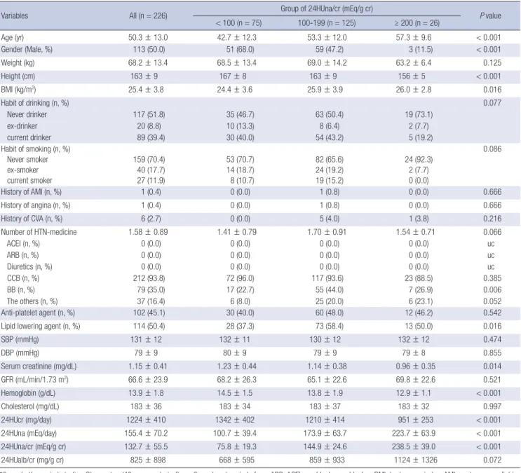
The mean age was 50.3 ± 13.0 yr at enrollment and 50% of the participants were male (Table 1). Seventy-five subjects (33.2%) were assigned to group 1 (24HUna/cr < 100 mEq/g), 125 sub- jects (55.3%) to group 2 (24HUna/cr 100-199 mEq/g), and 26 subjects (11.5%) to group 3 (24HUna/cr ≥ 200 mEq/g). Group 3 had the highest mean age, lowest proportion of men, highest BMI, and lowest levels of hemoglobin and 24-hr urine creati- nine. The 24HUalb/cr ratio in group 3 was higher than in group 1 (post hoc analysis with Fisher’s least significance difference (FLSD) method, P = 0.028). Blood pressures (BPs) and GFR were not different among groups. Other characteristics includ- ing medical history, drinking or smoking habits, medications, and serum cholesterol were not correlated with the amount of 24HUna/cr.
Relationship between baseline urinary sodium and albumin excretion
After univariate analysis, we found that systolic blood pressure, diastolic blood pressure, height, serum cholesterol, hemoglobin, GFR, uAGT/cr, uMCP1/cr showed the relationship with base- line 24HUalb/cr. Multiple linear regression analysis adjusted for these parameters and age showed that 24HUna/cr levels or group of 24HUna/cr had a positive correlation with 24HUalb/cr levels (Table 2).
Relationship between baseline urine sodium and cytokines
The levels of uAGT/cr and uMDA/cr were different between 24HUna/cr groups. Group 3 had the highest level of uAGT/cr and uMDA/cr and was different than group 1, although the uMCP1/
cr was not different among groups (Fig. 1). The uAGT/cr was 316 ± 284 pg/mg cr in group 1, 577 ± 744 pg/mg cr in group 2, and 840 ± 971 pg/mg cr in group 3 (P = 0.002), and the uMDA/cr was 0.17 ± 0.15 pM/mg cr in group 1, 0.20 ± 0.18 pM/mg cr in group 2, and 0.26 ± 0.19 pM/mg cr in group 3 (P = 0.044). When we divided participants according to the tertile value of each urine cytokine, group 3 was assigned to the highest tertile group of uAGT/cr or uMDA/cr, more frequently than other groups (Fig. 2).
With regard to the factors related to the level of each urine cy- tokine, independent factors for each cytokine were determined by an adjusted multiple linear regression analysis. 24HUna/cr levels had a positive correlation with uAGT/cr and uMDA/cr (Table 3). When we estimate uAGT/cr in the 24HUna/cr group by ANCOVA adjusted for 24HUalb/cr—which was another in- dependent factor for uAGT/cr—the uAGT/cr was 334 (95% CI, 184-483) pg/mg cr in group 1, 573 (95% CI, 457-689) pg/mg cr in group 2, and 708 (95% CI, 448-967) pg/mg cr in group 3 (P = 0.014) (data not shown). Furthermore, differences between group 1 and group 2 (P = 0.013) or group 3 (P = 0.015) were identified.
Similarly, uMDA/cr was estimated as 0.17 (95% CI, 0.14-0.21) pM/mg cr in group 1, 0.20 (95% CI, 0.17-0.23) pM/mg cr in group 2, and 0.27 (95% CI, 0.20-0.33) pM/mg cr in group 3 adjusted for beta-blocker medication (P = 0.054). There was a difference be- tween group 1 and 2 or 3 (P = 0.016) based on the LSD method (data not shown).
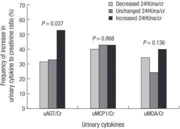
Change of 24HUna/cr and increase of urine cytokines The factors related to an increase in urine cytokine were deter- mined for each cytokine. The clinical parameters at baseline ex- amination were not related to an increase in all urinary cyto- kines. The patients with an increase in 24HUna/cr levels showed a higher frequency of increase in uAGT/cr levels during 16 weeks (P = 0.037, Fig. 3). The patients with increased 24HUna/
cr showed 2.441-fold (95% CI, 1.182-5.042) higher risk for in- crease in uAGT/cr during 16 weeks by multiple logistic regres- sion adjusted for age and gender (P = 0.040). The group of change
in 24HUna/cr was not related to an increase in uMCP1/cr or uMDA/cr.
DISCUSSION
In this prospective, open-label, randomized trial, we confirmed that the 24-hr urine sodium to creatinine ratio had a positive correlation to the amount of albuminuria. A high urinary excre- tion of sodium was related to high excretion of urinary angio- Table 1. Baseline characteristics of participants at 0-week*
Variables All (n = 226) Group of 24HUna/cr (mEq/g cr)
P value
< 100 (n = 75) 100-199 (n = 125) ≥ 200 (n = 26)
Age (yr) 50.3 ± 13.0 42.7 ± 12.3 53.3 ± 12.0 57.3 ± 9.6 < 0.001
Gender (Male, %) 113 (50.0) 51 (68.0) 59 (47.2) 3 (11.5) < 0.001
Weight (kg) 68.2 ± 13.4 68.5 ± 13.4 69.0 ± 14.2 63.2 ± 6.4 0.125
Height (cm) 163 ± 9 167 ± 8 163 ± 9 156 ± 5 < 0.001
BMI (kg/m2) 25.4 ± 3.8 24.4 ± 3.6 25.9 ± 3.9 26.0 ± 2.8 0.016
Habit of drinking (n, %) Never drinker ex-drinker current drinker
117 (51.8) 20 (8.8) 89 (39.4)
35 (46.7) 10 (13.3) 30 (40.0)
63 (50.4) 8 (6.4) 54 (43.2)
19 (73.1) 2 (7.7) 5 (19.2)
0.077
Habit of smoking (n, %) Never smoker ex-smoker current smoker
159 (70.4) 40 (17.7) 27 (11.9)
53 (70.7) 14 (18.7) 8 (10.7)
82 (65.6) 24 (19.2) 19 (15.2)
24 (92.3) 2 (7.7) 0 (0.0)
0.086
History of AMI (n, %) 1 (0.4) 0 (0.0) 1 (0.8) 0 (0.0) 0.666
History of angina (n, %) 1 (0.4) 0 (0.0) 1 (0.8) 0 (0.0) 0.666
History of CVA (n, %) 6 (2.7) 0 (0.0) 5 (4.0) 1 (3.8) 0.216
Number of HTN-medicine ACEI (n, %)
ARB (n, %) Diuretics (n, %) CCB (n, %) BB (n, %) The others (n, %)
1.58 ± 0.89 0 (0.0) 0 (0.0) 0 (0.0) 212 (93.8)
79 (35.0) 37 (16.4)
1.41 ± 0.79 0 (0.0) 0 (0.0) 0 (0.0) 72 (96.0) 17 (22.7) 6 (8.0)
1.70 ± 0.91 0 (0.0) 0 (0.0) 0 (0.0) 117 (93.6)
55 (44.0) 25 (20.0)
1.54 ± 0.71 0 (0.0) 0 (0.0) 0 (0.0) 23 (88.5)
7 (26.9) 6 (23.1)
0.066 uc uc uc 0.385 0.006 0.052
Anti-platelet agent (n, %) 102 (45.1) 30 (40.0) 60 (48.0) 12 (46.2) 0.542
Lipid lowering agent (n, %) 114 (50.4) 28 (37.3) 73 (58.4) 13 (50.0) 0.016
SBP (mmHg) 131 ± 12 132 ± 11 130 ± 12 132 ± 12 0.474
DBP (mmHg) 79 ± 9 80 ± 9 79 ± 9 79 ± 8 0.855
Serum creatinine (mg/dL) 1.15 ± 0.41 1.23 ± 0.44 1.14 ± 0.38 0.96 ± 0.35 0.014
GFR (mL/min/1.73 m2) 66.6 ± 23.9 68.2 ± 26.3 65.1 ± 22.6 69.8 ± 22.6 0.521
Hemoglobin (g/dL) 13.9 ± 1.8 14.5 ± 1.5 13.8 ± 1.9 12.9 ± 1.1 < 0.001
Cholesterol (mg/dL) 183 ± 36 183 ± 34 183 ± 37 183 ± 32 0.997
24HUcr (mg/day) 1224 ± 410 1342 ± 402 1210 ± 414 951 ± 253 < 0.001
24HUna (mEq/day) 155.4 ± 70.2 100.7 ± 39.4 173.9 ± 63.7 223.7 ± 63.9 < 0.001
24HUna/cr (mEq/g cr) 132.7 ± 55.5 75.8 ± 19.3 144.9 ± 24.6 238.5 ± 39.0 < 0.001
24HUalb/cr (mg/g cr) 825 ± 898 668 ± 595 859 ± 933 1124 ± 1326 0.072
*0-week, the period starting Olmesartan (40 mg per day) after a 8-wash out period of any ARB, ACEI, or aldosterone blocker. BMI, body mass index; AMI, acute myocardial in- farction; CVA, cerebrovascular accident including thrombotic cerebral infarction and intracranial hemorrhage; Number of HTN medications, number of medications to control hypertension; ACEI, angiotensin II converting enzyme inhibitor; ARB, angiotensin type I receptor blocker; CCB, calcium channel blocker; BB, beta-blocker; Anti-platelet agent, low dose aspirin, cilostazole, and clopidogrel; Lipid lowering agent, statins and fibrates; SBP, systolic blood pressure, DBP, diastolic blood pressure; GFR, glomerular filtration rate estimated using the original Modification of Diet in Renal Diease equation using serum creatinine values measured by the modified Jaffe reaction; 24HUcr, amount of daily urinary creatinine; 24HUna, amount of daily urinary sodium; 24HUna/cr, the ratio of 24-hr urine sodium to creatinine (mg/g creatinine); 24HUalb, amount of daily urinary albu- min; 24HUalb/cr, the ratio of 24-hr urine albumin to creatinine (mg/g creatinine); uc, unable to calculate. P values were calculated using the One-way ANOVA test among 24-hr urinary sodium/cr groups.
Table 2. The factors related to the 24-hr urine albumin to creatinine ratio at 0-week Parameters Coefficient B 95% CI of B t value P value 24HUna/cr* in
100 mEq/g cr 215.599 13.463 417.735 2.102 0.037 Group of 24HUna/cr 199.229 24.684 373.774 2.250 0.025
*24HUna/cr, 24-hr urine sodium to creatinine ratio. Each parameter was used in the multiple linear regression model adjusted with age and gender, systolic blood pres- sure, diastolic blood pressure, height, serum cholesterol, hemoglobin, GFR, urine MCP-1 to creatinine ratio, and urine angiotensinogen to creatinine ratio, which were the factors related to the 24-hr albumin to creatinine ratio.
tensinogen or MDA, and an increase of urinary sodium excre- tion predicted an increase in urinary angiotensinogen excretion.
This study reported the difference in urinary cytokines ac- cording to the level of urinary sodium excretion, to confirm the pathophysiology of high salt diet related to renal injuries previ- ously reported in animal experiments. Urinary excretion of an- giotensinogen was reported to reflect the status of the intrarenal
renin-angiotensin system in a previous animal experiment (24) and in patients with hypertension or glomerulonephritis (25, 26). High sodium intake increased renal angiotensin II levels and systolic blood pressure (SBP) in normal rats, salt sensitive hypertensive rats (27), and obese Zucker rats (28). Furthermore, it increased angiotensin converting enzyme (ACE) activity in obese rats (28), and activated angiotensin II type I receptor in Fig. 2. The frequency of tertile groups of cytokines according to 24HUna/cr groups.
The ratio of 24-hr urine sodium to creatinine (mg/g creatinine) by (A) uAGT/cr, urine angiotensinogen to creatinine ratio (pg/mg cr), (B) uMCP1/cr, urine monocyte che- moattractant protein-1 to creatinine ratio (pg/mg cr), (C) uMDA/cr: urine malondialde- hyde to creatinine ratio (pM/mg cr).
A
<100
100-199 ≥200
50 40 30 20 10 0
24HUna/cr (mEq/g cr) P = 0.019
3rd tertile 2nd tertile 1st tertile uAGT/cr
C
<100
100-199 ≥200
60
40
20
0
24HUna/cr (mEq/g cr) P = 0.031
3rd tertile 2nd tertile 1st tertile uMDA/cr
B
<100
100-199 ≥200
50 40 30 20 10 0
24HUna/cr (mEq/g cr) P = 0.180
3rd tertile 2nd tertile 1st tertile uMCP1/cr
Fig. 1. Urinary cytokines to creatinine ratio among 24HUna/cr groups. (A) AGT, angio- tensinogen. (B) MCP1, monocyte chemoattractant protein-1. (C) MDA, malondialde- hyde. 24HUna/cr, the ratio of 24-hr urine sodium to creatinine (mg/g creatinine). The P value was calculated by one-way ANOVA test. *Different from the group with 24HUna/cr<100 mEq/g cr. The bar means 95% confidence interval of each mean value.
The ratio of daily urinary AGT to creatinine (pg/mg cr)
< 100 100-199 ≥ 200
1,200 1,000 800 600 400 200 0
*
24HUna/cr (mEq/g cr) P = 0.002
A
The ratio of daily urinary MDA to creatinine (pM/mg cr)
< 100 100-199 ≥ 200
0.40
0.30
0.20
0.10
0.00
*
24HUna/cr (mEq/g cr) P = 0.044
C The ratio of daily urinary MCPI to creatinine (pg/mg cr)
< 100 100-199 ≥ 200
500
400
300
200
100
0
24HUna/cr (mEq/g cr) P = 0.312
B (%)
(%)
(%)
the renal tissue of spontaneously hypertensive rats associated with a rise in proteinuria and renal injury (16, 29), compared to animals on low salt diets. Increased RAS activity is associated with an increased risk of chronic kidney disease in rodent mod- els (30). However, Krikken reviewed that there were few reports about the direct effect of salt intake on renal RAS activation in patients and the available human data only suggests that a high sodium intake increases vascular ACE activity (2). In this study, we showed the direct relationship between the amount of salt intake estimated by the 24-hr urine sodium levels and renal RAS activity with a marker of urinary angiotensinogen in hyper- tensive CKD patients without RASI or diuretic medication. An increase in urinary sodium excretion could predict an increase in urinary angiotensinogen excretion and a decreased anti-pro- teinuric effect of ARB during 16 weeks. Recently, a randomized controlled trial and post hoc analysis of large trials studying the RAS effect on renal outcomes showed similar findings with re-
gard to the relationship between salt intake and proteinuria (3, 4, 6). A post hoc analysis including non-diabetic CKD patients enrolled in the REIN study (3) showed that baseline urinary so- dium/creatinine excretion was positively correlated with the urinary protein/creatinine ratio at baseline and follow-up, in- dependent from blood pressure. The antiproteinuric effect of RASI was significantly higher in low salt diet (LSD) patients compared with medium-salt diet (MSD) or high salt diet (HSD), after 3 months of treatment (3). Three months after the RASI treatment, the urinary protein/creatinine ratio was reduced by 31%, 25%, and 20% in LSD, MSD, and HSD patients, respective- ly (3). The incidence of ESRD was also the highest in HSD pa- tients (32.1%); however, in MSD patients (17.0%), the rate was not different from that of LSD (16.2%). In diabetic CKD patients, the effects of ARB treatment on reducing renal and cardiovas- cular events compared to the non-RASI treatment were lower with higher sodium intake (4). Taken together, proteinuria in- duced by a high salt diet was closely related to an increase of RAS activity in the kidney tissue, which also decreased the anti- proteinuric effect of the RASI treatment in hypertensive CKD patients.
In animal experiments, high salt intake increased the con- centration of renal cortical hydrogen peroxide (17), renal corti- cal NADH and NADPH oxidase activity, expression of gp91phox, p47phox (18), p22phox (16), and decreased renal expression of Mn- superoxide dismutase (SOD) (18) or Cu/Zn-SOD (16). Further- more, it increased urinary 8-isoprostane prostaglandin F2α and MDA, the end products of lipid oxidation (16). In human stud- ies, we could not find any direct evidence of the relationship between salt intake and these ROS production in the literature.
The positive relationship between urinary salt and MDA excre- tion was evident in patients without RASI or diuretics; however, the relationship was weaker than that between urinary salt and angiotensinogen excretion. In fact, a linear regression model including the urine angiotensinogen to creatinine ratio to esti- mate independent factors related to the urine MDA to creati- nine ratio revealed that the 24-hr urine sodium had no effect on continuous or categorical variables. However, the relationship between urinary sodium and angiotensinogen excretion was not affected by urinary MDA levels in the regression model.
Table 3. The group of 24HUna/cr as an independent factor to the level of urine cytokines at 0-week* by multiple linear regression
Factors Coefficient B 95% CI of B t value P value
For uAGT/cr Group of 24HUna/cr Level of 24HUalb/cr
203.395 0.110
65.228 0.013
341.563 0.207
2.901 2.245
0.004 0.026 For uMDA/cr Group of 24HUna/cr
Medication of BB
0.040 0.048
0.005 0.001
0.076 0.095
2.257 2.029
0.025 0.044
*0 week, starting week for Olmesartan (40 mg per day) after a 8-wash out period of any ARB, ACEI, or aldosterone blocker; 24HUna/cr group: group according to the ratio of 24-hr urine sodium to creatinine with the criteria of 100 and 200 mEq/g cr; 24HUalb/cr: the ratio of 24-hr urine albumin to creatinine (mg/g creatinine); uAGT/cr: urine angio- tensinogen to creatinine ratio (pg/mg cr); uMDA/cr: urine malondialdehyde to creatinine ratio (pM/mg cr); BB medication: beta-blocker medication; Linear regression for uAGT/
cr was adjusted for age, gender, hemoglobin, 24-hr urine albumin to creatinine ratio, GFR, and group of 24HUna/cr, which were the factors related to the urine angiotensinogen to creatinine ratio at 0-week. Linear regression for uMDA/cr was adjusted for age, gender, beta-blocker, serum total CO2, and group of 24HUna/cr.
Fig. 3. The relationship between the changes of 24-hr urine cytokines to creatinine ratio and urine sodium to creatinine ratio during 16 weeks after ARB medication. Fre- quency of increase in cytokine level: Frequency of increase in 24-hr urinary cytokine to creatinine ratio at 16-week compared to 0-week 25% or more. Increased 24HUna/
cr defined as increase of 24HUna/cr ratio 25% or more, Decreased 24HUna/cr de- fined as decrease of 24HUna/cr ratio 25% or more, and unchanged 24HUna/cr de- fined as the ratio of 24HUna/cr between -24.9% and 24.9%, at 16-week compared to 0-week. uAGT/Cr: 24-hr urine angiotensinogen to creatinine ratio (pg/mg cr), uMCP1/Cr: 24-hr urine monocyte chemoattractant protein-1 to creatinine ratio (pg/
mg cr), uMDA/Cr: 24-hr urine malondialdehyde to creatinine ratio (pM/mg cr).
Frequency of increase in urinary cytokine to creatinine ratio (%)
uAGT/Cr uMCP1/Cr uMDA/Cr
70 60 50 40 30 20 10 0
Urinary cytokines P = 0.037
P = 0.868
P = 0.136 Decreased 24HUna/cr Unchanged 24HUna/cr Increased 24HUna/cr
This finding suggested that the relationship between urinary ROS and urinary sodium might be confounded by urinary an- giotensinogen. A prolonged angiotensin II infusion in Sprague- Dawley rats increased the expression of gp91phox and p22phox in the renal cortex and increased urinary excretion of 8-isopros- tane prostaglandin F2α and MDA (18, 31). Therefore, high salt intake increases renal RAS activation, primarily, and affects the production of ROS in a direct or indirect manner through renal RAS activation.
This study had some limitations affecting generalization to all hypertensive CKD patients. The study did not intend to con- firm the relationship between salt intake and urinary cytokines;
hence, there might be confounding factors that were not con- sidered, regardless of the multivariate analysis. We used only one parameter for each signal of RAS, inflammation, and ROS, which reduces the accuracy of detecting the signal in the kid- ney. Although we standardized the urinary cytokines and sodi- um to urine creatinine levels to remove an error introduced by inappropriate urine collection, their accuracy could be affected by this error. Finally, urine samples were stored until the end of the study and the cytokines were measured 6 months after the first period of urine collection. During this period, the urine contents could have been altered in each sample, introducing an accuracy error.
In conclusion, our study confirms that high salt intake in hy- pertensive CKD patients is related to a high level of urine albu- min, based on the activation of the intrarenal RAS.
DISCLOSURE
The authors have no conflicts of interest to disclose.
ORCID
Jung Hwan Park http://orcid.org/0000-0002-8737-0084 Suhnggwon Kim http://orcid.org/0000-0001-6904-9126 Ho Jun Chin http://orcid.org/0000-0003-1185-2631
REFERENCES
1. Kim S, Lim CS, Han DC, Kim GS, Chin HJ, Kim SJ, Cho WY, Kim YH, Kim YS. The prevalence of chronic kidney disease (CKD) and the associ- ated factors to CKD in urban Korea: a population-based cross-sectional epidemiologic study. J Korean Med Sci 2009; 24: S11-21.
2. Krikken JA, Laverman GD, Navis G. Benefits of dietary sodium restric- tion in the management of chronic kidney disease. Curr Opin Nephrol Hypertens 2009; 18: 531-8.
3. Vegter S, Perna A, Postma MJ, Navis G, Remuzzi G, Ruggenenti P. Sodi- um intake, ACE inhibition, and progression to ESRD. J Am Soc Nephrol 2012; 23: 165-73.
4. Lambers Heerspink HJ, Holtkamp FA, Parving HH, Navis GJ, Lewis JB, Ritz E, de Graeff PA, de Zeeuw D. Moderation of dietary sodium poten-
tiates the renal and cardiovascular protective effects of angiotensin re- ceptor blockers. Kidney Int 2012; 82: 330-7.
5. Cook NR, Cutler JA, Obarzanek E, Buring JE, Rexrode KM, Kumanyika SK, Appel LJ, Whelton PK. Long term effects of dietary sodium reduction on cardiovascular disease outcomes: observational follow-up of the tri- als of hypertension prevention (TOHP). BMJ 2007; 334: 885-8.
6. McMahon EJ, Bauer JD, Hawley CM, Isbel NM, Stowasser M, Johnson DW, Campbell KL. A randomized trial of dietary sodium restriction in CKD. J Am Soc Nephrol 2013; 24: 2096-103.
7. Drüeke TB, Parfrey PS. Summary of the KDIGO guideline on anemia and comment: reading between the (guide)line(s). Kidney Int 2012; 82:
952-60.
8. The GISEN Group (Gruppo Italiano di Studi Epidemiologici in Nefrolo- gia). Randomised placebo-controlled trial of effect of ramipril on decline in glomerular filtration rate and risk of terminal renal failure in protein- uric, non-diabetic nephropathy. Lancet 1997; 349: 1857-63.
9. Ruggenenti P, Perna A, Loriga G, Ganeva M, Ene-Iordache B, Turturro M, Lesti M, Perticucci E, Chakarski IN, Leonardis D, et al. Blood-pres- sure control for renoprotection in patients with non-diabetic chronic re- nal disease (REIN-2): multicentre, randomised controlled trial. Lancet 2005; 365: 939-46.
10. De Nicola L, Minutolo R, Chiodini P, Zoccali C, Castellino P, Donadio C, Strippoli M, Casino F, Giannattasio M, Petrarulo F, et al. Global ap- proach to cardiovascular risk in chronic kidney disease: reality and op- portunities for intervention. Kidney Int 2006; 69: 538-45.
11. Sanders PW. Salt intake, endothelial cell signaling, and progression of kidney disease. Hypertension 2004; 43: 142-6.
12. Ying WZ, Aaron K, Sanders PW. Mechanism of dietary salt-mediated in- crease in intravascular production of TGF-beta1. Am J Physiol Renal Physiol 2008; 295: F406-14.
13. Ying WZ, Sanders PW. The interrelationship between TGF-beta1 and ni- tric oxide is altered in salt-sensitive hypertension. Am J Physiol Renal Physiol 2003; 285: F902-8.
14. Franco M, Martínez F, Quiroz Y, Galicia O, Bautista R, Johnson RJ, Ro- dríguez-Iturbe B. Renal angiotensin II concentration and interstitial in- filtration of immune cells are correlated with blood pressure levels in salt-sensitive hypertension. Am J Physiol Regul Integr Comp Physiol 2007; 293: R251-6.
15. Tian N, Gu JW, Jordan S, Rose RA, Hughson MD, Manning RD Jr. Im- mune suppression prevents renal damage and dysfunction and reduces arterial pressure in salt-sensitive hypertension. Am J Physiol Heart Circ Physiol 2007; 292: H1018-25.
16. Chandramohan G, Bai Y, Norris K, Rodriguez-Iturbe B, Vaziri ND. Ef- fects of dietary salt on intrarenal angiotensin system, NAD(P)H oxidase, COX-2, MCP-1 and PAI-1 expressions and NF-kappaB activity in salt- sensitive and -resistant rat kidneys. Am J Nephrol 2008; 28: 158-67.
17. Tian N, Moore RS, Braddy S, Rose RA, Gu JW, Hughson MD, Manning RD Jr. Interactions between oxidative stress and inflammation in salt- sensitive hypertension. Am J Physiol Heart Circ Physiol 2007; 293:
H3388-95.
18. Kitiyakara C, Chabrashvili T, Chen Y, Blau J, Karber A, Aslam S, Welch WJ, Wilcox CS. Salt intake, oxidative stress, and renal expression of NADPH oxidase and superoxide dismutase. J Am Soc Nephrol 2003; 14:
2775-82.
19. Nagase M, Matsui H, Shibata S, Gotoda T, Fujita T. Salt-induced ne-
phropathy in obese spontaneously hypertensive rats via paradoxical ac- tivation of the mineralocorticoid receptor: role of oxidative stress. Hyper- tension 2007; 50: 877-83.
20. Sharma K, Ziyadeh FN, Alzahabi B, McGowan TA, Kapoor S, Kurnik BR, Kurnik PB, Weisberg LS. Increased renal production of transforming growth factor-beta1 in patients with type II diabetes. Diabetes 1997; 46:
854-9.
21. Ellis D, Forrest KY, Erbey J, Orchard TJ. Urinary measurement of trans- forming growth factor-beta and type IV collagen as new markers of renal injury: application in diabetic nephropathy. Clin Chem 1998; 44: 950-6.
22. Yu W, Luying S, Haiyan W, Xiaomei L. Importance and benefits of di- etary sodium restriction in the management of chronic kidney disease patients: experience from a single Chinese center. Int Urol Nephrol 2012;
44: 549-56.
23. Zhou X, Zhang L, Ji WJ, Yuan F, Guo ZZ, Pang B, Luo T, Liu X, Zhang WC, Jiang TM, et al. Variation in dietary salt intake induces coordinated dynamics of monocyte subsets and monocyte-platelet aggregates in hu- mans: implications in end organ inflammation. PLoS One 2013; 8:
e60332.
24. Kobori H, Harrison-Bernard LM, Navar LG. Urinary excretion of angio- tensinogen reflects intrarenal angiotensinogen production. Kidney Int 2002; 61: 579-85.
25. Urushihara M, Kondo S, Kagami S, Kobori H. Urinary angiotensinogen accurately reflects intrarenal Renin-Angiotensin system activity. Am J Nephrol 2010; 31: 318-25.
26. Kobori H, Alper AB Jr, Shenava R, Katsurada A, Saito T, Ohashi N, Uru- shihara M, Miyata K, Satou R, Hamm LL, et al. Urinary angiotensino- gen as a novel biomarker of the intrarenal renin-angiotensin system sta- tus in hypertensive patients. Hypertension 2009; 53: 344-50.
27. Franco M, Martínez F, Quiroz Y, Galicia O, Bautista R, Johnson RJ, Ro- dríguez-Iturbe B. Renal angiotensin II concentration and interstitial in- filtration of immune cells are correlated with blood pressure levels in salt-sensitive hypertension. Am J Physiol Regul Integr Comp Physiol 2007; 293: R251-6.
28. Samuel P, Ali Q, Sabuhi R, Wu Y, Hussain T. High Na intake increases re- nal angiotensin II levels and reduces expression of the ACE2-AT(2)R- MasR axis in obese Zucker rats. Am J Physiol Renal Physiol 2012; 303:
F412-9.
29. Susic D, Frohlich ED, Kobori H, Shao W, Seth D, Navar LG. Salt-induced renal injury in SHRs is mediated by AT1 receptor activation. J Hypertens 2011; 29: 716-23.
30. Mills KT, Kobori H, Hamm LL, Alper AB, Khan IE, Rahman M, Navar LG, Liu Y, Browne GM, Batuman V, et al. Increased urinary excretion of angiotensinogen is associated with risk of chronic kidney disease.
Nephrol Dial Transplant 2012; 27: 3176-81.
31. Chabrashvili T, Kitiyakara C, Blau J, Karber A, Aslam S, Welch WJ, Wil- cox CS. Effects of ANG II type 1 and 2 receptors on oxidative stress, renal NADPH oxidase, and SOD expression. Am J Physiol Regul Integr Comp Physiol 2003;285: R117-24.