Risk Factors for Malignancy of Pheochromocytoma and Abdominal
Paraganglioma in Children:
Clinicopathologic Perspectives
Jihoon Chang, M.D., Soo-Hong Kim, M.D., Hye Sook Min, M.D.*, Hyun-Young Kim, M.D., Sung-Eun Jung, M.D., Kwi-Won Park, M.D., Seong-Cheol Lee, M.D.
Department of Pediatric Surgery, Seoul National University Children’s Hospital, Seoul, Korea
*Department of Pathology, Seoul National University Hospital, Seoul, Korea
Submission : 13 / 9 / 27 Acceptance : 13 / 11 / 13 Correspondence:Seong-Cheol Lee,, M.D.
Department of Pediatric Surgery, Seoul National University Children's Hospital 101, Daehang-Ro Jongro-Gu, Seoul 110-744, Korea
Tel : 02)2072-2338, Fax : 02)747-7730 E-mail: [email protected]
http://dx.doi.org/10.13029/jkaps.2013.19.2.108
INTRODUCTION
Pheochromocytoma (PHEO) and para- ganglioma (PGL) are rare neuroendocrine tumors in children. In 2004, World Health Organization (WHO) defined PHEO as a tumor arising from catecholamine- producing chromaffin cells in the adrenal medulla (an intra-adrenal PGL)1. Tumors arising from extra-adrenal sympathetic and parasympathetic paraganglia are defined as extra-adrenal PGLs, commonly found in the head, neck, and abdomen1. By WHO definition, PHEO and PGL are closely related, and are classified in terms
of location. In children, incidence of PHEO and PGL is estimated at 0.3 cases per million a year or less2, and approximately 10-20 % of PHEO and PGL are diagnosed during childhood at an average age of 11 years3.
Malignant PHEO and PGL are defined as the presence of distant metastasis1. Although the rate of malignancy is known as 10 %4, malignancy rate varies within the literature and may be as high as 26
%5. Because of unfavorable prognosis and lack of definite curative treatment for malignant PHEO and PGL, prediction of malignancy and early detection of metastasis are important for treatment and follow-up. But currently malignant PHEO and PGL lack histological or molecular markers that reliably distinguish them from benign tumors.
Thompson LD created the Pheochromo-
cytoma of the Adrenal gland Scaled Score (PASS), including 12 histologic features such as tumor necrosis, high mitosis rate, and vascular invasion as a scoring system to distinguish benign from malignant PHEO6. Strong et al proposed PASS as a prognostic indicator of malignant PHEO7. However, the patients enrolled in that study were mainly adults, and its applicability to pediatric patients has not been established.
Some immunohistochemical markers such as Ki-67, p53, bcl-2, mdm2, cyclin D1, p21, and p27 have been reported as potential tools to differentiate malignant from benign PHEO8-10. Since Ki-67 is found throughout the cell cycle except in resting cells (G0 phase)11, it can be used to evaluate the growth rate of tumors, and several studies have been demonstrated correlation between high Ki-67 positivity rate and malignancy9,12,13.
Because the features of diseases including clinicopathologic profiles, treatment, and clinical outcomes are poorly characterized in the pediatric literature, we designed this study to review clinical features of malignant PEHO and abdominal PGL (aPGL), to validate PASS as a prognostic indicator of malignant PHEO and aPGL in children, and to document risk factors of malignancy in patients with PHEO or aPGL.
PATIENTS AND METHODS
We reviewed the database from medical records of 20 patients (≤18 years of age) with pathologically confirmed PHEO or aPGL after surgical resection by 3 surgeons (14, 3, and 3 operations, respectively) at Seoul National University Children's Hospital between January 1990 and December 2010. With this prospectively collected database, demo- graphics, symptoms, signs, results of laboratory test, treatment information, pathologic results, and outcomes were obtained. Follow-up monitoring was accomplished through recent clinic visits and telephone correspondence. Patients were monitored until they died or we lost contact with them. Malignancy of PHEO and aPGL was defined as the presence of distant metastasis such as bone, liver, and lung1. The tumor which was recurred distantly after complete resection was also regarded as malignant. Only PHEO and PGL in the abdominal cavity were included, and PGL in the head and neck was excluded in this study.
To confirm diagnosis and to assess PASS score, all pathologic slides were reviewed by a single endocrinologic pathologist blinded to the clinical data.
Overall PASS score was calculated according to their microscopic features.
After obtaining five-micron sections from
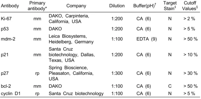
Table 1. Immunohistochemical Panel Antibody Primary
antibody* Company Dilution Buffer(pH)† Target Stain‡
Cutoff Values§ Ki-67 mm DAKO, Carpinteria,
California, USA 1:200 CA (6) N > 2 %
p53 mm DAKO 1:200 CA (6) N > 5 %
mdm-2 mm Leica Biosystems,
Heidelberg, Germany 1:100 EDTA (9) N > 50 %
p21 mm
Santa Cruz
biotechnology, Dallas, Texas, USA
1:200 CA (6) N > 10 %
p27 rp
Spring Bioscience, Pleasaton, California, USA
1:300 CA (6) N > 30 %
bcl-2 mm DAKO 1:100 CA (6) C > 50 %
cyclin D1 rp Santa Cruz biotechnology 1:100 CA (6) N > 5 %
*mm, mouse monoclonal; rp, rabbit polyclonal
†CA, citric acid; EDTA, Ethylenediaminetetraacetic acid
‡N, Nuclei stain; C, Cytoplasmic stain
§indicates high proliferative group in Ki-67; otherwise indicates group showed over- expression
the paraffin-embeded tissue block, we stained these sections with hematolxylin and eosin to identify representative areas of PHEO and aPGL in such tissue block.
After identification of representative area, core biopsies (2 mm in diameter) were taken from tissue block and arranged in a new recipient paraffin block using a tissue microarray device (Trephine Apparatus, Superbiochips Laboratories, Seoul, Korea).
The antibodies used for immuno- histochemistry included mouse human monoclonal antibodies to mdm-2, p21, p53, Ki-67, and bcl-2, and rabbit polyclonal antibodies to Cyclin D1 and p27. For all antibodies, heat-induced epitope retrieval was performed using selected buffer solution (Table 1). The immuno-
histochemical staining was processed with Leica BOND-MAXTM autostainer14.
An endocrinologic pathologist,who reviewed pathologic slides, also reviewed and scored each immunostain. The percentage of tumor cells showing characteristic staining was measured in a semiquantitative manner. Cutoff values for immunohistochemical stain were estab- lished (Table 1)15.
Descriptive statistics were used to provide a summary of the data. Fisher’s exact test, Student’s t-test, Mann-Whitney test were used for comparative statistical analysis. The Kaplan-Meier method to estimate overall survival and the log-rank test to compare overall survival were used between benign and malignant PHEO and
Table 2. Clinical Information of the Patients with PHEO and aPGL
Total (n=20) PHEO (n=14) aPGL (n=6) P value Age (months, mean±SD) 143.90 ± 40.87 141.93 ± 39.26 148.50 ± 19.60 .775*
Gender (M : F) 15 : 5 9 : 5 6 : 0 .260†
Location .829†
Right (%) 7 (35) 5 (35.7) 2 (33.3%)
Left (%) 6 (30) 5 (35.7) 1 (16.7%)
Bilateral (%) 7 (35) 4 (28.6) 3 (50.0%)
Greatest dimension (cm, mean±SD) 4.86 ± 1.50 4.84 ± 1.26 4.88 ± 2.08 .966†
Malignancy (%) 7 (35) 4 (28.6) 3 (50.0%) .613†
Hereditary Features (%) 4 (20) 4 (28.6) 0 .267†
Chemotherapy (%) 2 (10) 0 2 (33.3%) .079†
Radiotherapy (%) 6 (30) 3 (21.4) 3 (50.0%) .303†
Follow-up(months, mean±SD) 97.25 ± 55.67 95.64 ± 37.03 101.00 ± 90.47 .850*
Disease free survival
(months, mean±SD) 82.20 ± 58.25 82.43 ± 43.86 81.67 ± 88.82 .985*
Median survival (months, range) 86.20 (14-252) 86.50 (50-159) 82.50 (14-252) NA§
*Student’s t-test
†Fisher’s exact test
§Not applicable
aPGL. Confidence intervals of 95 % were generated for all positive findings. A P value < .05 was considered statistically significant. All analyses were conducted using the Statistical Package for the Social Sciences software (version 21.0;
Chicago, IL, USA).
RESULTS
Clinical Features of PHEO and aPGL
Among 20 patients, 14 patients had PHEO and 6 patients had aPGL.
Malignancy was found in 4 (28.6 %) and 3 (50 %) patients, respectively (p= .613).
There was no difference in age, gender distribution, location of primary tumor, size of tumor, hereditary features, adjuvant treatment modalities and follow-up period between PHEO and aPGL. In the aspect of hereditary disease, single case of neurofibromatosis and 3 cases of von Hippel-Lindau disease were found. For adjuvant radiotherapy, one patient with PHEO and 3 patients with aPGL got adjuvant external beam irradiation and two patients with PHEO got 131I- Metaiodobenzylguanidine treatment. During follow-up, 3 patients of PHEO and 2 patients of aPGL were dead (Table 2).
Analysis between benign and malignant tumors showed no difference in age,
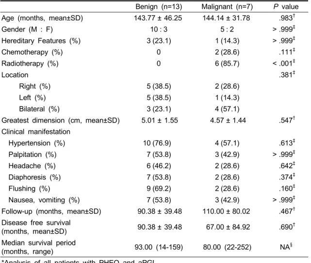
Table 3. Clinical Information of the Patients with Benign and Malignant Tumors*
Benign (n=13) Malignant (n=7) P value Age (months, mean±SD) 143.77 ± 46.25 144.14 ± 31.78 .983†
Gender (M : F) 10 : 3 5 : 2 > .999‡
Hereditary Features (%) 3 (23.1) 1 (14.3) > .999‡
Chemotherapy (%) 0 2 (28.6) .111‡
Radiotherapy (%) 0 6 (85.7) < .001‡
Location .381‡
Right (%) 5 (38.5) 2 (28.6)
Left (%) 5 (38.5) 1 (14.3)
Bilateral (%) 3 (23.1) 4 (57.1)
Greatest dimension (cm, mean±SD) 5.01 ± 1.55 4.57 ± 1.44 .547† Clinical manifestation
Hypertension (%) 10 (76.9) 4 (57.1) .613‡
Palpitation (%) 7 (53.8) 3 (42.9) > .999‡
Headache (%) 6 (46.2) 2 (28.6) .642‡
Diaphoresis (%) 7 (53.8) 2 (28.6) .374‡
Flushing (%) 9 (69.2) 2 (28.6) .160‡
Nausea, vomiting (%) 7 (53.8) 3 (42.9) > .999‡
Follow-up (months, mean±SD) 90.38 ± 39.48 110.00 ± 80.02 .467† Disease free survival
(months, mean±SD) 90.38 ± 39.48 67.00 ± 84.92 .690†
Median survival period
(months, range) 93.00 (14-159) 80.00 (22-252) NA§
*Analysis of all patients with PHEO and aPGL
†Student’s t-test
‡Fisher’s exact test
§Not applicable
gender distribution, location of primary tumor, size of tumor, hereditary features, and follow-up period except adjuvant radiotherapy (p< .001). Although clinical manifestations such as hypertension, palpitation, and headache, were relatively frequent in both benign and malignant tumors, there was no statistical difference between them. Whereas all patients with benign PHEO and aPGL survived, 5
patients with malignancy were dead (Table 3). Figure indicates Kaplan-Meier survival curves and benign PHEO and aPGL demonstrated significantly better prognosis.
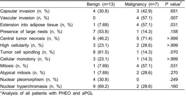
Microscopic and Immunohisto- chemical Features
In the analysis between benign and malignant PHEO and aPGL, vascular
Table 4. Microscopic Features of Benign and Malignant Tumors*
Benign (n=13) Malignancy (n=7) P value†
Capsular invasion (n, %) 4 (30.8) 3 (42.9) .651
Vascular invasion (n, %) 0 4 (57.1) .007
Extension into adipose tissue (n, %) 1 (7.69) 4 (57.1) .031 Presence of large nests (n, %) 7 (53.8) 1 (14.3) .158 Central tumor necrosis (n, %) 6 (46.2) 5 (71.4) >.999
High cellularity (n, %) 3 (23.1) 2 (28.6) >.999
Tumor cell spindling (n, %) 8 (61.5) 1 (14.3) .070
Cellular monotony (n, %) 3 (23.1) 1 (14.3) >.999
Mitosis (n, %) 1 (7.69) 4 (57.1) .031
Atypical mitosis (n, %) 1 (7.69) 2 (28.6) .270
Nuclear pleomorphism (n, %) 4 (30.8) 0 .249
Nuclear hyperchromasia (n, %) 9 (69.2) 2 (28.6) .160
*Analysis of all patients with PHEO and aPGL
†Fisher’s exact test
Table 5. Microscopic Features of Benign and Malignant PHEO*
Benign (n=10) Malignancy (n=4) P value†
Capsular invasion (n, %) 3 (30) 2 (50) .580
Vascular invasion (n, %) 0 3 (75) .033
Extension into adipose tissue (n, %) 0 3 (75) .003
Presence of large nests (n, %) 7 (70) 0 .023
Central tumor necrosis (n, %) 5 (50) 4 (100) .089
High cellularity (n, %) 3 (30) 1 (25) .857
Tumor cell spindling (n, %) 7 (70) 1 (25) .139
Cellular monotony (n, %) 3 (30) 0 .234
Mitosis (n, %) 1 (10) 3 (75) .019
Atypical mitosis (n, %) 0 1 (25) .114
Nuclear pleomorphism (n, %) 3 (30) 0 .234
Nuclear hyperchromasia (n, %) 8 (80) 0 .008
*Analysis of patients with PHEO only
†Fisher’s exact test
invasion, extension into peritumoral adipose tissue, and increased mitosis, were observed more frequently in malignant PHEO and aPGL (p= .007, .031, and .031, respectively). Difference in other pathologic profiles showed no statistical significance
(Table 4). In the subgroup analysis of patients with PHEO, these pathologic profiles were also significantly frequent in malignancy (p= .033, .003, and .019, re- spectively). Presence of large nests and nuclear hyperchromasia were more
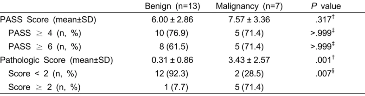
Table 6. Analysis of PASS Score and Pathologic Score between Benign and Malignant Tumor*
Benign (n=13) Malignancy (n=7) P value
PASS Score (mean±SD) 6.00 ± 2.86 7.57 ± 3.36 .317†
PASS ≥ 4 (n, %) 10 (76.9) 5 (71.4) >.999‡
PASS ≥ 6 (n, %) 8 (61.5) 5 (71.4) >.999‡
Pathologic Score (mean±SD) 0.31 ± 0.86 3.43 ± 2.57 .001†
Score < 2 (n, %) 12 (92.3) 2 (28.5) .007§
Score ≥ 2 (n, %) 1 (7.7) 5 (71.4)
*Analysis of all patients with PHEO and aPGL
†Student’s t-test
‡Mann-Whitney test
§Fisher’s exact test
Table 7. Immunohistochemistry*
Antibody Benign (n=13) Malignancy (n=7) P value†
Ki-67 (n, %) 2 (15.4) 4 (57.1) .122
p53 (n, %) 2 (15.4) 3 (42.9) .290
bcl-2 (n, %) 8 (61.5) 4 (57.1) >.999
mdm-2 (n, %) 2 (15.4) 3 (42.9) .290
cyclin D1 (n, %) 0 0 >.999
p21 (n, %) 3 (23.1) 3 (42.9) .613
p27 (n, %) 2 (15.4) 3 (42.9) .290
*Number of cases above cutoff values which are listed in Table 1.
†Fisher’s exact test
frequent in benign PHEO (p= .023 and p= .008, respectively) (Table 5). In the aPGL subgroup, there was no statistical difference in pathologic profiles between benign and malignant tumors.
Although mean PASS score of PHEO and aPGL was slightly higher in patients with malignancy, there was no significant difference in mean PASS score of PHEO and aPGL between benign and malignant patients (p= .317). Five of seven patients with malignant tumors and 10 of 13 patients with benign tumors showed PASS ≥ 4 (Table 6).
To predict malignancy of PHEO and aPGL in pediatric patients, a new pathologic score system was created using these significant pathologic profiles, vascular invasion (2 points), extension into adipose tissue (3 points), and increase in mitotic count (1 point). With this system, score of patients with malignancy was significantly higher (p= .001) (Table 6).
There is significant difference between benign and malignant tumor with cutoff value of 2, and its sensitivity is 71.4 % and specificity is 92.3 %.
In respect to immunohistochemical
Figure. The Kaplan-Meier survival curves between benign and malignant PHEO and aPGL (p = .007)
Figure. The Kaplan-Meier survival curves between benign and malignant PHEO and aPGL (p= .007).
markers, Ki-67, p53, bcl-2, mdm-2, cyclin D1, p21, and p27 did not show significant difference and failed to predict malignancy (Table 7). There was no over-expression of cyclin D1 in both benign and malignant groups. In the subgroup analysis of PHEO and aPGL, immunohistochemistry showed no difference between benign and malignant patients.
DISCUSSION
PHEO and PGL are rare neuroendocrine tumors in children and the majority is benign. Because the prognosis of malignant PHEO and PGL is poor, many studies have tried to demonstrate risk factors of malignancy. This study was designed to investigate the clinico-
pathologic features of malignant PHEO and aPGL, to validate PASS as a prognostic indicator of malignancy in pediatric patients, and to document risk factors of malignancy.
Clinical features showed no difference between PHEO and aPGL, and between benign and malignant tumors. Only postoperative adjuvant radiotherapy was significantly frequent in malignancy.
Malignancy rate in this study population was 35 %, and was higher than that for other pediatric population1 and for adults5,16. 28.6 % of PHEO and 50 % of aPGL were malignant, and these results corresponded well with those of the study reporting that sympathetic PGL showed greater risk of malignant transformation
4,17. Although hypertension, palpitation, and headache are the three most common symptoms and signs in this study, similar to the presentation in adults18,19, clinical manifestations demonstrate no difference between benign and malignant PHEO and aPGL. Primary tumor size and location showed no role in predicting malignancy, in contrast to the results of study reporting primary tumor size and location as prognostic indicators20.
Some pathologic profiles such as cellular atypia, capsular or vascular invasion, or areas resembling pediatric neuroblastoma have been proposed as prognostic factors in some literature1,21. In this study,
pathologic profiles, vascular invasion, extension into peritumoral adipose tissue, and increased mitotic count, indicate significant correlation with malignancy in PHEO and aPGL. Thompson LD6 and Strong et al7 have demonstrated PASS as a prognostic indicator of malignant PHEO.
Without prospective validation, PASS has been used to report pathologic results and to predict clinical prognosis. PASS was not validated in the study with pediatric population. PASS score more than four has suggested risk of malignant transformation6,7. However, this study revealed no difference in mean PASS score between benign and malignant PHEO and aPGL. Five of seven patients with malignant tumors and 10 of 13 patients with benign tumors showed PASS ≥ 4. Five of seven patients with malignant and 8 of 13 patients with benign tumors showed PASS ≥ 6.
With cutoff value of 4 and 6, PASS cannot predict malignancy in this pediatric population. Thompson LD6 and Strong et al7 demonstrated significance of PASS in mostly adult population, whereas our results did not show any difference in pediatric population. Interestingly, in the subgroup analysis of patients with PHEO, the pathologic profiles such as presence of large nests and nuclear hyperchromasia were more frequent in benign tumor.
These findings suggest that some of
pathologic profiles included in PASS system cannot be used to predict malignancy in certain pediatric population.
Moreover, Wu et al have found significant interobserver and intraobserver variation in assignment of PASS with variable interpretation of the underlying components22. Therefore, PASS system needs to be refined, prospectively validated, and strengthened by new markers of immunohistochemistry and molecular biology in pediatric population.
In this study, we created the new pathologic scoring system to predict malignancy of PHEO or aPGL. In the new pathologic score system using extension into peritumoral adipose tissue (3 points), vascular invasion (2 points), and increased mitosis (1 point), mean score is significantly higher in malignancy. This scoring system was made taking into account the frequency of appearance of pathologic profiles in malignancy and benign. Designing many models for this scoring system, this point system showed the best results. With a cutoff value of 2, its sensitivity and specificity to predict malignancy showed best results, which were 71.4 % and 92.3 %, respectively. Even though malignancy cannot be predicted clearly with this score system, the higher score can reflect malignancy.
With immunohistochemistry, some markers have been introduced to predict
prognosis of PHEO and PGL. Ki-67 has been considered as a proliferative marker and higher Ki-67 expression has been observed in malignant PHEO9,12,13. But high expression of Ki-67 is rarely observed in PHEO and sensitivity of Ki-67 is as low as 35 %9,12,13. Most malignant PHEOs can show Ki-67 positivity rate less than 1 %6. Such low Ki-67 expression rate indicates that PHEO and aPGL are growing slowly. Therefore, most authors have chosen a low cutoff value such as 2 %. In this study, 15.4 % of benign and 57.1 % of malignant PHEO and aPGL showed Ki-67 high expression with this cutoff value (p= .122). High expression rate of Ki-67 demonstrates that proliferation rate of pediatric malignant PHEO and aPGL is higher than that of malignant PHEO and aPGL in adults.
Bcl-2 was over-expressed in 61.5 % of benign and 57.1 % of malignant PHEO and aPGL (p> .999), and these findings differ from the study reporting a significant correlation between benign and malignant PHEO8. Whereas cyclin D1 has been known to have a role in PHEO tumorigenesis23, no over-expression of cyclin D1 was demonstrated in benign and malignant PHEO and aPGL. p53, mdm-2, p21, and p27 were unable to predict malignant PHEO and aPGL in this study.
The retrospective nature of this study has some limitations including selection
bias inherent in a tertiary referral hospital study. Limited number of cases can also give relatively low statistical power. The results of this study should be interpreted cautiously, and judicious introduction of the rates to general population may be necessary. Nevertheless, this study has multiple strengths. It is designed for pediatric patients, poorly described in the literature. PASS was validated with patients under age of 18 and its applicability of PASS to pediatric patients was established for the first time.
Furthermore, this study contains relatively large case series on pediatric PHEO and aPGL, and most patients have had long-term follow-up24-26.
CONCLUSION
Vascular invasion, extension into peritumoral adipose tissue, and increased mitotic count are significant risk factors for malignancy of PHEO and aPGL in children. The newly designed pathologic score system using these pathologic factors can be used as a potential prognostic factor of malignancy. Ki-67, p53, bcl-2, mdm-2, cyclin D1, p21, and p27 show no role in predicting malignancy.
REFERENCES
1. DeLellis RA: Pathology and genetics of tumours of endocrine organs. World Health Organization classification of tumours Lyon: IARC Press, 2004, Pp147-150
2. Spoudeas HA: Paediatric endocrine tumours. West Sussex UK: Novo Nordisk, 2005, Pp81-92
3. Waguespack SG, Rich T, Grubbs E, Ying AK, Perrier ND, Ayala-Ramirez M, Jimenez C: A current review of the etiology, diagnosis, and treatment of pediatric pheochromocytoma and paraganglioma. Journal of Clinical Endocrinology & Metabolism 95(5):
2023-2037, 2010
4. Eisenhofer G, Bornstein SR, Brouwers FM, Cheung NK, Dahia PL, de Krijger RR, Giordano TJ, Greene LA, Goldstein DS, Lehnert H, Manger WM, Maris JM, Neumann HP, Pacak K, Shulkin BL, Smith DI, Tischler AS, Young WF Jr:
Malignant pheochromocytoma: current status and initiatives for future progress.
Endocr Relat Cancer 11(3):423-36, 2004 5. Gimenez-Roqueplo AP, Favier J, Rustin
P, Rieubland C, Crespin M, Nau V, Khau Van Kien P, Corvol P, Plouin PF, Jeunemaitre X: Mutations in the SDHB gene are associated with extra-adrenal and/or malignant phaeochromocytomas.
Cancer research 63(17):5615-5621, 2003 6. Thompson LD: Pheochromocytoma of the
Adrenal gland Scaled Score (PASS) to separate benign from malignant neoplasms: a clinicopathologic and immunophenotypic study of 100 cases.
Am J Surg Pathol 26(5):551-66, 2002 7. Strong VE, Kennedy T, Al-Ahmadie H,
Tang L, Coleman J, Fong Y, Brennan
M, Ghossein RA: Prognostic indicators of malignancy in adrenal pheochromo- cytomas: clinical, histopathologic, and cell cycle/apoptosis gene expression analysis. Surgery 143(6):759-68, 2008 8. de Krijger RR, van der Harst E, van der
Ham F, Stijnen T, Dinjens WN, Koper JW, Bruining HA, Lamberts SW, Bosman FT: Prognostic value of p53, bcl-2, and c-erbB-2 protein expression in phaeo- chromocytomas. The Journal of pathology 188(1):51-55, 1999
9. August C, August K, Schroeder S, Bahn H, Hinze R, Baba HA, Kersting C, Buerger H: CGH and CD 44/MIB-1 immunohistochemistry are helpful to distinguish metastasized from nonmeta- stasized sporadic pheochromocytomas.
Modern pathology 17(9):1119-1128, 2004 10. Lam KY, Lo CY, Wat NM, Luk JM,
Lam KS: The clinicopathological features and importance of p53, Rb, and mdm2 expression in phaeochromocytomas and paragangliomas. Journal of clinical pathology 54(6):443-448, 2001
11. Gerdes J, Lemke H, Baisch H, Wacker HH, Schwab U, Stein H: Cell cycle analysis of a cell proliferation-associated human nuclear antigen defined by the monoclonal antibody Ki-67. The Journal of Immunology 133(4):1710-1715, 1984 12. Nagura S, Katoh R, Kawaoi A,
Kobayashi M, Obara T, Omata K:
Immunohistochemical estimations of growth activity to predict biological behavior of pheochromocytomas. Modern pathology: an official journal of the United States and Canadian Academy of Pathology, Inc 12(12):1107, 1999
13. Elder EE, Xu D, Höög A, Enberg U, Hou M, Pisa P, Gruber A, Larsson C, Bäckdahl M: KI-67 and hTERT expression can aid in the distinction
between malignant and benign pheochromocytoma and paraganglioma.
Modern pathology 16(3):246-255, 2003 14. Pinato DJ, Ramachandran R, Toussi STK,
Vergine M, Ngo N, Sharma R, Lloyd T, Meeran K, Palazzo F, Martin N, Khoo B, Dina R, Tan TM: Immunohisto- chemical markers of the hypoxic response can identify malignancy in phaeochro- mocytomas and paragangliomas and optimize the detection of tumours with VHL germline mutations. British Journal of Cancer (2013) 108, Pp.429-437
15. Stojadinovic A, Ghossein RA, Hoos A, Nissan A, Marshall D, Dudas M, Cordon-Cardo C, Jaques DP, Brennan MF: Adrenocortical carcinoma: clinical, morphologic, and molecular character- ization. Journal of Clinical Oncology 20(4):941-950, 2002
16. Pham TH, Moir C, Thompson GB, Zarroug AE, Hamner CE, Farley D, van Heerden J, Lteif AN, Young WF Jr:
Pheochromocytoma and paraganglioma in children: a review of medical and surgical management at a tertiary care center. Pediatrics 118(3):1109-1117, 2006 17. Burnichon N, Rohmer V, Amar L,
Herman P, Leboulleux S, Darrouzet V, Niccoli P, Gaillard D, Chabrier G, Chabolle F, Coupier I, Thieblot P, Lecomte P, Bertherat J, Wion-Barbot N, Murat A, Venisse A, Plouin PF, Jeunemaitre X, Gimenez-Roqueplo AP;
PGL.NET network: The succinate dehydrogenase genetic testing in a large prospective series of patients with paragangliomas. J Clin Endocrinol Metab 94(8):2817-2827, 2009
18. McNicol AM: Differential diagnosis of pheochromocytomas and paragangliomas.
Endocrine pathology 12(4):407-415, 2001 19. Manger WM, Eisenhofer G: Pheochromo-
cytoma: diagnosis and management update. Current hypertension reports 6(6):
477-484, 2004
20. Ayala-Ramirez M, Feng L, Johnson MM, Ejaz S, Habra MA, Rich T, Busaidy N, Cote GJ, Perrier N, Phan A, Patel S, Waguespack S, Jimenez C: Clinical risk factors for malignancy and overall survival in patients with pheochromocytomas and sympathetic paragangliomas: primary tumor size and primary tumor location as prognostic indicators. J Clin Endocrinol Metab 96(3):717-725, 2011
21. Tischler AS: Pheochromocytoma and extra-adrenal paraganglioma: updates.
Arch Pathol Lab Med 132(8):1272-1284, 2008
22. Wu D, Tischler AS, Lloyd RV, DeLellis RA, de Krijger R, van Nederveen F, Nosé V: Observer variation in the application of the Pheochromocytoma of the Adrenal Gland Scaled Score. The American journal of surgical pathology 33(4):599-608, 2009
23. Pollard PJ, El-Bahrawy M, Poulsom R, Elia G, Killick P, Kelly G, Hunt T, Jeffery R, Seedhar P, Barwell J, Latif F, Gleeson MJ, Hodgson SV, Stamp GW, Tomlinson IP, Maher ER: Expression of HIF-1α, HIF-2α (EPAS1), and their target genes in paraganglioma and pheochromocytoma with VHL and SDH mutations. Journal of Clinical Endocrinology
& Metabolism 91(11):4593-4598, 2006 24. Ein SH, Pullerits J, Creighton R, Balfe
JW: Pediatric pheochromocytoma. A 36-year review. Pediatr Surg Int 12(8):
595-598, 1997
25. Perel Y, Schlumberger M, Marguerite G, Alos N, Revillon Y, Sommelet D, De Lumley L, Flamant F, Dyon JF, Lutz P, Heloury H, Lemerle J: Pheochromo- cytoma and paraganglioma in children: a
report of 24 cases of the French Society of Pediatric Oncology. Pediatr Hematol Oncol 14(5):413-422, 1997
26. Reddy VS, O'Neill JA Jr, Ho GW 3rd, Neblett WW 3rd, Pietsch JB, Morgan
WM 3rd, Goldstein RE: Twenty-five-year surgical experience with pheochromo- cytoma in children. Am Surg 66(12):
1085-1092, 2000
소아에서 갈색세포종과 복강내 부교감신경절종의 악성화 예측인자
서울대학교어린이병원 소아외과
*서울대학교병원 병리과
장지훈・김수홍・민혜숙*・김현영・정성은・박귀원・이성철
목적
Pheochromocytoma of the Adrenal gland Scaled Score (PASS) 시스템과 면역화학염색 등 을 통한 갈색세포종 및 부교감신경절종의 악성화 예측인자가 제시되고 있으나 명확한 병리학적 또는 분 자생물학적 예측인자는 밝혀진 바 없다. 본 연구에서는 임상적, 병리학적 분석을 통해 갈색세포종 및 복 강내 부교감신경절종의 악성화 예측인자를 확인하고자 하였다.
대상 및 방법
1990년 1월부터 2010년 12월까지 서울대학교어린이병원에서 수술적 절제 후 병리학적으로 갈색세포종
및 복강내 부교감신경절종으로 확진된 20명의 18세 이하 소아 환자를 대상으로 임상적 특징을 분석하였
고, PASS 시스템에 따른 병리 슬라이드 판독하였다. 세포활성도를 반영한다고 알려진 유전자에 대한 항
체 중 Ki-67, p53, bcl-2, mdm-2, cycline D1, p21, p27을 이용해 면역 화학검사를 한 후 결과를 확인하였다.
결과
20명의 환자 중 갈색세포종은 14명, 복강내 부교감신경절종은 6명이었다. 악성화는 각각 4명, 3명에서 관찰되었다. 혈관 침범, 주변부 지방조직 침습, 세포분열 증가가 통계적으로 유의한 악성화 예측인자였으 며(각각 p= .007, .031, .031), 갈색세포종만 분석하였을 때도 통계적으로 유의하였다(각각 p= .033, .003, .019). PASS 시스템은 악성화를 예측하는데 있어 통계적으로 유의하지 않았으며, 혈관 침범, 주 변부 지방조직 침습, 세포분열 증가를 항목으로 하여 새롭게 만든 병리 스코어 시스템은 악성 환자군과 양성 환자군 사이에 통계적으로 유의한 차이를 보였다(p< .001). 악성과 양성 질환 사이의 면역화학염색 결과에서 유의한 차이는 없었다.
결론
소아에서 갈색세포종 및 복강내 부교감신경절종의 악성화 예측인자로 혈관 침범, 주변부 지방조직 침습,
세포분열 증가를 이용할 수 있다. 소아에서 PASS시스템으로 악성화를 예측할 수 없었으나, 새로운 병리
스코어 시스템으로 악성 환자군을 예측할 수 있었다. 면역화학검사 결과 세포 활성도를 반영하는 인자들 은 악성화를 예측할 수 없었다.
(J Kor Assoc Pediatr Surg 19(2):108~121), 2013.
Index Words:Pheochromocytoma, Pagaganglioma, Child, Risk Factors
교신저자:이성철
(110-744) 서울시 종로구 대학로 101 번지, 서울대학교어린이병원 소아외과 Tel : 02)2072-2338, Fax : 02)747-7730
E-mail : [email protected]