Advantage of urological experience with both transperitoneal and retroperitoneal laparoscopy in lymph node biopsy for malignant lymphoma diagnosis
Hiroaki Kawanishi1, Katsuhiro Ito1, Satoshi Kamido1, Yuka Kohno1, Toshihiro Uemura1, Keiji Kato1, Hirotsugu Uetsuki1, Hitoshi Ohno2, Kazuhiro Okumura1
Departments of 1Urology and 2Hematology, Tenri Hospital, Tenri, Japan
Purpose: Laparoscopic urologists are familiar with both transperitoneal and retroperitoneal approaches. That experience is an advantage when devising a strategy for intra-abdominal lymph node biopsy. We report the feasibility and effectiveness of lapa- roscopic biopsy using a urological laparoscopic technique for the treatment of patients with clinically suspected intra-abdominal lymphoma.
Materials and Methods: From October 2010 to April 2015, a total of 22 patients underwent laparoscopic biopsy for suspected intra-abdominal lymphoma. We adopted a retroperitoneal approach for paraaortic or paracaval masses, whereas we used a trans- peritoneal approach for mesenteric, iliac, or obturator masses. Whenever possible, an entire node was removed; otherwise, the biopsy consisted of wedge resection sized at least 1 cm3.
Results: Biopsy specimens were obtained from the following lymph node sites: 10 paraaortic, 5 paracaval, 3 mesenteric, 2 obtu- rator, 1 common iliac, and 1 perinephric fat. Laparoscopic lymph node biopsy was completed in all patients, and there were no conversions to open surgery. The median operating time was 97 minutes (range, 62–167 minutes). The estimated blood loss was
<50 mL in all cases. Postoperatively, one patient (4.5%) had symptomatic chylous lymphocele that required surgical intervention.
Precise diagnosis was established for all patients: malignant lymphoma in 20 patients and metastatic urothelial carcinoma and squamous cell carcinoma of unknown origin in 1 patient each. All lymphomas could be fully subclassified.
Conclusions: Appropriate use of the transperitoneal or retroperitoneal approach is safe and effective for laparoscopic lymph node biopsy in patients with suspected intra-abdominal lymphoma.
Keywords: Biopsy; Laparoscopy; Lymph nodes; Lymphoma
This is an Open Access article distributed under the terms of the Creative Commons Attribution Non-Commercial License (http://creativecommons.org/licenses/by-nc/4.0) which permits unrestricted non-commercial use, distribution, and reproduction in any medium, provided the original work is properly cited.
Received: 27 July, 2016 • Accepted: 21 September, 2016 Corresponding Author: Hiroaki Kawanishi
Department of Urology, Tenri Hospital, 200 Mishima, Tenri, Nara 632-8852, Japan TEL: +81-743-63-5611, FAX: +81-743-63-1530, E-mail: [email protected]
ⓒ The Korean Urological Association, 2016
www.icurology.org
Investig Clin Urol 2016;57:401-407.
https://doi.org/10.4111/icu.2016.57.6.401 pISSN 2466-0493 • eISSN 2466-054X
INTRODUCTION
An accurate subgroup classification of lymphomas is mandatory because the classification influences prognosis as well as treatment [1]. The current World Health Orga
nization classification for lymphomas is based on cli
nical information such as cytological, histological, and immunophenotypical features and other ancillary studies such as flow cytometry, fluorescence in situ hybridization, and genetic rearrangement testing [2,3]. Therefore, it is essential to obtain adequate tissue specimens for an accurate pathological diagnosis [4].
A patient presenting with an intraabdominal lym
phadenopathy is a common case scenario in clinical practice.
In those who have associated peripheral lymphadenopathy, a biopsy can be performed under local anesthesia. In the absence of peripheral lymphadenopathy, however, a tissue specimen for diagnosis is more difficult to obtain [5].
Up to the 1980s, laparotomy was the only effective method for the diagnosis of intraabdominal lymphoma.
However, laparotomy is an invasive procedure associated with prolonged hospital stay and subsequent recovery time.
In a series of 94 laparotomies for Hodgkin’s lymphoma, Muskat et al. [6] reported that early and late (greater than 30 days) morbidity were 17% and 5%, respectively.
Imageguided percutaneous biopsies have been used because of their low morbidity and acceptable diagnostic yield, but they are often unsuccessful in yielding a definitive diagnosis with complete subclassification [710]. Limitations are mostly related to the lack of adequate amount of tissue specimen for diagnosis.
Several studies have reported that laparoscopic intra
abdominal biopsy is a safe and effective procedure for providing adequate biopsy sampling. In most previous reports, the procedure was performed by general surgeons using the transperitoneal approach [4,1114]. However, a relatively high rate of conversion to laparotomy has been described, usually because of inadequate exposure, insufficient tissue, or postoperative adhesions. In fact, the transperitoneal approach sometimes seems somewhat troublesome, when the targeted retroperitoneal nodes are small in size or when excessive fatty tissue makes their identification difficult in obese patients [15]. A retroperitoneal approach that allows direct access to the proximity of large blood vessels seems more suitable in these cases.
Laparoscopic surgery in the urological field involves two different approaches, namely, transperitoneal or retroperitoneal. The availability of two approaches is a
significant advantage when planning the strategy for intra
abdominal lymph node biopsy.
Here, we report the feasibility and effectiveness of laparoscopic biopsy using the urological laparoscopic technique for the treatment of patients with clinically suspected intraabdominal lymphoma.
MATERIALS AND METHODS
From October 2010 to April 2015, a total of 22 patients underwent laparoscopic lymph node biopsy for suspected intraabdominal lymphoma. With the approval of our Institutional Review Board, we retrospectively analyzed the data collected from these patients. All patients were referred to our institution from the Department of Hematology or Internal Medicine with imaging studies including routine computed tomography (CT) and fluorine18 fluorodeoxyglucose positron emission tomography (18FFDG PET). An excisional biopsy of peripheral lymph nodes was performed in two cases, but failed to establish the diagnosis.
None of these patients underwent an imagingguided biopsy of the abdominal mass prior to laparoscopy.
We adopted a retroperitoneal approach for paraaortic or paracaval masses, because this approach presents direct access to the target organ. A retroperitoneal space was created by using a preperitoneal distension balloon (Tyco Healthcare, Mansfield, MA, USA) in a complete lateral position. Conversely, we used a transperitoneal approach for mesenteric, iliac, or obturator masses. For mesenteric masses, a right semilateral position was used, permitting mobilization of the small intestine to the right. For obturator or iliac masses, a 10o Trendelenburg position was used to retract the bowels cephalad. In most patients, three ports were required. In some cases, placement of a fourth port was necessary to provide retraction and improve exposure.
Whenever possible, an entire node was removed and the base of the node was then clipped; otherwise, the biopsy consisted of wedge resection sized at least 1 cm3. After the biopsy, hemostasis at the cut edges of the lymph node was secured by electrocautery. In some cases, fibrin glue was used to prevent lymphocele formation or to control bleeding.
The use of fibrin glue depended on the surgeons’ preference.
Specimens were sent for frozen and permanent sectioning by a pathologist. After confirming that an appropriate tissue specimen had been obtained, the surgery was completed. The tissue was divided as appropriate for routine, cytogenetic, and molecular studies. Molecular analyses that included immunoglobulin and Tcell receptor gene rearrangements and analysis of the EpsteinBarr virus genome were
performed in some samples to confirm the diagnosis.
RESULTS
A total of 11 men and 11 women, ranging in age from 51 to 85 years, were included. One patient had been treated for a previous lymphoma and visited us with suspicion of recurrence. In the other 21 cases, abdominal enlargement was highly suggestive of primitive lymphoma. Biopsy specimens were obtained from the following lymph node sites: 10 paraaortic, 5 paracaval, 3 mesenteric, 2 obturator, 1 common iliac, and 1 perinephric fat. We determined the laparoscopic approach according to the lymph node location.
Fifteen patients (9 paraaortic, 5 paracaval, and 1 perinephric fat) underwent surgery via the retroperitoneal approach (Fig. 1), whereas the remaining 7 patients (1 paraaortic, 3 mesenteric, 2 obturator, 1 common iliac) underwent surgery via the transperitoneal approach (Fig. 2).
All cases were completed laparoscopically. The median operating time was 97 minutes (range, 62–167 minutes). The estimated blood loss was <50 mL in all cases. The median postoperative length of stay was 4 days (range, 3–47 days).
Patients who were not discharged from the hospital were generally transferred to the Department of Hematology within 5 days for further examination or initiation of
treatment.
There were no periprocedural mortalities and we found only 1 important complication of symptomatic lymphocele.
A 77yearold woman developed abdominal fullness and pain on the 13th postoperative day after paraaortic lymph node biopsy. A CT scan revealed lymphocele in the retroperitoneal space, and a pigtail drainage tube was then placed, which revealed a large amount of serous milky fluid draining at the rate of 1,000 mL/day. After conservative management failed to stop the chylous lymphatic leakage, retroperitoneoscopic lymphostasis was performed on the 24th postoperative day. Fibrin glue was sprayed to pack the exposed paraaortic area until the obvious accumulation of lymphatic fluid disappeared. The drainage tube was finally removed 5 days after lymphostasis.
A precise diagnosis was obtained in all patients. Twenty patients were found to have malignant lymphoma and one each had metastatic urothelial carcinoma and squamous cell carcinoma of unknown origin. All lymphomas could be fully subclassified. The histopathological diagnosis for all patients is presented in detail in Table 1.
DISCUSSION
Urological laparoscopic surgery has evolved greatly since
A
B
C
Fig. 1. During evaluation of unintended weight loss and night sweats, a 63-year- old male patient was found to have paraaortic lymph node swelling and an enlarged spleen. (A) Coronal section of fluorine-18 fluorodeoxyglucose positron emission tomography. The maximum standardized uptake values were 19.1 and 14.7 at the paraaortic lymph nodes and spleen, respectively. (B) Axial section on computed tomography. The retroperi- toneal approach was used (arrow). (C) Laparoscopic image. The swollen lymph node is identifiable in front of the psoas muscle (arrowheads). The ureter is run- ning in contiguity with the lymph node (arrow). The histopathological diagnosis was the nodular sclerosis subtype of classical Hodgkin lymphoma bearing the Epstein-Barr virus.
the introduction of laparoscopic nephrectomy in 1991 [16].
Today, various types of laparoscopic urological surgeries, such as laparoscopic adrenalectomy, radical nephrectomy, nephroureterectomy, radical prostatectomy, and radical cystectomy, are performed daily by an increasing number of urologists in hospitals and medical centers. Utilizing this experience, it is easy to approach mesenteric lymph nodes, retroperitoneal lymph nodes below the renal hilar region, or pelvic lymph nodes. With new advances in technology and instrumentation, some advanced laparoscopic procedures are currently available in highvolume centers with adequate laparoscopic expertise, such as extended pelvic lymph node dissection for bladder cancer [17] and retroperitoneal lymph node dissection for testicular cancer [18].
B
C A
D E
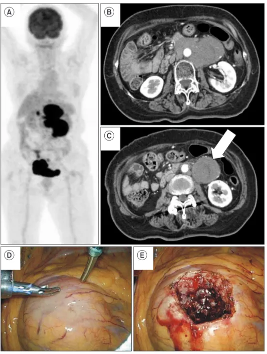
Fig. 2. During evaluation for abdominal pain, a 76-year-old female patient was found to have enlarged paraaortic and mesenteric lymph nodes. (A) Coronal section of fluorine-18 fluorodeoxyglu- cose positron emission tomography. (B, C) Axial section on computed tomog- raphy. The mesenteric lymph nodes were biopsied via the transperitoneal approach (arrow). (D, E) Laparoscopic im- age. The enlarged lymph node is directly visualized, when the first trocar is placed in a semilateral position. The biopsy was performed by wedge resection. The his- topathological diagnosis was adult T-cell leukemia/lymphoma.
Table 1. The histopathologic diagnosis for all patients
Histopathologic diagnosis No.
Lymphoma (n=20)
Hodgkin lymphoma 4
Non-Hodgkin lymphoma
Follicular lymphoma 9
Diffuse large B-cell lymphoma 5 Nodal marginal zone B-cell lymphoma 1 Adult T cell leukemia/lymphoma 1 Metastatic carcinoma (n=2)
Urothelial carcinoma 1
Squamous cell carcinoma 1
In the case of suspected intraabdominal lymphoma, laparoscopy and imageguided percutaneous biopsy are two reasonable means to open laparotomy to take a tissue specimen. Currently, there are no evidencebased guidelines recommending one technique over the other [10]. Multiple studies have shown percutaneous biopsy to be an adequately sensitive technique for identifying the presence of lymphoma; however, it is often unsuccessful in yielding a definitive diagnosis with complete subgroup classification [710]. In a series of 263 percutaneous image
guided biopsies used to diagnose suspected lymphoma, a diagnosis could be made in 90.1% of cases; however, only 75%
of lymphomas could be fully subclassified [9]. In a study comparing laparoscopic biopsy with percutaneous biopsy for the diagnosis of abdominal lymphoma, significantly more patients in the laparoscopic biopsy group had sufficient tissue for ancillary studies when needed than in the percutaneous biopsy group: 95.5% and 68.2%, respectively [10].
In light of this evidence, it has been our policy to perform laparoscopic lymph node biopsy at first presentation in the case of isolated abdominal lymphadenopathy with suspicion of lymphoma.
In 1988, Salky et al. [19] described the first laparoscopic biopsy in a case of retroperitoneal disease. From the late 1990s to the early 2000s, three large case series were reported by general surgeons [1,4,13]. The rate of conversion to open laparotomy, however, is relatively high (3.6%–17%), especially in the case of retroperitoneal lymph nodes. The primary reasons for conversion are previous surgery, obscure anatomy, inadequate explorations, inadequate sampling, and bleeding [4]. In these studies, the procedure was performed by using the transperitoneal approach. It may be possible to evade these causes of conversion by using a retroperitoneal approach. For mesenteric lymph nodes or interaortocaval lymph nodes, the transperitoneal approach may be safer and simpler than the retroperitoneal approach. There were no cases of conversion in our series. Although this was a small retrospective study, we consider that our study shows the feasibility of using two approaches appropriately in laparoscopic intraabdominal lymph node biopsy. The ability to choose the operative approach according to circumstances represents a clear advantage for urologists.
One patient (4.5%) had a symptomatic chylous lym
phocele, which was unsuccessfully managed by dietary intervention and needed surgical intervention. In this case, the retroperitoneal approach was applied for the paraaortic lymph node biopsy. Porte et al. [20] described surgical exploration of the retroperitoneal space through a direct vision endoscope without gas insufflation for retroperitoneal
lymphadenopathy. Lymphorrhea persisting for more than 5 days was observed in 5 of the 118 patients. The retroperitoneal procedure may be associated with a high risk of lymphorrhea because there is no intraperitoneal space to absorb the lymphatic fluid. In 9 patients, we used fibrin glue to reduce postoperative lymphorrhea, but it was not used in this case. Several prospective studies have addressed the use of a fibrin sealant to reduce lymphadenectomyrelated postoperative complications in patients undergoing dissection of the groin, axilla, or paraaortic lesions [2125]. However, these results remain controversial. Occurrence of these complications leads to a delay in the treatment of lymphoma.
Therefore, the use of fibrin glue may be recommended in the retroperitoneal approach for intraabdominal lymph node biopsy, especially when urgent chemotherapy is required for patients suspected to have highgrade lymphoma.
Previous laparoscopic biopsy experiences have shown a high frequency of falsenegative diagnoses (0%–14%) [1,4,11
14]. In our series, a precise diagnosis was established for all patients, and 20 lymphomas were completely subclassified.
To minimize falsenegative results, it is necessary to collect a sufficient quantity of a specimen tissue and to select an appropriate specimen as a target biopsy site. 18FFDG PET is currently considered the standard modality for not only staging but also evaluating targeted biopsy sites [26,27]. In an FDGavid tumor, it is important to select a high FDG uptake lesion as a target site. Furthermore, when multiple lesions are present, it is important to decide from which lesion we can easily and safely perform a biopsy. A thorough CT and PET evaluation by the surgical team is mandatory prior to laparoscopy, so as to select the surgical approach and the route from which to approach the node.
CONCLUSIONS
Appropriate use of a transperitoneal or a retroperitoneal approach is safe and effective for laparoscopic lymph node biopsy in patients with suspected intraabdominal lymphoma. Urologists should participate aggressively in this field, because these two approaches have become standard procedures in urological laparoscopy.
CONFLICTS OF INTEREST
The authors have nothing to disclose.
REFERENCES
1. Silecchia G, Raparelli L, Perrotta N, Fantini A, Fabiano P,
Monarca B, et al. Accuracy of laparoscopy in the diagnosis and staging of lymphoproliferative diseases. World J Surg 2003;27:
653-8.
2. Jaffe ES. The 2008 WHO classification of lymphomas: implica- tions for clinical practice and translational research. Hematol- ogy Am Soc Hematol Educ Program 2009:523-31.
3. Bakshi N, Maghfoor I. The current lymphoma classification:
new concepts and practical applications triumphs and woes.
Ann Saudi Med 2012;32:296-305.
4. Asoglu O, Porter L, Donohue JH, Cha SS. Laparoscopy for the definitive diagnosis of intra-abdominal lymphoma. Mayo Clin Proc 2005;80:625-31.
5. Bhandarkar DS, Shah RS, Katara AN, Shankar M, Chandira- mani VA, Udwadia TE. Laparoscopic biopsy in patients with abdominal lymphadenopathy. J Minim Access Surg 2007;3:14- 8.
6. Muskat PC, Johnson RA, Bowers GJ. Staging laparotomy in Hodgkin's lymphoma: 1979 to 1988. Am J Surg 1991;162:603- 6.
7. Vandervelde C, Kamani T, Varghese A, Ramesar K, Grace R, Howlett DC. A study to evaluate the efficacy of image-guided core biopsy in the diagnosis and management of lymphoma- -results in 103 biopsies. Eur J Radiol 2008;66:107-11.
8. Balestreri L, Morassut S, Bernardi D, Tavio M, Talamini R, Gloghini A, et al. Efficacy of CT-guided percutaneous needle biopsy in the diagnosis of malignant lymphoma at first presen- tation. Clin Imaging 2005;29:123-7.
9. Amador-Ortiz C, Chen L, Hassan A, Frater JL, Burack R, Nguyen TT, et al. Combined core needle biopsy and fine-nee- dle aspiration with ancillary studies correlate highly with tradi- tional techniques in the diagnosis of nodal-based lymphoma.
Am J Clin Pathol 2011;135:516-24.
10. Daly SC, Klairmont M, Arslan B, Vigneswaran Y, Roggin KF, Ujiki MB, et al. Laparoscopy has a superior diagnostic yield than percutaneous image-guided biopsy for suspected intra- abdominal lymphoma. Surg Endosc 2015;29:2496-9.
11. Casaccia M, Torelli P, Cavaliere D, Panaro F, Nardi I, Rossi E, et al. Laparoscopic lymph node biopsy in intra-abdominal lym- phoma: high diagnostic accuracy achieved with a minimally invasive procedure. Surg Laparosc Endosc Percutan Tech 2007;
17:175-8.
12. Gossot D, de Kerviler E, Brice P, Mariette X, Meignin V, Cazals-Hatem D, et al. Surgical endoscopic techniques in the diagnosis and follow-up of patients with lymphoma. Br J Surg 1998;85:1107-10.
13. Mann GB, Conlon KC, LaQuaglia M, Dougherty E, Moskowitz CH, Zelenetz AD. Emerging role of laparoscopy in the diagno- sis of lymphoma. J Clin Oncol 1998;16:1909-15.
14. Diulus L, Chalikonda S, Pitt T, Rosenblatt S. Efficacy of lapa-
roscopic mesenteric/retroperitoneal lymph node biopsy. Surg Endosc 2009;23:389-93.
15. Hara I, Tanaka K, Yamada Y, Miyake H, Takenaka A, Fujisawa M. Usefulness of laparo- or retroperitoneoscopic biopsy for retroperitoneal lymph node swelling of unknown origin. Int J Urol 2007;14:466-9.
16. Clayman RV, Kavoussi LR, Soper NJ, Dierks SM, Meretyk S, Darcy MD, et al. Laparoscopic nephrectomy: initial case re- port. J Urol 1991;146:278-82.
17. Yuan JB, Zu XB, Miao JG, Wang J, Chen MF, Qi L. Laparoscop- ic pelvic lymph node dissection system based on preoperative primary tumour stage (T stage) by computed tomography in urothelial bladder cancer: results of a single-institution pro- spective study. BJU Int 2013;112:E87-91.
18. Neyer M, Peschel R, Akkad T, Springer-Stöhr B, Berger A, Bartsch G, et al. Long-term results of laparoscopic retroperito- neal lymph-node dissection for clinical stage I nonseminoma- tous germ-cell testicular cancer. J Endourol 2007;21:180-3.
19. Salky BA, Bauer JJ, Gelernt IM, Kreel I. The use of laparoscopy in retroperitoneal pathology. Gastrointest Endosc 1988;34:227- 30.
20. Porte H, Copin MC, Eraldi L, Roumilhac D, Jaillard-Thery S, Puech P, et al. Retroperitoneoscopy for the diagnosis of infil- trating retroperitoneal lymphadenopathy and masses. Br J Surg 1997;84:1433-6.
21. Scholz HS, Petru E, Benedicic C, Haas J, Tamussino K, Winter R. Fibrin application for preventing lymphocysts after retro- peritoneal lymphadenectomy in patients with gynecologic ma- lignancies. Gynecol Oncol 2002;84:43-6.
22. Docimo G, Limongelli P, Conzo G, Gili S, Bosco A, Rizzuto A, et al. Axillary lymphadenectomy for breast cancer in elderly patients and fibrin glue. BMC Surg 2013;13 Suppl 2:S8.
23. Benevento R, Santoriello A, Pellino G, Sciaudone G, Candilio G, De Fatico GS, et al. The effects of low-thrombin fibrin seal- ant on wound serous drainage, seroma formation and length of postoperative stay in patients undergoing axillary node dissec- tion for breast cancer. A randomized controlled trial. Int J Surg 2014;12:1210-5.
24. Navarro-Rodríguez E, Gómez-Luque I, Díaz-Jiménez N, Rioja- Torres P, Bascuñana-Estudillo G, Ruiz-Rabelo JF, et al. Effec- tiveness of an absorbable fibrin sealant patch to reduce lym- phoceles formation after axillary lymphadenectomy for breast cancer: a matched-pair analysis. Am J Surg 2014;208:824-30.
25. Marchioni M, Ingrosso M, De Francesco P, Primiceri G, Man- co R, Tenaglia RL.The use of haemostatic agents and sealants for the prevention of lymphocele after urological surgery: a review of the literature. Surg Technol Int 2015;27:45-50.
26. D'souza MM, Jaimini A, Bansal A, Tripathi M, Sharma R, Mondal A, et al. FDG-PET/CT in lymphoma. Indian J Radiol
Imaging 2013;23:354-65.
27. Glaudemans AW, de Vries EF, Galli F, Dierckx RA, Slart RH, Signore A. The use of (18)F-FDG-PET/CT for diagnosis and
treatment monitoring of inflammatory and infectious diseases.
Clin Dev Immunol 2013;2013:623036.