https://doi.org/10.5468/ogs.2020.63.1.80 pISSN 2287-8572 · eISSN 2287-8580
Introduction
Polycystic ovary syndrome (PCOS) is a common endocrine disorder affecting 8–13% women of reproductive age [1,2].
Common clinical characteristics include chronic anovulation, hyperandrogenism (hyperandrogenemia or clinical hyper- androgenism), and polycystic ovaries on ultrasonography.
Infertility caused by chronic ovulatory dysfunction, abnormal gonadotropin secretion [3], and metabolic disturbances, such as central obesity, dyslipidemia, insulin resistance, and hyper- insulinemia, can be simultaneously encountered in PCOS [4].
The Rotterdam criteria have been widely used for the diag- nosis of PCOS [5]. PCOS is diagnosed in women with at least 2 of the following: oligo-anovulation, clinical and/or bio- chemical hyperandrogenism, and polycystic ovarian morphol- ogy (PCOM) on ultrasound (≥12 follicles measuring 2–9 mm in diameter in at least 1 ovary, and/or increased ovarian vol-
ume to >10 cm
3) after the exclusion of androgen excesses.
Anti-Müllerian hormone (AMH), produced in the granu- losa cells of small antral and preantral follicles, is a type of transforming growth factor β [6]. AMH plays a critical role in
Influence of combined oral contraceptives on polycystic ovarian morphology-related parameters in Korean
women with polycystic ovary syndrome
Chan-Hong Park, MD 1 , Sungwook Chun, MD, PhD 2
1
Health Care Division, The Provincial Office of Jeollanamdo, Muan;
2Department of Obstetrics and Gynecology, Haeundae Paik Hospital, Inje University College of Medicine, Busan, Korea
Objective
This study aimed to assess the effect of combined oral contraceptives (COCs) on polycystic ovarian morphology (PCOM) and serum anti-Müllerian hormone (AMH) levels in Korean women with polycystic ovary syndrome (PCOS).
Methods
This university hospital study enrolled 36 Korean women who were newly diagnosed with PCOS based on the Rotterdam criteria and were treated with COCs for at least 1 year. We retrospectively evaluated the ovarian volume and number of antral follicles using ultrasonography and assessed serum AMH levels at baseline and after 1 year of COC treatment.
Results
Significant decreases in ovarian volume and antral follicle count were observed after 1 year of COC treatment.
Compared to baseline, serum AMH levels were significantly decreased after 1 year.
Conclusion
COC treatment significantly affects ultrasound-assessed PCOM and serum AMH levels of patients with PCOS.
Keywords: Anti-Müllerian hormone; Combined oral contraceptives; Polycystic ovarian morphology; Polycystic ovary syndrome
Received: 2019.04.12. Revised: 2019.08.24. Accepted: 2019.09.26.
Corresponding author: Sungwook Chun, MD, PhD Department of Obstetrics and Gynecology, Inje University Haeundae Paik Hospital, 875 Haeun-daero, Haeundae-gu, Busan 48108, Korea
E-mail: [email protected] https://orcid.org/0000-0002-9948-0360
Articles published in Obstet Gynecol Sci are open-access, distributed under the terms of the Creative Commons Attribution Non-Commercial License (http://creativecommons.
org/licenses/by-nc/3.0/) which permits unrestricted non-commercial use, distribution, and reproduction in any medium, provided the original work is properly cited.
Copyright © 2020 Korean Society of Obstetrics and Gynecology
chronic anovulation by interrupting the initiation of primordi- al follicular growth [7] and promoting the arrest of follicular development [8]. AMH was recently recognized as a useful clinical marker for evaluating PCOS [9] and is considered promising for the accurate detection of PCOM [10].
Combined oral contraceptives (COCs), along with lifestyle modifications, are recommended for the primary manage- ment of PCOS to treat clinical symptoms such as irregular menstrual cycles, hirsutism, weight gain, and associated lipid, glucose, and testosterone disturbances [10]. However, in pre- vious studies, treatment effects varied in association with the heterogeneous composition of COCs, treatment duration, symptom severity/phenotype, and accessibility of the treat- ment regimen [11-14]. Reports on the effects of COCs on PCOM and AMH among Korean women are lacking.
Thus, we aimed to examine the effect of COC treatment on PCOM and serum AMH levels in Korean patients with PCOS.
Materials and methods
1. Patient population and study design
This retrospective study included Korean women aged 18–35 years who were diagnosed with PCOS during their first visit to Inje University Haeundae Paik Hospital between June 2010 and December 2014; these women were fol- lowed up with transvaginal (TV-US) or transrectal ultrasound (TR-US) and serum AMH levels after 1 year of COC treat- ment. PCOS was diagnosed according to the Rotterdam criteria [5]. PCOS is diagnosed in women with at least 2 of the following: oligo-anovulation, clinical and/or biochemical hyperandrogenism, and PCOM on ultrasonography; other pathologic conditions such as non-classical congenital adre- nal hyperplasia, androgen-producing tumors, hyperprolac- tinemia, Cushing’s syndrome, and thyroid dysfunction were ruled out. Oligomenorrhea is defined as an interval of >35 days between menstrual cycles, while amenorrhea is defined as an interval of >90 days between menstrual cycles. In our trial, clinical hyperandrogenism was defined as the presence of hirsutism based on a Ferriman-Gallwey score of >6, modi- fied for Korean women by Kim et al. [15]; biochemical hyper- androgenism was defined as a serum testosterone level ex- ceeding 95% confidence limits, as reported by Chae at al. [16]
(total testosterone level >0.68 ng/mL and/or free testoster- one level >1.72 pg/mL). PCOM was defined as follows: ≥12
follicles measuring 2–9 mm in diameter in each ovary and/
or an ovarian volume of >10 mL on TV-US or TR-US. Exclu- sion criteria were as follows: prior ovarian surgery, suspected ovarian malignancy, or known contraindications to estrogen therapy and use of oral contraceptives, ovulation-inducing drugs, glucocorticoids, or anti-androgenic agents, which can affect the hypophysis-pituitary-ovarian axis in the 6 months preceding the diagnosis. Finally, 36 patients were included in our study. All patients were given a 24-day regimen of COCs containing ethinylestradiol (EE) 20 µg and drospirenone (Yaz
®) 3 mg.
2. Assessment of ovarian volume and antral follicle count
All subjects were followed up with TV-US or TR-US to assess ovarian volume and antral follicle count. The TV-US or TR- US examination was performed using a Voluson S7 device (General Electric Systems, Seongnam, Korea) with a 7-MHz transvaginal probe. The baseline ultrasound was conducted at the early follicular phase, and the ultrasound after 1 year of treatment was performed during the pill-free interval (7–21 days) of the COC treatment. All the ultrasound ex- aminations were conducted by the same reproductive endo- crinologist in accordance with the international ultrasound definitions of PCOM [17]. The number of antral follicles mea- suring 2–9 mm in diameter was calculated using a continu- ous scanning method for each ovary from the inner to outer margins of a longitudinal cross-section. Ovarian volume was calculated according to a simplified formula for a prolate el- lipsoid (0.5×length×width×thickness) [17]. Total ovarian vol- ume (TOV) was calculated as the sum of the volume of the right ovary (ROV) and left ovary (LOV), and total antral follicle count (TFC) was calculated as the sum of the follicle count of the right ovary (RFC) and left ovary (LFC).
3. Measurement of serum anti-Müllerian hormone levels
Blood samples were collected in tubes without anticoagu- lants on the day of ultrasound examination, and the bio- chemical parameters of the serum were measured. Serum AMH levels were determined using an AMH Gen II assay (Beckman Coulter Inc., La Brea, CA, USA).
4. Statistical analysis
Data are expressed as mean±standard deviation and ana-
lyzed using SPSS version 25.0 (SPSS, Chicago, IL, USA). A paired samples t-test was used to compare parameters re- lated to ovarian follicle count (TFC, RFC, and LFC), ovarian volume (TOV, ROV, and LOV), and AMH levels at baseline and after 1 year of treatment. Statistical significance was set at P<0.05.
Results
Baseline clinical characteristics are shown in Table 1. Table 2 shows that the baseline ultrasound parameters related to PCOM were significantly decreased after 1 year of COC treat- ment. There was a significant decrease in TFC from baseline (73.83±43.14) to 1 year (44.11±28.09) (40.3% decrease, P<0.001). In addition, TOV decreased significantly after 1 year of treatment (from 24.57±8.39 to 14.13±5.92 cm
3; P<0.001). Serum AMH levels were decreased significantly from 11.09±4.6 ng/mL at baseline to 6.45±3.29 ng/mL after 1 year of COC treatment (P<0.001).
Discussion
PCOM is 1 of 3 items needed for diagnosis of PCOS. The
2 cardinal features of PCOM include increases in ovarian volume and AFC levels. The AMH level is correlated with the number of follicles that are 2–5 mm in diameter [18];
serum AMH level is useful for assessing PCOM in patients with PCOS. COCs are recommended as primary treatment for chronic anovulation and hyperandrogenism in PCOS [10]. However, adequate research on the effects of COCs on PCOM in women with PCOS is lacking, and to the best of our knowledge, no such research has been performed among Korean women. We found a significant decrease in baseline TFC, TOV, and AMH values after 1 year of COC treatment in Korean patients with PCOS.
Several studies have investigated whether COCs can modulate PCOM in patients with PCOS [13,19-25]. Accord- ing to Mes-Krowinkel et al. [19], the mean ovarian volume, mean ovarian follicle count, and AMH level remarkably de- creased in patients using COCs than in patients who had never used COCs after correction for confounding variables such as body mass index, age, waist-to-hip ratio, previous pregnancy, infertility treatment, and ethnicity. Another study [20] reported that ovarian volume (ROV, LOV, and TOV), ovarian follicle count (RFC, LFC, and TFC), and AMH level were significantly decreased after 3 months of COC treat- ment. The authors suggested that the AMH level decreased due to COC-induced suppression of ovarian function. They used cyproterone acetate (CPA) 2 mg/EE (Diane 35) 35 μg;
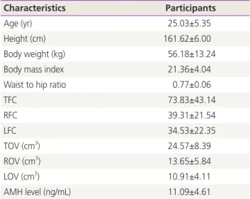
however, per current recommendations [10], this should not be first-line treatment because of adverse effects such as ve- nous thromboembolism. Moreover, the AMH assay kits used Table 1. Baseline patient characteristics (n=36)
Characteristics Participants
Age (yr) 25.03±5.35
Height (cm) 161.62±6.00
Body weight (kg) 56.18±13.24
Body mass index 21.36±4.04
Waist to hip ratio 0.77±0.06
TFC 73.83±43.14
RFC 39.31±21.54
LFC 34.53±22.35
TOV (cm
3) 24.57±8.39
ROV (cm
3) 13.65±5.84
LOV (cm
3) 10.91±4.11
AMH level (ng/mL) 11.09±4.61
Data was expressed as mean±standard deviation.
TFC, total follicle count; RFC, right ovarian follicle count; LFC, left ovarian follicle count; TOV, total ovarian volume; ROV, right ovarian volume; LOV, left ovarian volume; AMH, anti-Müllerian hormone.
Table 2. Serial changes in ultrasound parameters of ovarian size and serum anti-Müllerian hormone levels (n=36)
Parameters Basal Post 1 year P-value
a)TFC 73.83±43.14 44.11±28.09 <0.001
RFC 39.31±21.54 23.19±14.92 <0.001
LFC 34.53±22.35 20.91±13.78 <0.001
TOV (cm
3) 24.57±8.39 14.13±5.92 <0.001 ROV (cm
3) 13.65±5.84 7.53±3.43 <0.001 LOV (cm
3) 10.91±4.11 6.59±3.07 <0.001 AMH level (ng/mL) 11.09±4.61 6.45±3.29 <0.001 Data was expressed as mean±standard deviation.
TFC, total follicle count; RFC, right ovarian follicle count; LFC, left ovarian follicle count; TOV, total ovarian volume; AMH, anti-Mülleri- an hormone; LOV, left ovarian volume; ROV, right ovarian volume.
a)