급성
5
0
0
전체 글
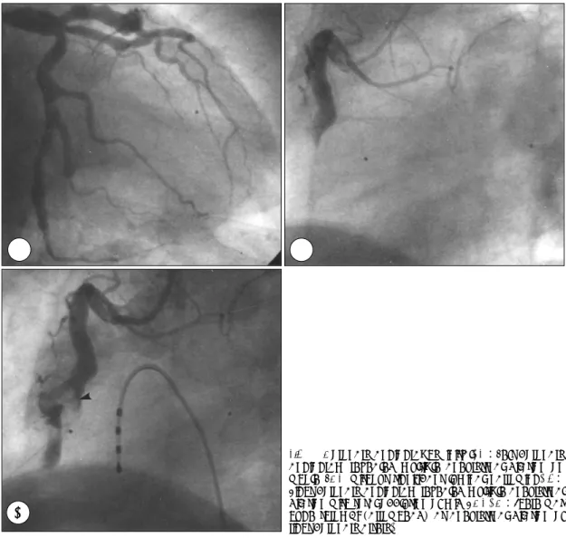
(2) 맥조영술 및 복부 대동맥조영술을 통해 진단한 다발성. 100회/분, 호흡수 14회/분, 체온은 36.7℃였고 급성병. 결절성 동맥염 1예를 경험하였기에 이를 문헌고찰과. 색을 띄었으나 정신상태는 양호하였다. 내원 10분 후. 함께 보고하는 바이다.. 수축기 혈압이 70 mmHg로 떨어지며 어지러움을 호소 하였으나 다량의 수액공급 후 곧 회복되었다. 흉부 청. 증. 례. 진상 호흡음은 깨끗하였고 심음은 규칙적이었으며 심 잡음은 청진되지 않았다. 간과 비장은 촉지되지 않았고. 평소 건강하게 지내던 65세 남자로 내원당일 새벽. 장음은 정상이었으며 사지부종은 관찰되지 않았다. 양. 갑작스럽게 발생한 격심한 흉통을 주소로 개인병원 방. 측 수부 및 족부에 다발성 홍반성 피부발진과 소포가. 문하여 급성 심근경색증 의심하에 본원 응급실을 경유. 관찰되었다. 흉부 X-선 검사상 좌측 폐첨부의 염증성. 하여 내원하였다. 과거력상, 30년전 고혈압으로 진단받. 변화가 관찰되었으며 심비대의 소견은 없었다. 심전도. 고 5년간 항고혈압제를 투약하였다. 흡연력은 40갑년. 검사상 lead Ⅱ, Ⅲ, aVF에서 ST분절의 상승과 aVL,. 이었으며 가족력상 특이사항은 없었다. 내원당시 문진. V2~6에서 상보적인 ST분절의 하강이 관찰되었다. 말. 상 특이소견은 없었으며, 혈압은 160/90 mmHg, 맥박. 초혈액 검사상 혈색소 11.3 g/dl, 적혈구용적 32.7%,. A. C. 228. B. Fig. 1. Coronary angiography, initial. A:Left coronary angiogram revealed multiple aneurysmal dilation on whole LCA without significant luminal narrowing. B: Right coronary angiogram revealed multiple aneurysmal dilation with total occlusion of mid-RCA. C:There was huge thrombi (arrowhead) at aneurysmal dilation of right coronary artery.. Korean Circulation J 2000;30(2):227-231.
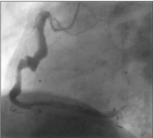
(3) 백혈구수 5,800/mm3(중성구 55.3%, 림프구 33.1%,. 44~166), calculated LDL-cholesterol 152 mg/dl,. 단핵구 9.2%, 호산구 2.0%, 호염구 0.4%), 혈소판수. lipoprotein(a) 22.6 mg/dl이었으며 C-reactive pro-. 3. 134,000/mm , 적혈구 침강속도 83 mm/hour였다. 혈. tein 1.35 mg/dl(정상≤0.8)였다. 면역혈청학적 검사상. 액화학 검사상 BUN 21.2 mg/dl, creatinine 1.3 mg/dl. anti-nuclear antibody(ANA) 1 : 40 음성, anti-. 였으며 AST 380 IU/L(정상 8~30), ALT 76 IU/L. DNA antibody 1:10 음성, anti-neutrophilic cyto-. (정상 8~30), CK 202 IU/L(정상 20~134), CK-. plasmic antibody(ANCA) 음성, rheumatic factor 음. MB 15.4 ng/ml(정상 2~8)로 증가되어 있었고 rapid. 성, C3 82 mg/dl(정상 45~86), C4 35 mg/dl(정상. troponin-T는 양성이었다. 혈청지질은 total cholest-. 11~47), CH50 50.2/ml(정상 30~45), circulating. erol 206 mg/dl(정상 100~220), HDL-cholesterol. immune complex(CIC) 1.24 ng/ml(정상≤1.23), anti-. 37 mg/dl(정상 30~80), triglyceride 86 mg/dl(정상. cardiolipin antibody IgG/IgM 음성, lupus anticoagulant 음성, HBs antigen 음성이었다. 신경전도검사 상 이상소견은 없었으며 피부생검은 시행하지 않았다. 심초음파상 우심실과 좌심실하벽에 중등도의 운동저하 소견이 관찰되었다. 심구출율은 55%였고 심방 및 심 실은 정상 크기였다. 관상동맥조영술 검사상 좌관상동 맥 전체에 다발성 동맥류 및 확장소견이 관찰되었으며 우관상동맥 근위부의 확장소견과 함께 중앙부 이하의 완전폐쇄 소견을 보였다(Fig. 1-A and B). 관상동맥의 직경이 5 mm이상으로 확장되어 있어 직경이 가장 굵 은 풍선도자로 풍선 확장술 후에도 우관상동맥 중앙부 동맥류에 많은 양의 혈전이 관찰되어(Fig. 1-C) 관상 동맥내로 urokinase 10만 단위를 두차례 투여하였다. 투여직후 시행한 조영술 소견상 관상동맥혈류의 일부. Fig. 2. Follow-up coronary angiogram, 2 month later. Right coronary angiogram revealed multiple aneurysmal dilation without significant luminal narrowing.. A. 호전을 보였으나 혈관 원위부까지 혈류가 진행하지는 못하였다. 시술을 중단하고 중환자실로 옮겨 heparin을. B. Fig. 3. Abdominal aortogram along with renal, mesenteric and splanchnic angiogram revealed multiple aneurysms (arrow-head) in the right renal (A) and superior mesenteric arteries (B).. 229.
(4) 지속적으로 정주하였으며. 입원 7일째 상태가 안정화되. Przybojewski는 불안정형 협심증으로 내원한 심근. 어 nicorandil, captopril, carvedilol, aspirin, ticlopi-. 경색의 과거력을 가진 29세 남자에서 관상동맥 조영술. dine을 투약하며 퇴원하였다. 2개월후 시행한 추적 관. 검사상 우측 관상동맥의 미만성 동맥류와 좌전하행지. 상동맥조영술 검사에서 우관상동맥의 완전폐쇄는 호전. 와 좌회선지의 심한 폐색소견과 함께 근육생검에서 결. 되었으며(Fig. 2) 확진을 위해 시행한 복부 대동맥조영. 체조직 질환에서 특징적인 근외막과 인접건막의 혈관. 술상 우신동맥 및 상장간막동맥의 분지에 동맥류가 관. 과 결체조직을 침범하는 섬유소양 괴사를 관찰하였다.. 찰되었다(Fig. 3-A and B). 다발성 결절성 동맥염 진. 입원기간중 환자는 심근하벽의 급성심근경색과 소화성. 단하에 일일 prednisolone 60 mg과 cyclophospha-. 궤양의 천공으로 사망하였다.4) Pick등은 급성심근경색. mide 125 mg로 면역억제치료를 시행하며 8개월째 증. 으로 내원한 26세 여자에서 관상동맥의 다발성 동맥류. 상없이 외래 추적 관찰중이다.. 를 관찰하였으며 근육생검 및 신장, 상장간막, 복강, 뇌, 하지혈관조영술상 이상소견은 없었으나 임상경과를 종. 고. 안. 합하여 다발성 결절성 동맥염으로 진단한 1예를 보고하 였다.5) Keith등은 급성심근경색으로 내원한 51세 여자. 다발성 결절성 동맥염에서 심장침범시 증상이 없는. 에서 관상동맥조영술상 좌전하행지에 자발성 혈관박리. 것이 보통이며 따라서 관상동맥의 침범이 사망전 관상. 외 다른 이상소견은 발견되지 않았으나 5일후 흉통이. 동맥조영술 검사에 의해 진단되는 경우는 매우 드물다.. 재발하여 시행한 조영술상 혈관박리의 진행과 좌회선지. 따라서 다발성 결절성 동맥염에서 심장의 침범에 대한. 의 폐색 및 우관상동맥의 후하행지에 동맥류가 발견되. 보고는 대부분 사망후 부검을 통해 후향적으로 이루어. 었고 상장간막동맥의 이차분지와 액와동맥, 위대망막동. 졌다. 1962년 Holsinger등이 1926년부터 1958년사. 맥 등에도 동맥류가 관찰되어 다발성 결절성 동맥염을. 이 Mayo Clinic에서 진단된 66예의 다발성 결절성 동. 진단후 면역억제치료를 시행한 1예를 보고하였다.6). 맥염 환자의 부검소견을 보고한 바에 따르면 관상동맥. 성인에서 발견되는 관상동맥의 다발성동맥류의 원인. 염과 심근경색이 각각 62%에서 관찰되었으며 심근경. 으로 다발성 결절성 동맥염 이외에 유년기 Kawasaki. 색이 있었던 환자의 88%에서 관상동맥염이 발견되었. 병의 후유증으로 인한 경우가 가장 많이 보고되고 있으. 다. 관상동맥염의 증상은 고혈압이 가장 흔하였으며 그. 나 이는 주로 젊은 성인에서 발생하며 상기 환자와 같. 외 빈맥과 심부전, 심잡음, 심비대, 심낭마찰음과 부정. 은 고연령에서 Kawasaki병은 보고된 바 없으며 상기. 3). 맥등이 있었다.. Schrader등은 1935년에서 1976년. 환자에서 Kawasaki병의 과거력을 찾을 수 없었다. 그. 사이 Johns Hopkins University에서 부검을 통해 다. 외 전신성 홍반성 낭창, 혈전성 혈소판감소성 자반증,. 발성 결절성 동맥염으로 진단된 환자 36명의 임상적. Takayasu 동맥염, Wegener 육아종증에서 관상동맥. 병리학적 소견을 보고하였으며 18명(50%)에서 활성. 의 다발성 동맥류가 보고되었으나 전신성 홍반성 낭창. 또는 치유된 관상동맥의 동맥염을 관찰하였다. 병변은. 과7)8) Takayasu 동맥염에서9-12) 동맥류 형성은 매우. 주로 심외막하 혈관에 위치하였으며 특징적으로 혈관. 드물며 일반적으로 이들 질환에 의한 동맥류는 크고 혈. 중막과 외막의 림프구의 침윤과 진행된 병변의 경우 혈. 관 근위부에 위치하는 것이외에 전신질환의 임상적 및. 관벽의 괴사와 혈관주위 결합조직을 포함한 혈관전층. 다른 혈청학적 표지자와 함께 나타나는 것이 보통이다.. 1). 의 침범을 관찰하였다.. 상기 환자는 전신성 홍반성 낭창, Kawasaki병, 혈전성. 다발성 결절성 동맥염의 확진은 생검에 의해 이루어. 혈소판감소성 자반증, Takayasu 동맥염, Wegener 육. 지며 생검이 용이하지 않은 경우 침범된 장기의 동맥조. 아종증의 합당한 혈청학적 표지자나 전신증상이 없었. 2). 영술 검사를 통해서도 진단할 수 있으나 저자들의 문. 으며 진단기준에도 부합하지 않았다. 따라서 상기환자. 헌고찰에 따르면 급성 관상동맥 증후군으로 내원한 환. 는 1990년 American College of Rheumatology에서. 자에서 동맥조영술을 통해 다발성 동맥류를 관찰하고. 제안한 다발성 결절성 동맥염의 진단기준을13) 만족하. 다발성 결절성 동맥염으로 진단하여 치료한 예는 3예. 지는 못하지만 고혈압과 관상동맥조영술 및 복부대동. 에 불과하며 국내에서는 아직 보고된 바 없다.. 맥조영술 소견등을 종합하여 다발성 결절성 동맥염에. 230. Korean Circulation J 2000;30(2):227-231.
(5) 가장 합당하다고 판단하였으며 관상동맥내 urokinase. 4) Przybojewski J. Polyarteritis nodosa in the adult. S Afr. 주입 및 보존적 치료를 통해 급성 심근경색을 치료후. 5) Pick R, Glover M, Viewer W. Myocardial infarction in a. Med J 1981;60:512-8.. 면역억제치료를 시행하며 증상없이 추적관찰중이다. 6). 요. 약. 이에 저자들은 급성심근경색으로 내원한 환자에서. 7). 관상동맥조영술 및 복부 대동맥조영술 검사를 통해 다 발성 결절성 동맥염을 진단하고 관상동맥내 풍선확장 술 및 urokinase 주입 그리고 보존적 치료를 병행하여 심근경색을 치료하고 이후 면역억제 치료를 성공적으 로 시행한 1예를 경험하였기에 이를 문헌고찰과 함께. 8) 9) 10). 보고하는 바이다.. 중심 단어:다발성 결절성 동맥염・급성 심근 경색. 11). REFERENCES 1) Schrader M, Hochman J, Bulkley B. The heart in polyarteritis nodosa: A clinicopathologic study. Am Heart J 1985;109:1353-9. 2) Fauci AS. The vasculitis syndromes. In: Harrison’s principles of internal medicine. Fauci AS, Braunwald E, Isselbacher KJ, Wilson JD, Martin JB, Kasper DL, et al. 14th ed. New York: McGraw Hill;1998. p.1910-22. 3) Holsinger D, Osmundson P, Edwards J. The heart in polyarteritis nodosa. Circulation 1962;25:610-8.. 12). 13). young women with isolated coronary arteritis. Chest 1982; 82:378-80. Keith HC, Frank JM, James CB, Ray H, Thomas H. Polyarteritis nodosa presenting as acute myocardial infarction with coronary dissection. Cathet Cardiovasc Diagn 1998; 44:320-4. Nobrega TP, Klodas E, Liggett SP, Higano ST, Reeder GS. Giant coronary aneurysms and myocardial infarction in a patient with systemic lupus erythematosis. Cathet Cardiovasc Diagn 1996;39:75-9. Wilson VE, Eck SL, Bates ER. Evaluation and treatment of acute myocardial infarction complicating systemic lupus erythematosis. Chest 1992;101:420-4. Amano J, Suzuki A. Coronary artery involvement in Takayasu’s arteritis. J Cardiovasc Surg 1991;102:554-60. Nakano T, Okano H, Konishi T, Takezawa H. Aneurysm of the left aortic sinus by Takayasu’s arteritis: Compression of the left coronary artery producing coronary insufficiency. J Am Coll Cardiol 1986;7:696-700. Kihara M, Kimura K, Yakuwa H, Minamisawa K, Hayashi S, Umemura S, et al. Isolated left coronary ostial stenosis as the sole arterial involvement in Takayasu’s disease. J Int Med 1992;232:353-5. Iannone L, Rayl K. Takayasu’s disease with axillary, right coronary artery and right internal mammary stenosis treated with angioplasty. Cathet Cardiovasc Diagn 1991;22: 42-4. Lightfoot RW, Michel BA, Bloch DA. The American college of rheumatology 1990 criteria for the classification of polyarteritis nodosa. Arthritis Rheum 1990;33:1088-93.. 231.
(6)
수치
관련 문서
Compromised CD4+ CD25(high) regulatory T-cell function in patients with relap sing-remitting multiple sclerosis is correlated with a reduced frequency of
No.. ramus insufficiency fracture, B) Thorax and abdomen CT showing multiple spine compression fracture.. ramus insufficiency fracture, B) After 4 months of
Shape memory alloy as second phase in composite materials :
Keywords □ acute coronary syndrome, discharge medication patterns, Korea, tertiary hospital, follow-up evaluation, troponin-T,
In order to verify whether the museum exhibition of intangible cultural heritage has a significant influence on the visitor's behavioral intention, a
Second, analyzing students' level of interest in the unit of Clothes and Life Style revealed that there was significant difference between genders and students in the two
(C-D) The hip anterioposterior and lateral radiograph shows bone ingrowth without subsidence or osteolysis after 62 months follow up after
First, the level of career barriers of visually impaired university students showed statistically significant differences only in their majors, but the level
