http://dx.doi.org/10.5468/ogs.2015.58.1.46 pISSN 2287-8572 · eISSN 2287-8580
Introduction
Osteoporosis is an important health problem and a major pre- disposing factor for fracture. As the population aging, the preva- lence of osteoporosis gets higher and the social burden increas- ing. So it has been an important subject to investigate the factors that influence the occurrence of osteoporosis. Some of them were modifiable factors such as smoking, alcohol consumption, low calcium intake and others were not modifiable factors such as aging, female gender, menopause [1-3].
Recently, oxidative stress or low circulating levels of antioxidants were proposed to be related with reduced bone mineral density (BMD) and caused osteoporosis by in vitro studies or animal
studies [4-6]. Uric acid, bilirubin and albumin has been known as natural antioxidants. Actually, it has been reported that higher
The association between oxidative stress and bone mineral density according to menopausal status of Korean women
Young Joo Lee, Ji Yun Hong, Seung Chul Kim, Jong Kil Joo, Yong Jin Na, Kyu Sup Lee
Department of Obstetrics and Gynecology, Pusan National University Hospital, Pusan National University School of Medicine, Busan, Korea
Objective
The aim of this study is to investigate the association between oxidative stress and bone mineral density (BMD) according to menopausal status of Korean women.
Methods
A total of 2,232 women who visited to the health promotion center at Pusan National University Hospital between 2010 and 2014 were included in this cross-sectional study. Laboratory tests, such as uric acid, albumin, total bilirubin, which were evaluated as a natural antioxidants. Homocysteine also was evaluated as a factor associated with oxidative stress. Correlation analyses and partial correlation coefficient between BMD scores and laboratory parameters associated with oxidative stress according to menopausal status were performed with Pearson test.
Results
By correlation analysis, uric acid had only positive correlation with femur and lumbar BMD in premenopausal and postmenopausal group. But albumin and bilirubin, which were the other natural antioxidants, had no correlation with BMD except total bilirubin with femur BMD in postmenopausal group. Homocysteine had negative correlation with femur BMD in postmenopausal group. But there were different results in partial correlation coefficient adjusted by age and BMI. In premenopausal group, uric acid was still positive correlation with femur and lumbar BMD, whereas in postmenopausal group homocysteine had no correlation with femur BMD, total bilirubin and uric acid had no correlation with lumbar BMD. At the multiple logistic regressions, only age and menopause status, uric acid had correlation with BMD.
Conclusion
In this study, homocysteine had no correlation with BMD. But in natural antioxidant, uric acid had only positive correlation with BMD.
Keywords: Bone density; Homocysteine; Menopause; Uric acid
Received: 2014.5.26. Revised: 2014.8.11. Accepted: 2014.9.1.
Corresponding author: Jong Kil Joo
Department of Obstetrics and Gynecology, Pusan National University Hospital, Pusan National University School of Medicine, 179 Gudeok- ro, Seo-gu, Busan 602-739, Korea
Tel: +82-51-240-7287 Fax: +82-51-248-2384 E-mail: [email protected]
Articles published in Obstet Gynecol Sci are open-access, distributed under the terms of the Creative Commons Attribution Non-Commercial License (http://creativecommons.
org/licenses/by-nc/3.0/) which permits unrestricted non-commercial use, distribution, and reproduction in any medium, provided the original work is properly cited.
Copyright © 2015 Korean Society of Obstetrics and Gynecology
uric acid levels were linearly associated with higher lumbar spine BMD in perimenopausal and postmenopausal women. It might be due to the major role of uric acid in free radical scavenger activity [7]. Bilirubin suppressed oxidation and albumin was an important contributor to maintain total antioxidant status [8,9].
So, the antioxidant activity by these factors could be influence the BMD. On the contrary, homocysteine was a factor associated with oxidative stress in vivo [8,10]. But, the relationship between homocysteine and BMD is still unclear. Some studies reported that high homocysteine was related with increased bone turn- over and fracture risk in elderly [11,12], but other studies didn’t [13,14]. Homocysteine level might be changed with endogenous sex steroids levels [15]. So, the change of homocysteine levels through the menopausal transition may have influence on the decrease of BMD or development of osteoporosis in postmeno- pausal women. The research for the relationship of natural anti- oxidants and BMD could confirm the influence of oxidative stress for development of osteoporosis and the natural antioxidants levels could be used as variables predicting the occurrence of osteoporosis. The aim of this study is to investigate the associa- tion between oxidative stress and BMD according to menopausal status of Korean women
Materials and methods
1. Study population and anthropometric measurements
A total of 2,232 women who visited to the health promotion center at Pusan National University Hospital between 2010 and 2014 were included in this cross-sectional study. Demographic data were collected at the time of the visit. Information on menstrual history, lifestyle, disease history and medication his- tory were obtained with self-report questionnaires and inter- views with healthcare providers. Body weight and height were
measured when standing barefoot, up to 0.1 kg and 0.1 cm, respectively. Body mass index (BMI) was calculated as weight in kilograms divided by height in meters squared.
We conducted our research based on self-report question- naires. We included patients prepared our self-report question- naires without exception and excluded patients who had been taking a steroid medicine for asthma, arthritis, rheumatic disease, which could affect BMD and also excluded patients taking a bisphosphonate and selective estrogen-receptor modulator. But we included 390 hypertension patients, 95 diabetes patients, 353 hyperlipidemia patient, all these patients had been taking a medication, 115 smokers. We also included patients had been taking a vitamin D or calcium medication on our research.
2. Blood sampling and laboratory analysis
Bloods were obtained from antecubital vein from all subjects between 8:30 and 10:00 a.m., after fasting for at least eight hours. Laboratory tests were evaluated, which consisted of uric acid, albumin, total bilirubin as a natural antioxidants and ho- mocysteine as a factor associated with oxidative stress.
BMD was measured by dual-energy X-ray absorptiometry (Hologic QDR-4500A, Bedford, MA, USA) at the lumbar spine (L1–L4), femur neck and femur total. BMD results were classi- fied into three groups according to World Health Organization criteria (normal BMD, T-score ≥-1; osteropenia, -2.5< T-score
<-1; and osteroporosis, T-score ≤-2.5, respectively) [16]. Intraas- say and interassay coefficients of variation of uric acid were 1.0% and 1.3%, albumin were 1.6% and 0.9%, total bilirubin were 2.7% and 2.6%, homocysteine were 4.4% and 3.3%, BMD were 2.3% and 2.7%.
3. Statistical analysis
PASW ver. 18.0 (SPSS Inc., Chicago, IL, USA) was used for sta- tistical analysis. All data were entered into a database and were
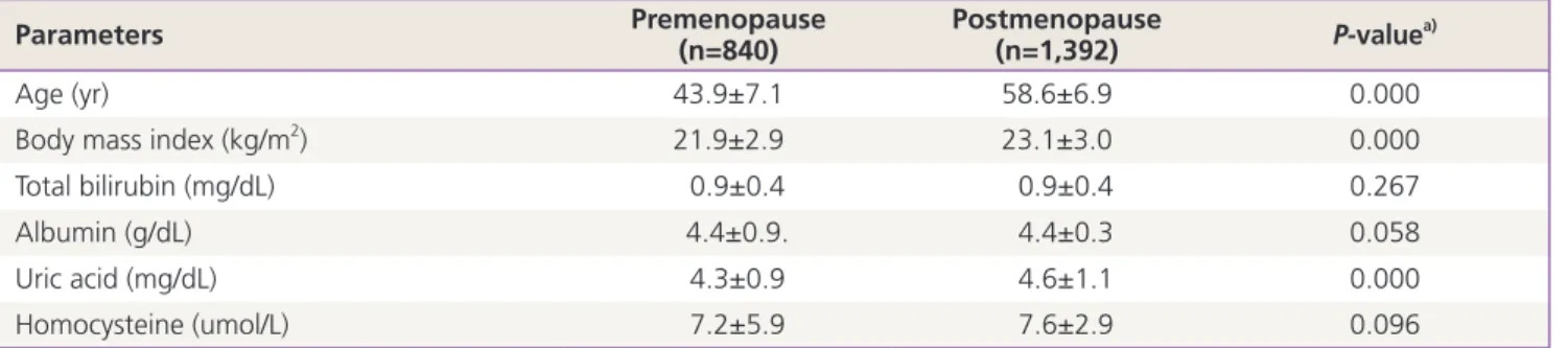
Table 1. Demographics and laboratory parameters of study population
Parameters Premenopause
(n=840) Postmenopause
(n=1,392) P-valuea)
Age (yr) 43.9±7.1 58.6±6.9 0.000
Body mass index (kg/m2) 21.9±2.9 23.1±3.0 0.000
Total bilirubin (mg/dL) 0.9±0.4 0.9±0.4 0.267
Albumin (g/dL) 4.4±0.9. 4.4±0.3 0.058
Uric acid (mg/dL) 4.3±0.9 4.6±1.1 0.000
Homocysteine (umol/L) 7.2±5.9 7.6±2.9 0.096
Data are presented as the mean±standard deviation.
a)Student’s t-test.
verified by a second independent person.
Data were presented as mean±standard deviation for normally distributed variables (age, BMI, albumin, total bilirubin, homocys- teine, and uric acid). The study population was divided into two groups, premenopause and postmenopause. Menopause group was categorized according to self questionnaires. If the patient had a hysterectomy, menopause was diagnosed by serum follicle stimulating hormone level. Serum follicle stimulating hormone more than 40 IU/mL was used for diagnosis of menopause.
The differences in baseline characteristics between groups were analyzed by Student’s t-test. Correlation analyses and par- tial correlation coefficients were performed with Pearson test.
Logistic regression analysis was performed to identify significant independent related factors for osteoporosis. Two-sided values of P<0.05 were considered as statistically significant.
Results
The demographics and laboratory parameters of the study participants were presented in Table 1. There were statistically significant differences on age, BMI, uric acid level between two groups (P< 0.05).
The correlation analysis results between oxidative stress mark-
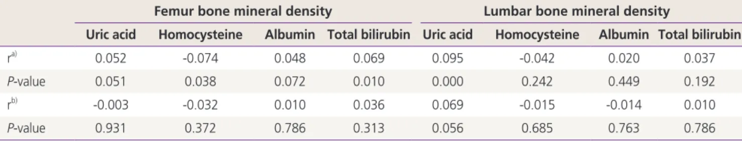
ers with BMD scores according to menopausal status were sum- marized in Tables 2 and 3. Uric acid was a sole parameter that had significantly positive correlation with femur and lumbar BMD in premenopausal group with strongly, but the magnitude of correlation was small (r=0.143, P=0.000). In postmenopausal group, uric acid was positively correlated with lumbar BMD (r=0.095, P=0.000). Homocysteine had negative correlation significantly with femur BMD (r=-0.074, P=0.038). A positive correlation was found between total bilirubin level and femur BMD scores (r=0.069, P=0.010). But there were different re- sults in partial correlation coefficient adjusted by age and BMI.
In premenopausal group, uric acid was still positive correlation with femur and lumbar BMD, whereas in postmenopausal group homocysteine had no correlation with femur BMD (r=- 0.032, P=0.372), total bilirubin and uric acid had no correlation with lumbar BMD.
Table 4 showed the results of multiple logistic regression anal- yses. BMD was calculated as continuous variable parameters and we investigated correlation of serial BMD score and several factors (uric acid, homocysteine, albumin, and total bilirubin).
Cumulative logistic regression analyses revealed that age (odds ration [OR], 0.934; 95% confidence interval [CI], 0.914 to 0.954;
P<0.000), menopause (OR, 0.323; 95% CI, 0.214 to 0.491;
P<0.000) and uric acid (OR, 1.208; 95% CI, 1.100 to 1.302;
Table 2. Correlation coefficients between bone mineral density scores and laboratory parameters associated with oxidative stress in pre- menopausal women
Femur bone mineral density Lumbar bone mineral density
Uric acid Homocysteine Albumin Total bilirubin Uric acid Homocysteine Albumin Total bilirubin
ra) 0.162 -0.046 -0.057 -0.021 0.143 -0.040 -0.057 -0.003
P-value 0.000 0.305 0.096 0.538 0.000 0.372 0.096 0.963
rb) 0.127 -0.036 -0.003 -0.013 0.092 -0.032 0.050 0.026
P-value 0.005 0.421 0.954 0.766 0.042 0.478 0.273 0.561
a)Pearson’s correlation coefficient; b)Partial correlation coefficient adjusted by age and body mass index.
Table 3. Correlation coefficients between bone mineral density scores and laboratory parameters associated with oxidative stress in post- menopausal women
Femur bone mineral density Lumbar bone mineral density
Uric acid Homocysteine Albumin Total bilirubin Uric acid Homocysteine Albumin Total bilirubin
ra) 0.052 -0.074 0.048 0.069 0.095 -0.042 0.020 0.037
P-value 0.051 0.038 0.072 0.010 0.000 0.242 0.449 0.192
rb) -0.003 -0.032 0.010 0.036 0.069 -0.015 -0.014 0.010
P-value 0.931 0.372 0.786 0.313 0.056 0.685 0.763 0.786
a)Pearson’s correlation coefficient; b)Partial correlation coefficient adjusted by age and body mass index.
P<0.000) were independent variables associated with increas-
ing lumbar and femur BMD.
Discussion
Oxidative stress has been proposed as an underlying mecha- nism of many diseases such as cancer, atherosclerosis, rheu- matoid arthritis and osteoporosis [17]. Oxidative stress may cause osteoporosis by altering the function of osteoclast and osteoblast. Architecture of bone is maintained by continuous destruction and renewal of bone. These continuous remodel- ing of bone are regulated by balanced action of osteoclast and osteoblast. In osteoporosis patients, the ratio of superoxide dis- mutase and glutathione peroxidase, which were oxidative stress biological markers, were increased and it favors the increase in H
2O
2levels [18,19]. High H
2O
2levels developed the differentia- tion of osteoblastic cells to osteoclasts and inhibit the differen- tiation of osteoblastic cells to osteoblasts [20,21].
In addition, there were some reports that explained the effect of antioxidants to the development of osteoporosis. Low serum albumin reflected significant systemic inflammation and bone resorption had been found to be increased by systemic inflam- mation as a result of increased number of various cytokines [22]. There have been several epidemiological analyses that had shown a significant inverse association between total bilirubin
and BMD in patients with or without underlying liver disease [23-25]. Low intake of antioxidant vitamins increased the risk of hip fracture in smoker [26]. Maggio et al. [27] also showed possible link between plasma antioxidants and BMD in osteo- porotic women.
Despite of this clear causal relationship between oxidative stress and osteoporosis, factors that affect natural antioxidant have not been well identified. One of suggested factors is ho- mocysteine. Homocysteine caused the production of reactive oxygen species by autooxidation [28]. Theoretically, homocyste- ine has negative effect on antioxidant and bone. But conflicting results have been reported about the relationship of homocys- teine and bone.
Bucciarelli et al. [29] reported that total plasma homocysteine was negatively associated with the variance of BMD of the total femur. The association was clinically relevant but the contribu- tion of homocysteine to BMD was small (2% of the total vari- ance). On the contrary, Dhonukshe-Rutten et al. [30] and Flem- ing et al. [31] showed no relation between homocysteine and BMD. The transition of homocysteine level after menopause is also unclear. It has been reported that homocysteine was lower in premenopausal as compared to postmenopausal women [32].
However other researchers reported that menopause did not affect the homocysteine levels [33-35].
In this study, the average level of homocysteine was higher in the postmenopausal women but there was no statistical differ-
Table 4. Cumulative logistic regression analysis results of the possible correlates for lumbar (L1–L4) and femur bone mineral density95% confidence interval
P-valuea)
Odds ratio Lower Upper
Lumbar bone mineral density
Age 0.934 0.914 0.954 0.000
Menopause 1.676 1.509 1.786 0.000
Uric acid 1.208 1.100 1.302 0.000
Homocysteine 0.992 0.954 1.028 0.660
Albumin 1.046 0.440 1.417 0.851
Total albumin 1.017 0.617 1.302 0.920
Femur bone mineral density
Age 0.939 0.920 0.958 0.000
Menopause 1.394 1.115 1.585 0.010
Uric acid 1.165 1.054 1.263 0.005
Homocysteine 0.991 0.960 1.021 0.550
Albumin 0.670 0.463 1.174 0.238
Total albumin 0.817 0.364 1.145 0.309