Veterinary Science
†The first and second author contributed equally to this work.
*Corresponding author
Tel: +82-2-450-4140; Fax: +82-2-444-4396 E-mail: [email protected]
Canine model of ischemic stroke with permanent middle cerebral artery occlusion: clinical and histopathological findings
Byeong-Teck Kang
1,†, Jong-Hwan Lee
2,†, Dong-In Jung
1, Chul Park
3, Su-Hyun Gu
1, Hyo-Won Jeon
1, Dong-Pyo Jang
5, Chae-Young Lim
1, Fu-Shi Quan
2, Young-Bo Kim
5, Zang-Hee Cho
5, Eung-Je Woo
4, Hee-Myung Park
1,*
1
Department of Veterinary Internal Medicine, and
2Department of Anatomy, College of Veterinary Medicine, Konkuk University, Seoul 143-701, Korea
3
Acupuncture & Meridian Science Research Center and
4College of Electronics and Information, Kyunghee University, Yongin 446-701, Korea
5
Neuroscience Research Institute, Gachon University of Medicine and Science, Incheon 405-760, Korea
The aim of the present study was to assess the clinical and histopathological findings in a canine model of ische- mic stroke. Cerebral ischemic stroke was induced by mid- dle cerebral artery occlusion in four healthy beagle dogs using silicone plugs. They showed neurological signs of forebrain dysfunction such as reduced responsiveness, head turning, circling, postural reaction deficits, percep- tual deficits, and hemianopsia. These signs gradually re- gressed within 4 weeks without therapy. On magnetic res- onance imaging, T2 hyperintensity and T1 hypointensity were found in the cerebral cortex and basal ganglia. These lesions were well-defined and sharply demarcated from adjacent brain parenchyma with a homogenous appea- rance. No abnormalities of the cerebrospinal fluid were observed. At necropsy, atrophic and necrotic lesions were observed in the cerebral cortex. The cerebral cortex, basal ganglia, and thalamus were partially unstained with tri- phenyl-tetrazolium chloride. Histopathologically, typical features of infarction were identified in cortical and thala- mic lesions. This study demonstrates that our canine mod- el resembles the conditions of real stroke patients.
Key words: dog, histopathology, ischemic stroke, MCAO, MRI
Introduction
Strokes have been reported rarely in dogs and cats [1,6,7,11,13,16,21]. They are likely underdiagnosed be- cause of a lack of clinical suspicion, unavailability of mag- netic resonance imaging (MRI) or computed tomography
(CT), and the wide array of presenting clinical signs [9].
However, strokes are now being recognized with greater frequency in dogs due to the increased availability of MRI [13]. Until recently, most reports of strokes in dogs have been limited to postmortem investigations of dogs that died or were euthanized due to the severity of their brain infarct or the suspected underlying cause for infarction [6]. Thus, the true incidence and prognosis of strokes in dogs are unknown. There is no proven preventive or treatment for stroke in dogs [10].
Even though suspected cases of stroke in dogs can now be studied antemortem, necropsy must be performed for de- finitive diagnosis. It is also difficult to find effective diag- nostic or therapeutic methods for stroke in clinical patients during short periods because of presentation related to time of onset, the limited number of cases, and restriction of continuous examination and monitoring after the improve- ment of neurological signs. In human medicine, to over- come these difficulties, many animal models of stroke have been created using various techniques [23]; among these, some canine models have been developed [4,8,12,14, 15,18-20,24].
The aims of this study were to create a canine cerebral in- farction model by modifying previous methods [15,19]
and to describe the clinical presentation, MRI features, prognosis, and histopathological findings of experimen- tally embolized dogs.
Materials and Methods
Four healthy mature beagle dogs (all males, 3-4 years old, weighing 10 to 15 kg), which had been reared in a farm for 6 months after importation from a commercial laboratory animal company (Harlan Interfauna, UK), were studied.
Prior to arrival in our facility, all dogs were tested for ca-
nine distemper virus by reverse transcription polymerase chain reaction (RT-PCR), and for Toxoplasma and Neospora by IgG antibody titer test, and were only ac- cepted after testing seronegative. They were screened for metabolic diseases by complete blood count and serum chemistry analysis, and for external and internal parasites related to neurological diseases (ticks and Dirofilaria im- mitis). After arrival, they were adapted and assessed daily for neurological or behavioral abnormalities and general health status for 2 weeks. Each dog was housed in a single cage and fed twice a day with commercial dry food at a well-ventilated facility. The surgical procedures and the experimental protocol were approved by the Institutional Animal Care and Use Committee (Konkuk University, Korea). The approved study endpoint was 4 months fol- lowing middle cerebral artery occlusion (MCAO). All dogs were euthanized at this point. Criteria for early eutha- nization included: serious neurological or clinical com- promise and inability of the animal to care for itself, inabil- ity to self-feed after the initial recovery period, and in- activity and lack of alertness for a continuous 24-h period.
Animal preparation and monitoring
Dogs were restricted for 12 h prior to the induction of anesthesia. They were premedicated with atropine (Je-Il Pharm, Korea) (0.02 mg/kg body weight, subcutaneously [SC]) and acepromazine (Samu Median, Korea) (0.2 mg/kg body weight, intramuscularly [IM]), and were then anesthetized 30 min later using propofol (Hana Pharm, Korea) (5 mg/kg body weight, intravenously [IV]), orally intubated, and mechanically ventilated. Anesthesia was maintained with isoflurane (Minrad, USA) at 2 to 3% in- spired volume during surgery. The tidal volume and ven- tilatory frequency were adjusted to maintain a partial pres- sure of arterial oxygen (PaO
2) of 150 ± 50 mm Hg and a partial pressure of arterial carbon dioxide (PaCO
2) of 40 ± 5 mmHg. Blood gases, glucose, and hematocrit were meas- ured before, during, and after MCAO. The fluid balance was maintained by intravenous administration of 0.9% so- dium chloride (Dai Han Pharm, Korea). Rectal temper- ature was monitored continuously and maintained at 36-38
oC throughout the surgery.
Embolus preparation
The embolus was made as described previously [15,19], with some modifications. In brief, a silk suture (4-0) (B.Braun Medical Industries, Malaysia) was passed into the tip of a 20-gauge venous catheter (Becton Dickinson Korea, Korea), lopped at the hub, and passed back out of the tip. Using a 3-ml syringe, silicone rubber (Dow Corning, USA) with catalyst was injected into the hub of the suture-containing catheter and cured for 24 h. After curing, the catheter was dissected from the silicone-at- tached suture. The embolus was made by cutting the cured
silicone to a length of 7 mm. It was inserted into the tip of an 18-gauge venous catheter; the suture was passed out of the hub and then coiled in a 25-ml syringe. The syringe was connected to the embolus-containing catheter, and was then filled with 20 ml of physiological saline. The plunger was then placed in the syringe.
Surgical procedure
Cerebral ischemia was induced by MCAO as described previously [15,19], with some modifications. Animals were positioned in right lateral recumbency. Hair from the neck area that was to be surgically exposed was shaved, and the skin was thoroughly prepared with povidone-io- dine and alcohol scrub. A strict, sterile surgical technique was utilized in all cases. A cervical incision was made to expose carotid arteries. By using blunt dissection and pal- pating the carotid pulse, the carotid sheath was exteriorized at the level of bifurcation under the sternomastoideus muscle. The common carotid artery was separated from the vagosympathetic trunk. The internal and external carotid arteries were identified. The common carotid artery was temporarily elevated using umbilical tape. A 16-gauge ve- nous catheter was directly inserted into the left internal car- otid artery through the carotid bulb. The 18-gauge cathe- ter/25-ml syringe loaded with an embolus was inserted through the 16-gauge catheter. The embolus was flushed into the internal carotid artery and up to the origin of the middle cerebral artery (MCA) by applying moderate force to the syringe plunger. The saline was injected at a total volume of 20 ml, at a rate of 2 ml per second. Delivery of the embolus was confirmed by arterial back-flow in the syringe. The traction on the common carotid artery was re- moved, and the catheters were also removed. The neck in- cision was then sutured, exposing the remaining suture of the neck. All dogs were permanently occluded until they were euthanized.
Recovery
After surgery, the dogs were woken up, extubated, and then returned to the cages in the animal recovery room.
Butorphanol (Myungmoon Pharm, Korea) (0.4 mg/kg
body weight, IM) and ampicillin (Unibiotech, Korea) (20
mg/kg body weight, IV) were administered to the dogs for
1 week to control pain and bacterial infection. A floor heat-
ing lamp was placed in front of each cage, and the radiant
heat was directed to one side of the cage (not directly at the
animal). Animals were observed continuously until they
had fully recovered, for about 4 h in total. The next day,
they were transported to the holding area and periodically
observed until euthanasia. The incision was cleaned daily
with chlorohexidine flush solution and bandaged for 1
week. Diuretic therapy with mannitol (Daehan Pharm,
Korea) (1 g/kg body weight, constant rate of IV infusion
for 30 min) was also continued for the next 1-3 days
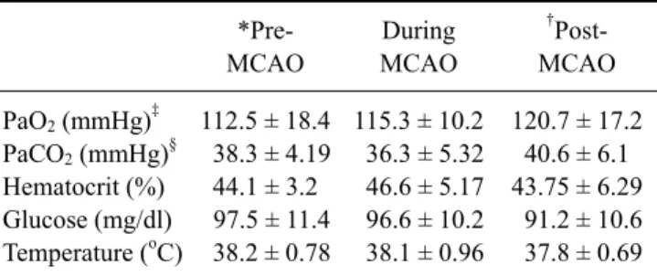
Table 1. Physiological parameters
*Pre- MCAO
During MCAO
†
Post- MCAO PaO
2(mmHg)
‡PaCO
2(mmHg)
§Hematocrit (%) Glucose (mg/dl) Temperature (
oC)
112.5 ± 18.4 38.3 ± 4.19 44.1 ± 3.2 97.5 ± 11.4 38.2 ± 0.78
115.3 ± 10.2 36.3 ± 5.32 46.6 ± 5.17 96.6 ± 10.2 38.1 ± 0.96
120.7 ± 17.2 40.6 ± 6.1 43.75 ± 6.29 91.2 ± 10.6 37.8 ± 0.69
*Examined at 1 h before the surgery; †examined at 1 h after the surgery. ‡PaO2: partial pressure of arterial oxygen; §PaCO2: partial pressure of arterial carbon dioxide.