© 2017 The Korean Ophthalmological Society
This is an Open Access article distributed under the terms of the Creative Commons Attribution Non-Commercial License (http://creativecommons.org/licenses /by-nc/3.0/) which permits unrestricted non-commercial use, distribution, and reproduction in any medium, provided the original work is properly cited.
Original Article
Intraoperatively Observed Lacrimal Obstructive Features and Surgical Outcomes in External Dacryocystorhinostomy
Min Joung Lee1, Sang In Khwarg2, In Hyuk Kim3, Jeong Hoon Choi4, Youn Joo Choi5, Namju Kim6, Ho-Kyung Choung7
1Department of Ophthalmology, Hallym University Sacred Heart Hospital, Anyang, Korea
2Department of Ophthalmology, Seoul National University Hospital, Seoul, Korea
3SW Bright Eye Clinic, Pocheon, Korea
4Department of Ophthalmology, Korean Armed Forces Capital Hospital, Seongnam, Korea
5Department of Ophthalmology, Kangdong Sacred Heart Hospital, Hallym University Medical Center, Seoul, Korea
6Department of Ophthalmology, Seoul National University Bundang Hospital, Seongnam, Korea
7Department of Ophthalmology, Seoul Metropolitan Government Seoul National University Boramae Medical Center, Seoul, Korea
Purpose: To analyze the features of lacrimal drainage system obstruction confirmed during external dacryo- cystorhinostomy surgeries and report the surgical outcomes.
Methods: We reviewed the medical records of a total of 769 cases who underwent external dacryocystorhi- nostomy for primary lacrimal drainage obstruction between 2005 and 2014. Data about detailed location and extent of obstruction were collected intraoperatively. The sites of obstruction were classified into nasolacri- mal duct obstruction (NLDO), common canalicular obstruction (CCO), and canalicular obstruction. Lacrimal sac mucosa and lumen were grossly inspected, and the frequency of lacrimal sac changes, such as signifi- cant inflammation or fibrosis, was analyzed in cases of CCO or canalicular obstruction. The surgical success rate was also evaluated including effect of lacrimal sac status in the CCO and canalicular obstruction groups.
Results: Of 769 cases, primary NLDO with patent canaliculi was diagnosed intraoperatively in 432 cases (56.2%), CCO in 253 (32.9%), and canalicular obstruction in 84 (10.9%). Of 253 cases with CCO, 122 (48.2%) showed clear lacrimal sac lumen, and the other 131 (51.8%) showed significant inflammation or fibrosis of the lacrimal sac. In cases with canalicular obstruction, 35 of 84 (41.7%) showed a clear lacrimal sac, and the other 49 cases (58.3%) cases revealed mucosal changes of the lacrimal sac. The functional success rate was 87.5% for primary NLDO, 75.5% for CCO, and 72.6% for canalicular obstruction. In the CCO group, the functional success rate was lower in cases with significant lacrimal sac change (p = 0.044).
Conclusions: Even in patients with CCO or canalicular obstruction, a large number of cases have lacrimal sac changes, and those changes were associated with lower functional success rate.
Key Words: Dacryocystorhinostomy, Lacrimal duct obstruction, Nasolacrimal duct
Epiphora is one of the most common problems encoun- tered by ophthalmologists, and accurate evaluation of the lacrimal drainage system is crucial in making a diagnosis and establishing an effective treatment plan. Although
Received: September 12, 2016 Accepted: December 26, 2016
Corresponding Author: Sang In Khwarg, MD. Department of Ophthal- mology, Seoul National University Hospital, #101 Daehak-ro, Jongno-gu, Seoul 03080, Korea. Tel: 82-2-2072-2879, Fax: 82-2-741-3187, E-mail:
there are various in-office examinations and imaging stud- ies for the lacrimal drainage system, the location and ex- tent of lacrimal obstruction are sometimes elusive. This can be especially true if patients with epiphora symptoms have common canalicular obstruction (CCO) or canalicular obstruction; it is usually very difficult to accurately assess the status of the lacrimal sac using preoperative tests such as lacrimal irrigation, lacrimal probing, or dacryocystogra- phy. Therefore, research using these examinations seems to not be adequate to evaluate the obstruction site of the lacrimal drainage system [1]. We observed that the status of the lacrimal sac was variable in cases with CCO or can- alicular obstruction during external dacryocystorhinosto- my (DCR); some cases had a clear lacrimal sac, while oth- ers showed mucosal swelling, erythema, and contracture of the lacrimal sac by intraluminal fibrosis and luminal stricture of the proximal nasolacrimal duct. However, there have been few studies that have conducted a detailed ex- amination of the obstruction characteristics of the lacrimal drainage system in patients with epiphora.
External DCR is the standard management option for most cases with lacrimal drainage obstruction including nasolacrimal duct obstruction (NLDO), CCO, and distal canalicular obstruction [2-4]. In addition to the excellent surgical outcomes, external DCR has another advantage over endonasal DCR, as the external approach offers direct visualization of the lacrimal sac. During the procedure, the medial wall of the lacrimal sac is incised, and the lumen and internal punctum of the lacrimal sac can be clearly vi- sualized. Thus, the detailed features of the lacrimal drain- age obstruction can be assessed more precisely during ex- ternal DCR.
The purpose of this study was to classify the locational patterns of lacrimal drainage obstruction and the status of the lacrimal sac in CCO or canalicular obstruction cases based on intraoperative observation during external DCR surgery. Recently, we reported the surgical outcomes of 10 years of external DCR and the risk factors associated with functional failure [5]. Anatomical success was achieved in 98.8% of cases (760 / 769) and functional success in 81.9%
(630 / 769). When analyzing 760 anatomically successful DCRs, CCO (odds ratio [OR], 1.752; p = 0.014) and canalic- ular obstruction (OR, 2.058; p = 0.015) were independent risk factors for functional failure. In subgroup analysis of patients with primary NLDO, patients with a small lacri- mal sac had a significantly higher risk of functional fail-
ure. In that report, the surgical outcomes according to ob- struction site were not specifically described, and the lacrimal sac status in CCO or canalicular obstruction pa- tients was not evaluated. In this study, we investigated the surgical outcomes according to obstruction site and effect of lacrimal sac status on surgical outcomes in CCO and canalicular obstruction groups.
Materials and Methods
Institutional review board approval was obtained for this study. A retrospective review of the electronic medical re- cords of all patients who had undergone external DCR at Seoul National University Hospital between 2005 and 2014 was performed. Patients who were presumed to have NLDO, CCO, or distal canalicular obstruction preopera- tively were indicated for external DCR. Patients with sec- ondary NLDO who had a history of ocular adnexal tu- mors, failed DCR, traumatic NLDO, or previous nasal cavity surgery and patients with events or diseases that can affect the lacrimal drainage system such as facial nerve palsy or lower lid ectropion were excluded. Patients who had a history of chemotherapy or radioiodine therapy were included in this study if the lacrimal obstructive feature indicated DCR surgery. Preoperatively, all patients under- went an evaluation of the lacrimal drainage system includ- ing the lacrimal syringing test and probing. Dacryocys- tography or lacrimal scintigraphy was not routinely performed.
All surgeries were performed by a single, experienced oculoplastic surgeon (SIK) under general anesthesia. The detailed surgical procedure was described in our previous reports [6-8]. A Bowman’s probe was inserted through the upper and lower puncta, and the lacrimal sac was opened with a vertical incision along its medial wall. During the procedure, lacrimal obstructive features were carefully in- spected and described in detail. The obstruction site was determined by canalicular irrigation and probing and in- spection of the internal punctum of the lacrimal sac (com- mon internal ostium) during the surgical procedure. If up- per, lower, and common canaliculus were patent and the lacrimal sac was easily tented, primary NLDO was de- fined. CCO was defined when the canalicular irrigation showed an opposite punctal reflux but canalicular probing showed a soft stop with impossible tenting of the lacrimal
sac. When the tip of the probe was visible under the thin membrane at the internal punctum on canalicular probing after opening the medial wall of the lacrimal sac, thin membranous CCO was diagnosed. When the tip of the probe was not visible at the internal punctum on canalicu- lar probing, thick CCO was diagnosed. Also, the status of the inside of the lacrimal sac was inspected whether the lacrimal sac lumen showed a clean, normal appearance or showed intraluminal swelling, erythema, or scarring. In most cases, a silicone bicanalicular tube (solid tube, 0.064- cm diameter, C-line canaliculus intubation set 8590450;
Medtronic Ophthalmics, Jacksonville, FL, USA) was in- serted. In cases with CCO or canalicular obstruction, dou- ble silicone intubation was performed after internal exci- sion of the obstructed portion of the canaliculus.
Postoperatively, all patients were followed up at 1 week, 1 month, and between 4 and 6 months postoperatively and then variably thereafter. The silicone tube was removed 4 to 6 months after surgery, and only patients who were fol- lowed up more than 4 months were included in this study.
The surgical outcome was assessed anatomically and func-
tionally based on records from the last visit. Anatomical success was defined as good passage without significant reflux on the lacrimal syringing test. Functional success was defined as absence of a tearing symptom as assessed using Munk score. Statistical analyses were performed us- ing IBM SPSS ver. 21.0 (IBM Corp., Armonk, NY, USA).
Chi-square test, t-tests, and Fisher exact tests were used to determine statistical significance.
Results
A total of 769 eyes of 603 patients received external DCR for primary lacrimal drainage obstruction during the enrollment period. The mean age of the patients was 57.7 ± 12.2 years (range, 3 to 84 years), and 468 patients (77.6%) were female. The mean duration of tearing symptom was 62.8 ± 92.3 months (range, 1 to 720 months). Table 1 shows the distribution of the detailed lacrimal obstructive fea- tures observed during external DCR surgery. Primary NLDO accounted for 56.2% (432 / 769) of primary lacri-
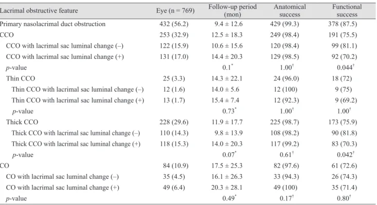
Table 1. Distribution of the detailed lacrimal obstructive features observed during external dacryocystorhinostomy for patients with primary lacrimal outflow obstruction and the associated anatomical and functional success rates
Lacrimal obstructive feature Eye (n = 769) Follow-up period
(mon) Anatomical
success Functional success Primary nasolacrimal duct obstruction 432 (56.2) 9.4 ± 12.6 429 (99.3) 378 (87.5)
CCO 253 (32.9) 12.5 ± 18.3 249 (98.4) 191 (75.5)
CCO with lacrimal sac luminal change (–) 122 (15.9) 10.6 ± 15.6 120 (98.4) 99 (81.1) CCO with lacrimal sac luminal change (+) 131 (17.0) 14.4 ± 20.3 129 (98.5) 92 (70.2)
p-value 0.1* 1.00† 0.044†
Thin CCO 25 (3.3) 14.3 ± 22.1 24 (96.0) 18 (72)
Thin CCO with lacrimal sac luminal change (–) 12 (1.6) 14.0 ± 5.6 12 (100) 9 (75) Thin CCO with lacrimal sac luminal change (+) 13 (1.7) 15.4 ± 7.4 12 (92.3) 9 (69.2)
p-value 0.73* 1.00† 1.00†
Thick CCO 228 (29.6) 11.9 ± 17.7 225 (98.7) 173 (75.9)
Thick CCO with lacrimal sac luminal change (–) 110 (14.3) 9.8 ± 13.9 108 (98.2) 90 (81.8) Thick CCO with lacrimal sac luminal change (+) 118 (15.3) 14.0 ± 20.3 117 (99.2) 83 (70.3)
p-value 0.07* 0.61† 0.042†
CO 84 (10.9) 17.5 ± 25.3 82 (97.6) 61 (72.6)
CO with lacrimal sac luminal change (–) 35 (4.5) 16.1 ± 26.3 33 (94.3) 26 (74.3) CO with lacrimal sac luminal change (+) 49 (6.4) 20.3 ± 28.1 49 (100) 35 (71.4)
p-value 0.49* 0.17† 0.80†
Values are presented as number (%) or mean ± standard deviation.
CCO = common canalicular obstruction; CO = canalicular obstruction.
*Student t-test; †Chi-square test or Fisher exact test.
mal drainage obstruction. CCO was diagnosed in 32.9%
(253 / 769); thin membranous CCO accounted for 3.3% (25 / 769), thick CCO accounted for 29.6% (228 / 769), and canalicular obstruction was diagnosed in 10.9% (84 / 769).
Significant lacrimal sac changes, such as intraluminal swelling, erythema, or fibrosis, were present in 51.8% (131 of 253 eyes) of the CCO group and 58.3% (49 of 84 eyes) of the canalicular obstruction group. The frequency of lacri- mal sac changes was similar between thin CCO (52%, 13 of 25 eyes) and thick CCO (51.8%, 118 of 228 eyes).
The overall anatomical and functional success rate was 98.8% (760 of 769 eyes) and 81.9% (630 of 769 eyes). The anatomical success rate was not significantly different among primary NLDO, CCO, and canalicular obstruction groups (p = 0.484), but the functional success rate was sig- nificantly higher in the primary NLDO group than in the CCO or canalicular obstruction group (p = 0.004 and p = 0.001, respectively). The subgroup analyses for success rate according to the status of the lacrimal sac are described in Table 1. When there were lacrimal sac luminal changes, the functional success rate significantly decreased in the CCO group (p = 0.044). Subgroup analysis revealed that the functional success rate was higher in cases without lacri- mal sac changes than in those with lacrimal sac changes in the thick CCO group, while the functional success rate was not different regardless of the status of the lacrimal sac in the thin CCO group. In the canalicular obstruction group, the functional success rate was not different according to the status of the lacrimal sac.
Discussion
This study analyzed the distribution of lacrimal drainage system obstruction patterns in patients who required DCR surgeries based on intraoperative findings of the canalicu- lus and lacrimal sac. In 769 cases with primary lacrimal drainage obstruction, 56.2% had primary NLDO with pat- ent canaliculi; 32.9% had CCO; and 10.9% had canalicular obstruction. Approximately half of the cases of CCO and canalicular obstruction (131 of 253 eyes and 49 of 84 eyes) demonstrated simultaneous lacrimal sac mucosal changes, such as inflammation or fibrosis.
The level of lacrimal drainage obstruction can be rough- ly estimated preoperatively using several examinations.
However, in cases with upper lacrimal drainage system
obstruction, the precise status of the lower lacrimal drain- age system cannot be accurately assessed using these tests.
Consequently, lacrimal drainage obstruction is only able to be broadly classified as NLDO, CCO, or canalicular ob- struction. In this study, we describe the detailed character- istics of primary lacrimal drainage obstruction based on intraoperative observations of the canaliculus and lacrimal sac at the step of lacrimal sac incision during the external DCR procedure. When a lacrimal probe could not be in- serted into the lacrimal sac, CCO or canalicular obstruc- tion was diagnosed according to the soft stop level. CCO can be subdivided into thin membranous CCO and thick CCO based on the nature of the soft tissue at the internal punctum of the lacrimal sac. In cases with CCO or cana- licular obstruction, if the incised lacrimal sac showed sig- nificant intraluminal swelling, erythema, or scarring, we assumed that there were concomitant lacrimal sac luminal changes. This allowed us to make a more detailed classifica- tion of primary lacrimal drainage obstruction; primary NLDO with patent canaliculus (56.2%), CCO without lacri- mal sac luminal change (15.9%), CCO with lacrimal sac lu- minal change (17.0%), canalicular obstruction without lacri- mal sac luminal change (4.5%), and canalicular obstruction with lacrimal sac luminal change (6.4%) (Table 1).
Even though we often observed gross changes of the lac- rimal sac mucosa during DCR, these changes have been rarely noted in other studies. It is well known that chronic inflammation and fibrosis are the most common histo- pathologic changes in lacrimal sacs of patients with NLDO [9-11]. We speculated that the cases with CCO or canalicu- lar obstruction, who showed intraluminal lacrimal sac changes, presumed to have simultaneous NLDO. In these patients, CCO or canalicular obstruction might develop secondary to a chronic inflammatory reaction of the lacri- mal sac. In a study using dacryoendoscopy, the authors tried canalicular incision and tube intubation in patients with CCO and reported that there was simultaneous NLDO in one-quarter of patients, which makes intubation difficult, supporting our concept [12]. In the present study, approximately half of the cases with CCO or canalicular obstruction (53.4%, 180 / 337 cases) showed intraluminal mucosal swelling, erythema, or fibrosis in the lacrimal sac.
Our findings support the assertion that DCR is a more suitable option for management of CCO or canalicular ob- struction rather than canalicular trephination or canalicu- loplasty, considering the possibility of combined lacrimal
sac changes. In terms of success rate of external DCR, the functional success rate was higher in the primary NLDO group than in CCO or canalicular obstruction, in agree- ment with previous studies [5,13,14]. In cases of thick CCO, the status of the lacrimal sac affected the functional suc- cess rate, and this is a novel finding that has not been pre- viously reported. The difference was not significant in the thin CCO group or the canalicular obstruction group. We think that the number of cases in the thin CCO group was too small to allow a statistical conclusion. In canalicular obstruction, canalicular obstruction itself is a strong, unfa- vorable factor for functional success, so the influence of lacrimal sac status may be limited.
In conclusion, lacrimal sac mucosal changes were con- firmed intraoperatively during external DCR in a signifi- cant proportion (53.4%) of cases with CCO or canalicular obstruction. In the CCO group, those changes were associ- ated with lower functional success rate.
Conflict of Interest
No potential conflict of interest relevant to this article was reported.
References
1. Beigi B, Uddin JM, McMullan TF, Linardos E. Inaccuracy of diagnosis in a cohort of patients on the waiting list for dacryocystorhinostomy when the diagnosis was made by only syringing the lacrimal system. Eur J Ophthalmol 2007;17:485-9.
2. Huang J, Malek J, Chin D, et al. Systematic review and me- ta-analysis on outcomes for endoscopic versus external da- cryocystorhinostomy. Orbit 2014;33:81-90.
3. Pandya VB, Lee S, Benger R, et al. External dacryocysto- rhinostomy: assessing factors that influence outcome. Orbit
2010;29:291-7.
4. Erdol H, Akyol N, Imamoglu HI, Sozen E. Long-term fol- low-up of external dacryocystorhinostomy and the factors affecting its success. Orbit 2005;24:99-102.
5. Lee MJ, Khwarg SI, Kim IH, et al. Surgical outcomes of ex- ternal dacryocystorhinostomy and risk factors for functional failure: a 10-year experience. Eye (Lond) 2017;31:691-7.
6. Lee MJ, Khwarg SI, Choung HK, Kim N. Associated fac- tors of functional failure of external dacryocystorhinosto- my. Can J Ophthalmol 2014;49:40-4.
7. Choung HK, Khwarg SI. Selective non-intubation of a sili- cone tube in external dacryocystorhinostomy. Acta Oph- thalmol Scand 2007;85:329-32.
8. Hwang SW, Khwarg SI, Kim JH, et al. Bicanalicular dou- ble silicone intubation in external dacryocystorhinostomy and canaliculoplasty for distal canalicular obstruction. Acta Ophthalmol 2009;87:438-42.
9. Salour H, Hatami MM, Parvin M, et al. Clinicopathologi- cal study of lacrimal sac specimens obtained during DCR.
Orbit 2010;29:250-3.
10. Lee-Wing MW, Ashenhurst ME. Clinicopathologic analy- sis of 166 patients with primary acquired nasolacrimal duct obstruction. Ophthalmology 2001;108:2038-40.
11. Paulsen FP, Thale AB, Maune S, Tillmann BN. New in- sights into the pathophysiology of primary acquired da- cryostenosis. Ophthalmology 2001;108:2329-36.
12. Sasaki T, Sounou T, Sugiyama K. Dacryoendoscopic sur- gery and tube insertion in patients with common canalicu- lar obstruction and ductal stenosis as a frequent complica- tion. Jpn J Ophthalmol 2009;53:145-50.
13. Choi JC, Jin HR, Moon YE, et al. The surgical outcome of endoscopic dacryocystorhinostomy according to the ob- struction levels of lacrimal drainage system. Clin Exp Oto- rhinolaryngol 2009;2:141-4.
14. Konuk O, Kurtulmusoglu M, Knatova Z, Unal M. Unsuc- cessful lacrimal surgery: causative factors and results of surgical management in a tertiary referral center. Ophthal- mologica 2010;224:361-6.