pISSN: 1011-8942 eISSN: 2092-9382
© 2015 The Korean Ophthalmological Society
This is an Open Access article distributed under the terms of the Creative Commons Attribution Non-Commercial License (http://creativecommons.org/licenses /by-nc/3.0/) which permits unrestricted non-commercial use, distribution, and reproduction in any medium, provided the original work is properly cited.
138
Bilateral Macula-involving Meta- static Infection Resulting from Sep- tic Embolization
Dear Editor,
Retinal features associated with septicemia usually do not involve the macula. However, there have been a small number of reports regarding macular hole cases secondary to septic emboli [1,2]. We report a case of bilateral macu- la-involving metastatic infection rather than macular hole resulting from septic embolization.
A 29-year-old man who had initially presented to the emergency room for altered mentality and been diagnosed with gram-positive cocci bacteremia and meningoenceph- alitis after work-up was referred for decreased bilateral vi- sion, noted about 3 days after admission. Methicillin-sensi- tive Staphylococcus aureus was observed on blood culture.
The patient began a 4-week course of intravenous penicil- lin G (4 million units every 4 hours). On initial examina- tion, his best-corrected visual acuities (BCVA) were 20 / 40 in right eye and 20 / 100 in left eye. Fundus examina- tion showed small white infiltration with hemorrhage in the juxtafovea of both eyes (Fig. 1A). Fluorescein angiog- raphy showed no active leakage (Fig. 1B). Spectral do- main-optical coherence tomography images showed dis- ruption at the junction of the photoreceptor inner and outer segments (IS/OS) junction and cone outer segment and hy- perreflective intraretinal deposits involving both the inner and outer retinal layers at the fovea (Fig. 1C). The area of IS/OS junction and cone outer segment layer disruption was much wider and more extensive in the left eye, in which there was also a focal area of near total retinal tissue defect close to the shape of an impending macular hole in the juxtafovea of the left eye (Fig. 1D).
On the second visit 2 weeks after appropriate intrave- nous antibiotic therapy, his BCVA had improved to 20 / 22 in right eye and 20 / 30 in left eye. Fundus examination showed complete resolution of the hyperreflective intraret- inal deposits in both eyes (Fig. 1E). Spectral domain-opti- cal coherence tomography showed near full restoration of the previously disrupted IS/OS junction and cone outer segment lines except for a small focal defect of the IS/OS junction in the left eye. The hyperreflective intraretinal de- posits in both eyes were no longer present (Fig. 1F). Three months later, his BCVA were 20 / 20 in right eye and 20 / 40 in left eye; however, the focal defect of the IS/OS junc- tion in the left eye was nearly unchanged, and the continu- ity of each retinal layer remained incomplete.
Although the incidence of macula-involving metastatic infection secondary to bacterial septic embolism is rare, careful observation is required when performing fundus examination for possible metastatic infection in bacteremia patients. Metastatic septic emboli may involve both the outer and inner retinal layers at the bilateral fovea and may cause a near total retinal tissue defect close to that of an impending macular hole. Our case showed that a macu- la-involving metastatic infection can involve reversible ret- inal structural change with no functional sequelae and im- provement of visual acuity after suitable antibiotic therapy.
Junyoung Park
Department of Ophthalmology, Seoul National University College of Medicine, Seoul, Korea
Tae Wan Kim, Jeeyun Ahn
Department of Ophthalmology, SMG-SNU Boramae Medical Center, Seoul National University College of Medicine, Seoul, Korea
E-mail: [email protected] Korean J Ophthalmol 2015;29(2):138-139
http://dx.doi.org/10.3341/kjo.2015.29.2.138
Correspondence
139
Conflict of Interest
No potential conflict of interest relevant to this article was reported.
References
1. Beatty S, Harrison RJ, Roche P. Bilateral macular holes re- sulting from septic embolization. Am J Ophthalmol 1997;
123:557-9.
2. Yoon CH, Park KH, Woo SJ. Macular hole formation sec- ondary to bacterial septic embolism demonstrated by serial spectral-domain optical coherence tomography imaging.
Ocul Immunol Inflamm 2013;21:163-5.
A
C
F
B
D
E
Nasal
OD
OS
Temporal
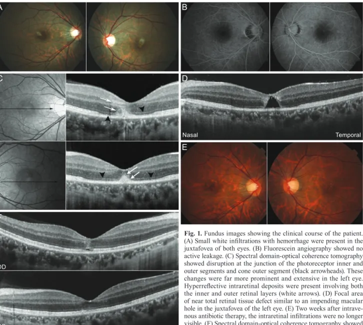
Fig. 1. Fundus images showing the clinical course of the patient.
(A) Small white infiltrations with hemorrhage were present in the juxtafovea of both eyes. (B) Fluorescein angiography showed no active leakage. (C) Spectral domain-optical coherence tomography showed disruption at the junction of the photoreceptor inner and outer segments and cone outer segment (black arrowheads). These changes were far more prominent and extensive in the left eye.
Hyperreflective intraretinal deposits were present involving both the inner and outer retinal layers (white arrows). (D) Focal area of near total retinal tissue defect similar to an impending macular hole in the juxtafovea of the left eye. (E) Two weeks after intrave- nous antibiotic therapy, the intraretinal infiltrations were no longer visible. (F) Spectral domain-optical coherence tomography showed near full restoration of inner and outer segments junction and cone outer segment lines except for small focal defects at the fovea. Hy- perreflective intraretinal infiltrations had decreased substantially.
OD = right eye; OS = left eye.