Author contributions: P.K.R. did literature survey and wrote the article. A.B.
helped in literature survey and writing article. J.K.V. edited the manuscript.
A.S.J. conceived the idea, arranged the manuscript and did the final revi- sion.
This is an Open Access article distributed under the terms of the Creative Commons Attribution Non-Commercial License, which permits unrestricted non-commercial use, distribution, and reproduction in any medium, provided the original work is properly cited.
Copyright © Korean J Physiol Pharmacol, pISSN 1226-4512, eISSN 2093-3827
INTRODUCTION
Cardiac conditioning is a therapeutic approach to induce an endogenous protective adaptive response in the heart for coun- teracting sustained ischemia-reperfusion injury [1,2]. In this ap- proach, heart or other remote organ is subjected to short period of ischemia and reperfusion (conditioning stimulus) either before or after sustained ischemia. Based on the period of delivery of
the conditioning stimulus (before, during or after sustained isch- emia), and site of delivery (to the target organ or remote organ), the treatment approach is classified into ischemic precondition- ing [3], ischemic perconditioning, ischemic post-conditioning [4], remote preconditioning [5] and remote postconditioning [6].
Pharmacological preconditioning is a treatment approach where- in the administration of various pharmacological agents elicits preconditioning like cardioprotective effects [7].
Review Article
Conditioning-induced cardioprotection: Aging as a confounding factor
Puneet Kaur Randhawa
1, Anjana Bali
2, Jasleen Kaur Virdi
1, and Amteshwar Singh Jaggi
1,*
1Department of Pharmaceutical Sciences and Drug Research, Punjabi University, Patiala 147002 India, 2Akal College of Pharmacy and Technical Education, Mastuana Sahib, Sangrur 148002, India
ARTICLE INFO
Received November 22, 2017 Revised March 28, 2018 Accepted May 15, 2018
*Correspondence Amteshwar Singh Jaggi
E-mail: [email protected] Key Words
Adenosine Aging Conditioning Oxidative stress
ABSTRACT The aging process induces a plethora of changes in the body including
alterations in hormonal regulation and metabolism in various organs including the
heart. Aging is associated with marked increase in the vulnerability of the heart to
ischemia-reperfusion injury. Furthermore, it significantly hampers the development
of adaptive response to various forms of conditioning stimuli (pre/post/remote condi-
tioning). Aging significantly impairs the activation of signaling pathways that mediate
preconditioning-induced cardioprotection. It possibly impairs the uptake and release
of adenosine, decreases the number of adenosine transporter sites and down-regu-
lates the transcription of adenosine receptors in the myocardium to attenuate ade-
nosine-mediated cardioprotection. Furthermore, aging decreases the expression of
peroxisome proliferator-activated receptor gamma co-activator 1-alpha (PGC-1a) and
subsequent transcription of catalase enzyme which subsequently increases the oxida-
tive stress and decreases the responsiveness to preconditioning stimuli in the senes-
cent diabetic hearts. In addition, in the aged rat hearts, the conditioning stimulus fails
to phosphorylate Akt kinase that is required for mediating cardioprotective signaling
in the heart. Moreover, aging increases the concentration of Na
+and K
+, connexin
expression and caveolin abundance in the myocardium and increases the susceptibil-
ity to ischemia-reperfusion injury. In addition, aging also reduces the responsiveness
to conditioning stimuli possibly due to reduced kinase signaling and reduced STAT-3
phosphorylation. However, aging is associated with an increase in MKP-1 phosphory-
lation, which dephosphorylates (deactivates) mitogen activated protein kinase that
is involved in cardioprotective signaling. The present review describes aging as one
of the major confounding factors in attenuating remote ischemic preconditioning-
induced cardioprotection along with the possible mechanisms.
The aging process induces a plenty of changes in the composi- tion of body by altering the energy balance and may increase the susceptibility of individuals to the cardiovascular diseases [8,9].
Despite the advances in prophylactic and preventive therapies, cardiovascular disorders remain one of the main causes of deaths in the elderly cohorts [10]. There have been abundant preclini- cal reports indicating that aging increases the vulnerability of the heart to ischemia-reperfusion injury [11,12]. Furthermore, various researchers have demonstrated that aging reduces the development of adaptive cardioprotective response on being subjected to conditioning stimuli, including hypoxic or ischemic preconditioning [13-15], ischemic postconditioning [16] and re- mote ischemic preconditioning [17]. The present review describes aging as one of the major confounding factors that may attenuate conditioning-induced cardioprotection along with the possible mechanisms.
EVIDENCES
Aging exaggerates ischemia-reperfusion injury
Aging is one of the major prognostic determinants of myocar- dial infarction and renders the aged heart more susceptible to coronary artery disease [18-20]. Lesnefsky et al. reported that ag- ing increases the susceptibility of the isolated heart to ischemia- reperfusion injury. The authors reported that aging significantly reduces the recovery of left ventricular developed pressure (LVDP) following ischemic injury in aged rats (24 months). Furthermore, tissue damage in terms of lactate dehydrogenase (LDH), creatine kinase (CK) release and total protein release was much higher in the aged rat hearts [21]. Similarly, Tani and coworkers reported a gradual decline in ischemic tolerance with increasing age in iso- lated rat hearts. Sustained ischemia and thereafter reperfusion led to greater impairment in functional contractility, enhanced LDH and CK release in the aged rat hearts (50-100 weeks) in compari- son to the young hearts (12 weeks) [22].
Scientists have also reported that there is enhancement of ischemia reperfusion-induced myocardial injury due to ageing in mice. Azhar et al. demonstrated that left anterior coronary artery ligation followed by reperfusion led to greater myocardial necro- sis, DNA fragmentation, and mitochondrial disruption in the old mice hearts (22-24 months) in comparison to the young mice hearts (6-8 months) [23]. Other studies have also reported that se- nescence substantially reduces the ischemic tolerance in the mice hearts (18-28 months) [11,24]. Furthermore, it also shown that the deleterious effect of co-morbidity on ischemia reperfusion injury is significantly increased in aged animals. Whittington et al.
reported that the aged diabetic Goto-Kakizaki rats (18 months) are more prone to ischemic injury in comparison to the young diabetic rat hearts (3 months) [15].
Apart from the studies on animals, Mariani et al. reported
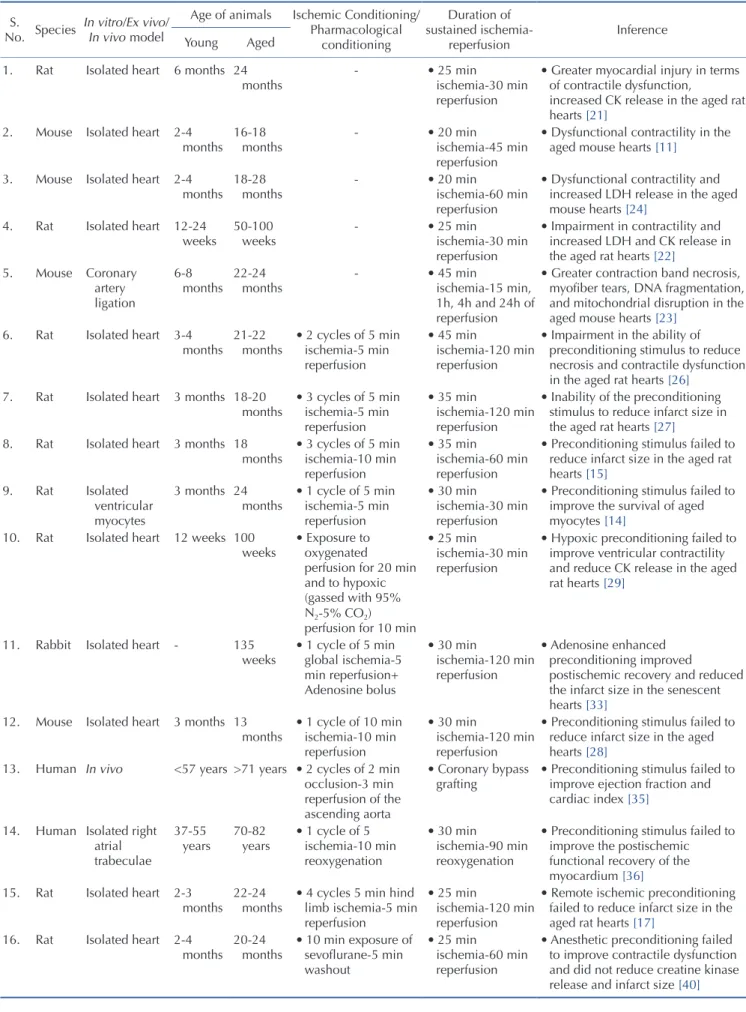
that aged human myocardium also loses the capacity to recover following hypoxic injury. The authors reported the lesser degree of recovery (in terms of development of force) in the atrial pecti- nate trabeculae (dissected from the right atrial appendages of the patients, 34-89 years) isolated from the old cohorts (around 89 years) in comparison to the young cohorts (around 34 years) in response to hypoxic injury (Table 1) [25].
Aging attenuates the protective effects of ischemic preconditioning
In animals: Apart from studies showing per se deleterious effects of aging on ischemia reperfusion injury, studies have also shown that aging attenuates the cardioprotective effects of ischemic preconditioning [14,15,26,27]. Fenton and co-workers reported that two episodes of ischemic preconditioning (5 min ischemia-5 min reperfusion) prior to ischemic insult (45 min ischemia-120 min reperfusion) significantly reduced the devel- opment of necrosis and remarkably improved the post-ischemic functional recovery in the isolated young adult (3-4 months) rat hearts (Fischer 344). However, ischemic preconditioning stimulus failed to attenuate the development of necrosis and impaired the contractile function in the aged hearts (21-22 months) suggest- ing that aging impairs the ability of preconditioning stimulus to reduce necrosis and contractile dysfunction in the rat hearts [26]. Schulman et al. reported that preconditioning stimulus led to reduction of infarct size in the young rat hearts (3 months).
However, the senescent hearts (18-20 months) were refractory to ischemic and pharmacological preconditioning with 2-chloro- N(6)-cyclopentyladenosine (adenosine A
1receptor agonist), 1,2-dioctanoyl-sn-glycerol (the protein kinase C agonist) and diazoxide (mitochondrial K
ATP-channel opener). This indicates that aging induces alterations within ischemic preconditioning signaling cascade to reduce its ability to produce cardioprotection [27]. Tani et al. reported that hypoxic preconditioning stimulus improves post-ischemic recovery and reduces CK release selec- tively in the young rat hearts, but not in the senescent hearts [28].
Besides, Boengler and co-workers also reported that ischemic preconditioning (10 min ischemia and 10 min reperfusion) sig- nificantly reduced ischemia-reperfusion-induced myocardial infarct size in the young hearts (<3 months), without producing protective effects in the aged mice hearts (>13 months) [29].
McCully et al. reported the novel protective protocol termed
as “adenosine-enhanced ischemic preconditioning” in which
adenosine was given along with ischemic preconditioning to en-
hance the cardioprotection [30]. These authors reported that the
combination of adenosine with ischemic preconditioning signifi-
cantly produced cardioprotection in senescent rabbit hearts and
also produced functional recovery in these hearts. Based on this,
it may tentatively proposed that decrease in adenosine levels due
to aging may possibly contribute in attenuating the cardioprotec-
tive effects of ischemic preconditioning. Therefore, addition of
Table 1. Conditioning-induced protection in aged vs. young hearts S.
No. Species In vitro/Ex vivo/
In vivo model
Age of animals Ischemic Conditioning/
Pharmacological conditioning
Duration of sustained ischemia-
reperfusion Inference
Young Aged
1. Rat Isolated heart 6 months 24
months - • 25 min
ischemia-30 min reperfusion
• Greater myocardial injury in terms of contractile dysfunction,
increased CK release in the aged rat hearts [21]
2. Mouse Isolated heart 2-4
months 16-18
months - • 20 min
ischemia-45 min reperfusion
• Dysfunctional contractility in the aged mouse hearts [11]
3. Mouse Isolated heart 2-4
months 18-28
months - • 20 min
ischemia-60 min reperfusion
• Dysfunctional contractility and increased LDH release in the aged mouse hearts [24]
4. Rat Isolated heart 12-24
weeks 50-100
weeks - • 25 min
ischemia-30 min reperfusion
• Impairment in contractility and increased LDH and CK release in the aged rat hearts [22]
5. Mouse Coronary artery ligation
6-8
months 22-24
months - • 45 min
ischemia-15 min, 1h, 4h and 24h of reperfusion
• Greater contraction band necrosis, myofiber tears, DNA fragmentation, and mitochondrial disruption in the aged mouse hearts [23]
6. Rat Isolated heart 3-4
months 21-22
months • 2 cycles of 5 min ischemia-5 min reperfusion
• 45 min
ischemia-120 min reperfusion
• Impairment in the ability of preconditioning stimulus to reduce necrosis and contractile dysfunction in the aged rat hearts [26]
7. Rat Isolated heart 3 months 18-20
months • 3 cycles of 5 min ischemia-5 min reperfusion
• 35 min
ischemia-120 min reperfusion
• Inability of the preconditioning stimulus to reduce infarct size in the aged rat hearts [27]
8. Rat Isolated heart 3 months 18
months • 3 cycles of 5 min ischemia-10 min reperfusion
• 35 min
ischemia-60 min reperfusion
• Preconditioning stimulus failed to reduce infarct size in the aged rat hearts [15]
9. Rat Isolated ventricular myocytes
3 months 24
months • 1 cycle of 5 min ischemia-5 min reperfusion
• 30 min
ischemia-30 min reperfusion
• Preconditioning stimulus failed to improve the survival of aged myocytes [14]
10. Rat Isolated heart 12 weeks 100
weeks • Exposure to oxygenated perfusion for 20 min and to hypoxic (gassed with 95%
N2-5% CO2) perfusion for 10 min
• 25 min
ischemia-30 min reperfusion
• Hypoxic preconditioning failed to improve ventricular contractility and reduce CK release in the aged rat hearts [29]
11. Rabbit Isolated heart - 135
weeks • 1 cycle of 5 min global ischemia-5 min reperfusion+
Adenosine bolus
• 30 min
ischemia-120 min reperfusion
• Adenosine enhanced preconditioning improved
postischemic recovery and reduced the infarct size in the senescent hearts [33]
12. Mouse Isolated heart 3 months 13
months • 1 cycle of 10 min ischemia-10 min reperfusion
• 30 min
ischemia-120 min reperfusion
• Preconditioning stimulus failed to reduce infarct size in the aged hearts [28]
13. Human In vivo <57 years >71 years • 2 cycles of 2 min occlusion-3 min reperfusion of the ascending aorta
• Coronary bypass
grafting • Preconditioning stimulus failed to improve ejection fraction and cardiac index [35]
14. Human Isolated right atrial trabeculae
37-55
years 70-82
years • 1 cycle of 5 ischemia-10 min reoxygenation
• 30 min
ischemia-90 min reoxygenation
• Preconditioning stimulus failed to improve the postischemic functional recovery of the myocardium [36]
15. Rat Isolated heart 2-3
months 22-24
months • 4 cycles 5 min hind limb ischemia-5 min reperfusion
• 25 min
ischemia-120 min reperfusion
• Remote ischemic preconditioning failed to reduce infarct size in the aged rat hearts [17]
16. Rat Isolated heart 2-4
months 20-24
months • 10 min exposure of sevoflurane-5 min washout
• 25 min
ischemia-60 min reperfusion
• Anesthetic preconditioning failed to improve contractile dysfunction and did not reduce creatine kinase release and infarct size [40]
adenosine in ischemic preconditioning protocol produced cardio- protection in aged hearts [31].
Cells of animals: The studies performed on cardiomyocytes have also revealed that cardioprotective effects of ischemic pre- conditioning are lost due to aging. Brien and Howlett reported that ischemic preconditioning of ventricular myocytes isolated from young adult Fischer 344 rats significantly abolished post- ischemic contractile dysfunction, diastolic contracture and markedly improved the survival of myocytes. Moreover, ischemic preconditioning also increased the amplitude of Ca
2+transients reperfusion and diastolic Ca
2+during reperfusion. Although, ischemic preconditioning increased the amplitude of Ca
2+tran- sients, improved contractile function, reduced the marked rise in diastolic Ca
2+in the aged myocytes during reperfusion, yet it failed to improve the survival of these aged myocytes. This indicates that increased availability of intracellular Ca
2+during preconditioning possibly improves the contractile dysfunction.
However, the decrease in survival of the aged myocytes can be possibly due to age-dependent changes in the structure and func- tioning of the isolated myocytes [14].
Preconditioning stimulus tends to reduce platelet adhesion and aggregation in models of myocardial infarction and recurrent thrombosis. The anti-platelet effect of ischemic preconditioning is possibly due to enhanced discharge of adenosine and subsequent activation of adenosine A
2receptors on the platelet surface [32].
Przyklenk and Whittaker demonstrated that exogenous adminis- tration of CGS 21680 (A
2receptor agonist) in young adult rabbits led to reduction of platelet aggregation in an in vitro model of platelet aggregation. However, CGS 21680 administration failed
to reduce platelet aggregation in the old cohort indicating age- dependent reduction in responsivity of the platelets to adenosine preconditioning [33].
Humans and human tissues: Apart from this, various re- searchers have reported that ischemic preconditioning does not produce cardioprotective effects in the senescent human myocar- dium [34-36]. The existence of angina before acute myocardial in- farction serves as preconditioning stimulus (physiological precon- ditioning) and exerts cardioprotective effect in the adult patients.
However, the protection afforded by antecedent angina is lost in the senescent myocardium indicating absence of preconditioning like protective effects in the old cohorts [34]. Furthermore, Wu et al. reported that ischemic preconditioning stimulus protects the heart against ischemia-reperfusion injury in adult patients undergoing coronary artery bypass grafting in terms of reduced cardiac troponin I release and improvement in ejection fraction and cardiac index. However, preconditioning stimulus failed to reduce cardiac troponin I release, improve ejection fraction and cardiac index in the aged patients undergoing coronary artery bypass grafting. This again indicates that the protective effect of preconditioning stimuli diminishes with age [35]. The loss of protective effects of ischemic preconditioning with increasing age is further supported by a study of showing that ischemic precon- ditioning did not improve functional recovery of right atrial tra- beculae of old patients (more than 70 years age) in comparison to adult patients (less than 55 years) [36]. Another study reports that aging significantly reduces ischemic preconditioning-induced protective effects on endothelial cells. The authors reported that ischemic preconditioning selectively preserved flow-mediated
Table 1. Continued S.
No. Species In vitro/Ex vivo/
In vivo model
Age of animals Ischemic Conditioning/
Pharmacological conditioning
Duration of sustained ischemia-
reperfusion Inference
Young Aged
17. Mouse Isolated heart 10-14
months 24-26
months • Acute infusion of morphine
• Chronic exposure to morphine
• 25 min ischemia-45 min reperfusion
• Preconditioning with acute morphine infusion failed to improve ventricular contractility and did not reduce LDH release
• Chronic exposure to morphine improved contractile dysfunction and reduced LDH release [41]
18. Rat Coronary artery ligation model
3-5
months 20-24
months • 30 min exposure of
isoflurane • 30 min
ischemia-120 min reperfusion
• Isoflurane exposure failed to reduce infarct size in the old rats [42]
Ischemic Postconditioning 19. Mouse Isolated heart 3-4
months 20-24
months • 3/6, 10 second cycles of stuttered reflow
• 30 min
ischemia-120 min reperfusion
• Postconditioning failed to reduce the infarct size in the old mouse hearts [16]
20. Mouse Isolated heart 3 months 13
months • 3 cycles of
ischemia-reperfusion
• 5 cycles of
ischemia-reperfusion
• 30 min
ischemia-120 min reperfusion
• 3 cycles of postconditioning failed to reduce the infarct size in the aged mouse hearts
• 5 cycles of postconditioning reduced the infarct size in the aged mouse hearts [45]
dilation in the brachial artery in young men (20-25 years), but not in the elderly patients (68-77 years) suggesting that aging reduces the ability of ischemic preconditioning to protect against isch- emia-reperfusion injury associated endothelial dysfunction [37].
On the contrary, a study of Loubani et al. on right atrial tissues isolated from hearts undergoing elective heart surgery indicated that aging does not influence the protective effects of ischemic preconditioning and ischemic preconditioning was shown to pro- tect atrial tissues of all age groups against ischemia-reperfusion injury (Table 1) [38].
Aging abolishes remote ischemic preconditioning- induced cardioprotection
It has also been documented that aging is a confounding factor in reducing the effectiveness of remote ischemic preconditioning- induced adaptive response against ischemia-reperfusion injury in rodents [17]. Behmenburg et al. reported that four cycles of remote ischemic preconditioning (5 min hind limb ischemia-5 min reperfusion) prior to sustained ischemia (25 min) and 120 min reperfusion led to significant reduction in the infarct size in young adult rats (2-3 months). However, remote ischemic preconditioning-dependent reduction in infarct size was absent in senescent heart (22-24 months). This indicates that the efficacy of remote ischemic preconditioning to reduce infarct size reduces with age [17]. Moreover, Hausenloy et al. reported that remote ischemic preconditioning failed to improve clinical outcomes in patients (n=1612) undergoing coronary artery bypass graft surgery. The authors reported that the incidences of myocardial infarction, coronary revascularization, stroke and death in older adult patients (76.3±7.0 years) were not reduced even after remote preconditioning. Furthermore, remote ischemic preconditioning did not reduce troponin T levels, inotropic score, acute kidney injury and total stay period in the intensive care unit suggesting that the efficacy of remote ischemic preconditioning may not be observed in older patients (Table 1) [39].
Negative influence of aging on pharmacological preconditioning and postconditioning
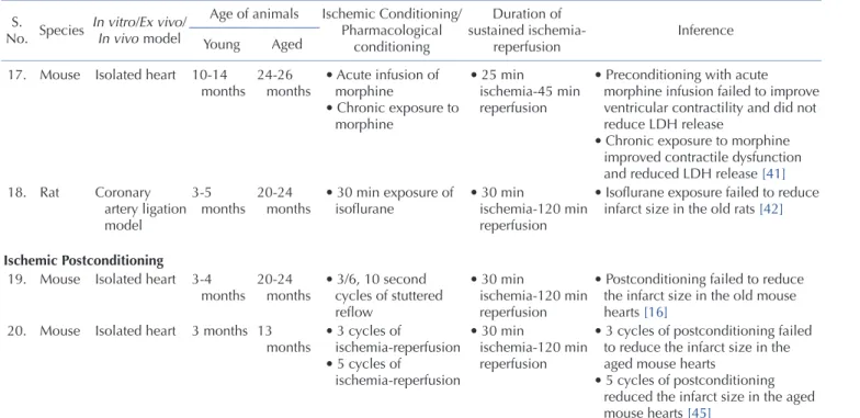
As per reports of ischemic and remote preconditioning, the effects of pharmacological preconditioning and postcondition- ing are also abolished with increasing age. Sniecinski and Liu reported that sevoflurane preconditioning significantly reduced CK release, infarct size and improved left ventricular developed pressure following 25 min of ischemia and 60 min of reperfusion in young rats, but not in the aged rats [40]. Peart and Gross dem- onstrated that acute infusion of morphine significantly improved left ventricular developed pressure and end diastolic pressure in the young (10-14 months), but not in the senescent mouse hearts (24-26 months) [41]. Nguyen et al. reported that pharmacological preconditioning with isoflurane, an anesthetic, abolished coro-
nary artery occlusion-induced increase in myocardial infarct size in the young rats 0 (3-5 months) in comparison to old rats (20-24 months) [42]. Similarly, Li et al. reported that sevoflurane post- conditioning followed by 30 min ischemia and 120 min reperfu- sion did not reduce infarct size in senescent rats (20-24 months) in comparison to the young rats (3-4 months) [43]. On the contrary, Jiang and co-workers reported that sevoflurane postconditioning significantly improved coronary flow, functional recovery, re- duced mitochondrial lesion severity and apoptosis in the aged rat hearts (Table 1 ) [44].
Aging as a confounder in ischemic postconditioning
Przyklenk et al. reported that repeated cycles of ischemic post- conditioning reduce the infarct size in isolated adult mouse hearts (3-4 months), while it failed to reduce the infarct size in the aged mouse hearts (20-24 months). This suggests that aging reduces the effectiveness of ischemic postconditioning in providing pro- tection against myocardial necrosis [16]. Boengler et al. reported that three cycles of ischemic postconditioning reduced the infarct size in the young mice (3 months), without any significant effect in aged mice (>13 months). However, augmented ischemic post- conditioning i.e., five cycles (instead of three cycles) of ischemic postconditioning reduced infarct size in the aged mice hearts (>13 months). This indicates that an increase in the number of cycles of ischemic postconditioning stimulus may overcome the con- founding effects of aging (Table 1 ) [45].
MECHANISMS RESPONSIBLE FOR AN INCREASE IN ISCHEMIC INJURY DUE TO AGING
Estrogen deficiency
Estrogen status of a female rat determines the susceptibility of the heart to ischemia-reperfusion injury [46,47]. Aging leads to a significant decline in the estrogen levels in the females and increases the susceptibility of the heart to ischemia-reperfusion injury [48,49]. Xu et al. reported that chronic in vivo adminis- tration of estrogen in aged ovariectomized rats (12-14 months) markedly improved functional recovery after 25 min ischemia-40 min reperfusion [48]. In corroboration with the above study, Novotny et al. described that selective estrogen receptor-a activa- tion in aged female rats partly reduces infarct size [50]. A previ- ous study has also indicated that administration of 17b-estradiol significantly reduces myocardial infarct size in canine hearts [51].
Furthermore, a study has indicated that administration of 17b- estradiol in ovariectomized female rats significantly increased the number of viable myocytes and reduced mitochondrial and ultra- structural damage to the myocytes isolated from the hearts [46].
Apart from these, clinical trials have shown that estrogen levels
decline with age [52] and postmenopausal hormone replacement therapy tends to reduce mortality after myocardial infarction [53]. Researchers have shown that estradiol prevents the ischemia- induced release of cytochrome c from mitochondria, prevents caspase activation and apoptosis [54]. Furthermore, Kam et al. re- ported that estrogen confers cardioprotective effects possibly via suppressing the b
1-adrenoreceptor expression [55]. Based on these studies, it may proposed that a decrease in estrogen levels with ag- ing may contribute in augmenting ischemic injury by promoting myocardial apoptosis and increasing the expression of excitatory b
1-adrenoreceptors on heart.
Reduction in STAT3 phosphorylation
Aging markedly inhibits the activation of signal transducer and activator of transcription (STAT) signaling pathway and increases the susceptibility to ischemia-reperfusion injury in the heart.
STAT3 are transcription factors that play pivotal role in cytopro- tective signaling and are activated in the myocardium exposed to ischemia-reperfusion injury [56-58]. Hilfiker-Kleiner revealed that STAT3 deficient mice exhibit increased susceptibility to myocardial ischemia-reperfusion injury [59]. Furthermore, Os- hima and co-workers reported that transgenic mice constitutively expressing active STAT3 gene had reduced ischemia-reperfusion injury. Meanwhile, formation of reactive oxygen species (ROS) was significantly decreased in transgenic mice and the expression of metallothionein 1 and metallothionein 2 (ROS scavengers) was up-regulated in the transgenic hearts. However, homozygous deletion of metallothionein1 and metallothionein2 markedly reduced the cardioprotective effect of STAT3 against ischemia- reperfusion injury. This indicates that activation of STAT3 pro- tects the heart against ischemia-reperfusion injury probably via metallothionein 1 and metallothionein 2 [60]. Therefore, it may be hypothesized that aging may decrease the phosphorylation of STAT3, which may in turn decrease the levels of metallothionein 1 and metallothionein 2 and increase the levels of reactive oxygen species to augment ischemia-reperfusion induced myocardial in- jury.
Impaired mitochondrial function, autophagy and increased ROS formation
Autophagy has a critical role in maintaining cardiac structure and function [61]. Mitochondria regulate biological signals for au- tophagy and maintain a healthy pool of mitochondria by remov- ing damaged mitochondria through autophagy [62,63]. Dutta and co-workers reported that aging results in progressive decline in the cardiomyocyte mitochondrial function and reduces macroau- tophagy, which leads to accumulation of damaged mitochondria within the cardiomyocytes. This is accompanied by reduced ATP production and increased formation of ROS [64]. In corrobora- tion with the above report, Fernandez-Sanz et al. reported that
aged hearts display greater ischemia-reperfusion injury and there is a rapid decline in the mitochondrial membrane potential in comparison to the young hearts. Furthermore, the authors re- ported a reduction in mitochondrial Ca
2+uptake and increased cytosolic Ca
2+overload in the aged cells [65]. This contention is supported by a previous study conducted by Duicu et al. indicat- ing that aging significantly reduces mitochondrial membrane po- tential and increases ROS production. Furthermore, the authors reported that aging markedly increases the mitochondrial sensi- tivity to Ca
2+-induced mPTP opening [66]. Accordingly, it may be proposed that aging may decrease the process of autophagy, which may lead to accumulation of damaged mitochondria. It may produce mitochondrial dysfunction, which may be mani- fested in the form of an increase in production of reactive oxygen species, induction of apoptosis and increase in cytosolic calcium levels to augment myocardial injury.
Reduced glycogen synthase kinase-3b
phosphorylation (GSK-3b) and protein kinase C (PKC) signaling
Glycogen synthase kinase-3 is a key regulatory enzyme for glu- cose metabolism which is inactivated on phosphorylation and ex- ists as an active protein kinase in the basal state [67]. An increase in GSK-3b phosphorylation and subsequent inhibition of GSK-3b activity protects the young myocardium against sustained isch- emia-reperfusion injury [68]. However, aging tends to reduce the ischemic tolerance possibly due to decreased GSK-3b phosphory- lation and decrease in the levels of p70s6K (pro-survival signal- ing kinase, which inhibits GSK-3b) in aged murine myocardium [69]. The above contention is further supported by the fact that administration of GSK-inhibitor in the aged rats failed to induce cardioprotective effects in comparison to the younger rats [70].
Furthermore, authors reported that senescence possibly reduces PKC signaling to decrease the ischemic tolerance [50,71]. It is pos- sible that there may be a decrease in levels of pro-survival kinases, which may decrease GSK-3b phosphorylation and increase GSK- 3b activity to augment myocardial injury.
Reduction in thioredoxin activity
Thioredoxin is an antioxidant redox protein that is an im- portant antiapoptotic molecule and tends to reduce myocardial ischemia-reperfusion injury [72]. Aging significantly decreases thioredoxin activity and increases the susceptibility to myocar- dial ischemia-reperfusion injury [73]. Furthermore, thioredoxin expression and thioredoxin nitration (posttranslational modifica- tion that inhibits thioredoxin activity) was markedly increased in the aging heart [73]. Therefore, it is hypothesized that reduction in thioredoxin activity potentially contributes in increasing the risk to ischemia-reperfusion injury.
Based on all these proposed mechanisms, it may be proposed
that aging may promote the production of free radicals and in- duce apoptotic cell death due to mitochondrial dysfunction, de- crease in estrogen, decrease in STAT3 phosphorylation, decrease in antioxidants including metallothionein and thioredoxin to exaggerate ischemia-reperfusion-induced myocardial injury.
MECHANISM RESPONSIBLE FOR
REDUCED EFFECTIVENESS OF DIFFERENT CONDITIONING STIMULI
Ischemic preconditioning
Aging leads to progressive loss of mechanisms underlying isch- emic preconditioning-induced cardioprotection.
Impairment in adenosine functioning: It has been well docu- mented that preconditioning stimulus induces adenosine release, which subsequently activates myocardial adenosine receptors to produce cardioprotection [24,74,75]. The catabolism of ATP re- leases adenosine into the interstitial compartment of the myocar- dium, which is then partitioned between the intracellular com- partment and interstitial space of the heart via Na
+-dependent and Na
+-independent transporters [76]. Lorbar et al. revealed that aging reduces the uptake of adenosine into the cardiomyo- cytes from the interstitial compartment [77]. In corroboration to the above study, Dobson and Fenton also reported that aging increases interstitial concentration of adenosine in the heart. The authors also reported that an increase in the adenosine levels in interstitium exerts anti-adrenergic action and may reduce the re- sponsiveness to adrenergic stimulation [78].
Apart from the decrease in uptake of adenosine into cardio- myocytes, there may be the reduction in the availability of mi- tochondrial AMP to produce adenosine [79]. The loss of cardiac conditioning response in the aged hearts may also be due to in- sensitivity of adenosine A
1and A
3receptors [69]; down-regulation of transcription and expression of adenosine A
1, A
2and A
3recep- tors [11,80] and reduced coupling between A
1receptors and G- proteins [81]. Indeed, it has been reported that A
1over-expression overcomes age related failure in signaling and restores adenosine mediated cardioprotection [82]. It is also documented that acti- vation of adenosine receptors reduces free radical formation via opening of K
ATPchannels [83]. Therefore, it is possible that the reduction in adenosine release or its impaired functioning may increase the generation of free radicals to attenuate the cardiopro- tective effects of ischemic preconditioning during aging.
Increase in oxidative stress: As discussed in previous sections, aging is associated with increased formation of free radicals in the myocardium [84]. Whittington et al. reported a significant decline in the expression of PGC-1a (Peroxisome proliferator- activated receptor-gamma coactivator) and catalase (anti-oxidant enzyme) in the aged diabetic rat hearts during ischemic precondi- tioning [15]. PGC-1a regulates the transcriptional activity of anti-
oxidant genes in including catalase [85]. Thus, increased oxidative stress in the form of reactive oxygen species/reactive nitrogen species may contribute to aging dependent loss in responsiveness to the conditioning stimuli in the senescent diabetic rat hearts [15]. Lucas and Szweda reported that aging increases the con- centration of 4-hydroxy-2-nonenal, HNE (major product of lipid peroxidation) in the cardiac tissue upon exposure to ischemia and reperfusion, which modifies proteins and mitochondrial respiration [86]. In concordance with the above study, Lesnefsky and co-workers revealed that aging increases the rate of oxidative phosphorylation and the activity of electron transport complex 3 in the cardiac mitochondria of aged rats indicating that aging increases oxidative stress [87]. Therefore, an increase in oxidative stress through multiple mechanisms may contribute in attenuat- ing ischemic preconditioning-induced cardioprotection during aging.
Reduction in connexin 43 content in the myocardium: Con- nexin 43 is the major constituent protein of myocardial gap junc- tions, and it is implicated in mediating ischemic preconditioning- induced cardioprotection [28,88]. Boengler and co-workers demonstrated that ischemic preconditioning significantly in- creases the mitochondrial connexin 43 content in the atrial and ventricular tissue homogenates of the young mouse hearts, but not in the aged mouse hearts [28]. Furthermore, Schwanke et al.
reported that ischemic preconditioning did not induce cardio- protective effects in the cardiomyocytes isolated from connexin 43 deficient mice indicating that connexin 43 significantly con- tributes in inducing in vivo cardioprotective effects of ischemic preconditioning [89,90]. In addition, Li et al. reported that besides maintaining intercellular communication through gap junctions, connexin 43 also maintain volume homeostasis [91] and are nec- essary for providing preconditioning-induced cardioprotection.
Therefore, it may suggested that aging may impair the process of augmenting the connexin 43 levels in the myocardium, which may contribute in attenuating the cardioprotective effects of isch- emic preconditioning.
Kinase signaling: The activation of pro-survival kinases in
response to the conditioning stimuli is essential to provide car-
dioprotection and counteract sustained ischemia-reperfusion
injury [92]. Studies have strongly indicated the enhancement
of Akt dependent signaling cascade [93,94] in eliciting adaptive
conditioning-induced cardioprotective response against ischemia
reperfusion injury. Whittington et al. revealed that ischemic pre-
conditioning stimulus fails to up-regulate Akt phosphorylation
in the aged diabetic rat hearts in comparison to the young rat
hearts [15]. Furthermore, Ledvenyiova and co-workers reported
that gender and age also influence the activation of Akt signaling
pathway during ischemic preconditioning [95]. Tani and co-work-
ers reported that ischemic preconditioning fails to translocate
protein kinase C (PKC) isoform from the cytosolic/membrane
to the perinuclear region [96]. In corroboration with the above
study, Peart et al. reported that aging leads to dysfunction of car-
diac PKC signaling [69]. This indicates that aging possibly results in reduced phosphorylation of pro-survival kinases such as Akt and PKC, which is manifested in the form of decreased cardio- protective effects of ischemic preconditioning.
Moreover, aging also markedly inhibits the activation of JAK- STAT (Janus kinase and two signal transducer and activator of transcription) signaling cascade. STAT 3 is involved in the trans- duction of stress signals during ischemia-reperfusion injury from the plasma membrane to the nucleus to induce transcriptional changes in the genes [97]. Hattori and co-workers demonstrated that ischemic preconditioning increases the phosphorylation of JAK2 and STAT3; increases the expression of bcl-2 (antiapop- totic gene) and down-regulates bax (pro-apoptotic gene). These changes were significantly abolished in the presence of tyrphostin AG490, a selective JAK2 inhibitor suggesting that the activation of JAK-STAT signaling pathway during ischemic preconditioning may prevent myocardial apoptosis during ischemia-reperfusion injury [98]. The above contention was further supported by the results showing the attenuated effects of ischemic precondition- ing in STAT3-deficient cardiomyocytes [99]. However, experi- mental studies are required to confirm that there is decrease in STAT3 expression in aged hearts, which directly attenuates the cardioprotection offered during ischemic preconditioning.
Alterations in ionic balance: The cardiac action potential relies on the ionic gradients set up by ion channels, and cardiac ionic balance disturbance may increase the vulnerability to ischemia- reperfusion injury [22,100]. Tani et al. documented that aged rats are more susceptible to ischemia-reperfusion injury possibly due to alterations in the myocardial ionic balance. Aging significantly increases the intracellular levels of Na
+and K
+in the myocar- dium, which potentially decreases the cardioprotective response to hypoxic preconditioning in the senescent heart [29].
Increase in caveolin: Caveolae are cholesterol and sphingolipid
enriched invaginations of the plasma membrane. Caveolins are the structural proteins required for caveolae formation and these exist in three isoforms viz. caveolin-1, caveolin-2 and caveolin-3 [101]. Caveolae can sense extracellular stress such as ischemia, flow-induced mechanical stretch and activate multiple signaling pathways [102]. Ajmani and co-workers reported that ischemic preconditioning fails to provide cardioprotection in diabetic rat hearts possibly due to up-regulation of caveolin and subsequent, reduction in eNOS activity [103]. Similarly, another study has reported that ischemic preconditioning fails to provide cardio- protection in ovariectomized rats possibly due to up-regulation of caveolin and decline in eNOS activity [104]. Moreover, it is also documented that the levels of caveolin-1 and caveolin-3 are in- creased with aging in the cardiomyocytes [105]. Therefore, it may be hypothesized that aging may increase the caveolin content in heart, which may reduce NO signaling to attenuate the cardio- protective effects of ischemic preconditioning. However, experi- mental studies are required to support this hypothesis.
Increase in phosphatase activity: Ischemic preconditioning promotes phosphorylation of signal transduction proteins to in- duce cardioprotective effects. However, aging tends to reduce the phosphorylation of these critical proteins in the aged rat myocar- dium (21-22 month). Fenton et al. reported that inhibition of en- dogenous protein phosphatases using okadaic acid (serine/threo- nine phosphatase inhibitor) restores preconditioning-dependent cardioprotective effects in the aged hearts. Furthermore, the authors reported that protein phosphatase 2A activity is greater in the aged ventricular myocardium in comparison to the young hearts (3-4 month) emphasizing the fact that aging-dependent protein dephosphorylation is responsible for reduction in isch- emic preconditioning-induced cardioprotective effects [106].
Fig. 1. Aging attenuates conditioning- induced cardioprotection and de- creases the responsiveness to the con- ditioning stimuli due to impairment in the cellular signaling. Aging reduces the uptake and release of adenosine, decreases the number of transporter sites and the transcription of adenosine re- ceptors in the heart to ultimately reduce adenosine functioning. Furthermore, aging reduces the expression of PGC-1a and reduces catalase activity, which sub- sequently increases the oxidative stress in the heart. Aging also tends to reduce ERK and AKT phosphorylation which indi- cates decreased kinase signaling and re- duced cardioprotection. Aging also leads to cardiac imbalance which attenuates conditioning-induced protective effects.
Ischemic postconditioning
Kinases: As described in ischemic preconditioning section, ischemic postconditioning also significantly enhances STAT- 3 phosphorylation in young mice to produce cardioprotection.
However, these effects are abolished in the presence of AG-490 (JAK-2 Inhibitor) indicating that JAK phosphorylates and ac- tivates STAT-3 to induce cardioprotection. Moreover, ischemic postconditioning does not elicit cardioprotection in STAT-3 deficient hearts again suggesting the critical role of STAT-3 in ischemic postconditioning. However, STAT-3 phosphorylation is remarkably reduced in the aged mouse hearts [45], which em- phasizes that reduction in STAT3 with aging may contribute in attenuating cardioprotection offered by ischemic postcondition- ing. Moreover, it has been shown that ischemic postconditioning stimulus fails to produce cardioprotection in old cohorts possibly due to decrease in ERK (a type of MAP kinase) and increase in MKP-1 (Mitogen-activated protein (MAP) kinase phosphatase-1) expression. MKP-1 is a protein phosphatases that deactivate mitogen-activated protein kinases by dephosphorylating both phospho-threonine and phospho-tyrosine residues. However, administration of sodium orthovanadate (non-specific MKP inhibitor) reduced MKP-1 expression, increased ERK expres- sion and restored postconditioning-dependent cardioprotection (Fig. 1 ) [16]. Based on these, it may be proposed that decrease in JAK-STAT3 activity, increase in MAP kinase phosphatase and decrease in MAP kinase activities play an important role in at- tenuating cardioprotective effects of ischemic postconditioning in aged animals.
SUMMARY
Aging is one the major confounding factors that increases the susceptibility of the heart to ischemia-reperfusion injury. It is unlikely to assume that aging affects a single factor; rather mul- tiple mechanisms may contribute in producing deleterious effects during aging. Aging tends to reduce the ischemic tolerance in the hearts due to multiple mechanisms including decline in estrogen levels in the females [48,49]; decrease in autophagy, accumulation of damaged mitochondria, increase in reactive species forma- tion, induction of apoptosis in the cardiomyocytes [64]; reduction in JAK-STAT-3 activity [59]; decrease in GSK-3b phosphoryla- tion, increase in GSK-3b activity [70]; decrease in PKC signaling [50,71] and decrease in antioxidants such as thioredoxin [73] and metallothionein [60].
Furthermore, it is manifested that aging decreases the devel- opment of adaptive response to various forms of conditioning stimuli (pre/post/remote conditioning). Adenosine release and subsequent activation of adenosine receptors during ischemic preconditioning exerts potent protective effects in the myocardi- um [2]. However, aging markedly impairs the ability of adenosine
in preventing tissue damage during sustained ischemia through multiple mechanisms including impairment in the uptake and re- lease of adenosine, decrease in the number adenosine transporter sites, down-regulation of the transcription of adenosine receptors in the myocardium and uncoupling of adenosine receptors with G-proteins [11,77]. Decrease in adenosine functioning during ischemic preconditioning may trigger enhanced production of reactive oxygen species. Ischemic preconditioning stimulus also possesses the potential to activate antioxidant defense machinery to reduce the oxidative stress in the tissue [15]. However, aging tends to reduce the capability of the preconditioning stimuli to decrease the oxidative stress in the senescent hearts by decreas- ing the expression of antioxidant genes such as PGC-1a [15]. In addition, decrease in JAK-STAT-3 activity [98]; increase in MAP kinase phosphatase and decrease in MAP kinase expression;
alteration in ionic balance (Na
+and K
+) inside cardiomyocytes [29]; reduction in myocardial gap junction protein, connexin 43 content [28]; caveolin abundance and decrease in NO signaling [103,104] and increases phosphatase activity leading to decrease in phosphorylation of cell signaling proteins [106] may contribute in attenuating ischemic preconditioning-induced cardioprotec- tion in senescent hearts.
CONCLUSION
Aging markedly abolishes the potential of the preconditioning stimuli to induce cardioprotection due to impairment in adenos- ine functioning, increased oxidative stress, decrease in connexin 43 expression, caveolin abundance, reduced activation of kinases, STAT-3 and alteration in cardiac ionic balance. Aging also reduc- es the effectiveness of postconditioning stimuli possibly due to defective kinase signaling and reduced STAT-3 phosphorylation.
ACKNOWLEDGEMENTS
The authors are thankful to Department of Science and Tech- nology F. No. SB/SO/HS/0004/2013, New Delhi for their grate- fulness for providing us financial assistance and Department of Pharmaceutical Sciences and Drug Research, Punjabi University, Patiala, India for supporting us.
CONFLICTS OF INTEREST
The authors declare no conflicts of interest.
REFERENCES
1. Randhawa PK, Bali A, Jaggi AS. RIPC for multiorgan salvage in
clinical settings: evolution of concept, evidences and mechanisms.
Eur J Pharmacol. 2015;746:317-332.
2. Randhawa PK, Jaggi AS. Unraveling the role of adenosine in re- mote ischemic preconditioning-induced cardioprotection. Life Sci.
2016;155:140-146.
3. Murry CE, Jennings RB, Reimer KA. Preconditioning with isch- emia: a delay of lethal cell injury in ischemic myocardium. Circula- tion. 1986;74:1124-1136.
4. Vinten-Johansen J, Shi W. Perconditioning and postconditioning:
current knowledge, knowledge gaps, barriers to adoption, and fu- ture directions. J Cardiovasc Pharmacol Ther. 2011;16:260-266.
5. Randhawa PK, Jaggi AS. Gadolinium and ruthenium red attenuate remote hind limb preconditioning-induced cardioprotection: pos- sible role of TRP and especially TRPV channels. Naunyn Schmie- debergs Arch Pharmacol. 2016;389:887-896.
6. Crimi G, Pica S, Raineri C, Bramucci E, De Ferrari GM, Klersy C, Ferlini M, Marinoni B, Repetto A, Romeo M, Rosti V, Massa M, Raisaro A, Leonardi S, Rubartelli P, Oltrona Visconti L, Ferrario M. Remote ischemic post-conditioning of the lower limb during primary percutaneous coronary intervention safely reduces enzy- matic infarct size in anterior myocardial infarction: a randomized controlled trial. JACC Cardiovasc Interv. 2013;6:1055-1063.
7. Minou AF, Dzyadzko AM, Shcherba AE, Rummo OO. The influ- ence of pharmacological preconditioning with sevoflurane on in- cidence of early allograft dysfunction in liver transplant recipients.
Anesthesiol Res Pract. 2012. doi: 10.1155/2012/930487.
8. Gallagher D, Allen A, Wang Z, Heymsfield SB, Krasnow N. Smaller organ tissue mass in the elderly fails to explain lower resting meta- bolic rate. Ann N Y Acad Sci. 2000;904:449-455.
9. See Hoe LE, May LT, Headrick JP, Peart JN. Sarcolemmal depen- dence of cardiac protection and stress-resistance: roles in aged or diseased hearts. Br J Pharmacol. 2016;173:2966-2991.
10. Roth GA, Forouzanfar MH, Moran AE, Barber R, Nguyen G, Feigin VL, Naghavi M, Mensah GA, Murray CJ. Demographic and epide- miologic drivers of global cardiovascular mortality. N Engl J Med.
2015;372:1333-1341.
11. Ashton KJ, Nilsson U, Willems L, Holmgren K, Headrick JP. Effects of aging and ischemia on adenosine receptor transcription in mouse myocardium. Biochem Biophys Res Commun. 2003;312:367-372.
12. Juhaszova M, Rabuel C, Zorov DB, Lakatta EG, Sollott SJ. Protection in the aged heart: preventing the heart-break of old age? Cardiovasc Res. 2005;66:233-244.
13. O’Brien JD, Ferguson JH, Howlett SE. Effects of ischemia and reperfusion on isolated ventricular myocytes from young adult and aged Fischer 344 rat hearts. Am J Physiol Heart Circ Physiol.
2008;294:H2174-2183.
14. O’Brien JD, Howlett SE. Simulated ischemia-induced precondition- ing of isolated ventricular myocytes from young adult and aged Fischer-344 rat hearts. Am J Physiol Heart Circ Physiol. 2008;295:
H768-777.
15. Whittington HJ, Harding I, Stephenson CI, Bell R, Hausenloy DJ, Mocanu MM, Yellon DM. Cardioprotection in the aging, dia- betic heart: the loss of protective Akt signalling. Cardiovasc Res.
2013;99:694-704.
16. Przyklenk K, Maynard M, Darling CE, Whittaker P. Aging mouse hearts are refractory to infarct size reduction with post-condition- ing. J Am Coll Cardiol. 2008;51:1393-1398.
17. Behmenburg F, Heinen A, Bruch LV, Hollmann MW, Huhn R. Car- dioprotection by remote ischemic preconditioning is blocked in the aged rat heart in vivo. J Cardiothorac Vasc Anesth. 2017;31:1223- 1226.
18. Tofler GH, Muller JE, Stone PH, Willich SN, Davis VG, Poole WK, Braunwald E. Factors leading to shorter survival after acute myo- cardial infarction in patients ages 65 to 75 years compared with younger patients. Am J Cardiol. 1988;62:860-867.
19. Abete P, Cioppa A, Calabrese C, Pascucci I, Cacciatore F, Napoli C, Carnovale V, Ferrara N, Rengo F. Ischemic threshold and myocar- dial stunning in the aging heart. Exp Gerontol. 1999;34:875-884.
20. Boengler K, Schulz R, Heusch G. Loss of cardioprotection with age- ing. Cardiovasc Res. 2009;83:247-261.
21. Lesnefsky EJ, Gallo DS, Ye J, Whittingham TS, Lust WD. Aging in- creases ischemia-reperfusion injury in the isolated, buffer-perfused heart. J Lab Clin Med. 1994;124:843-851.
22. Tani M, Suganuma Y, Hasegawa H, Shinmura K, Ebihara Y, Hayashi Y, Guo X, Takayama M. Decrease in ischemic tolerance with aging in isolated perfused Fischer 344 rat hearts: relation to increases in intracellular Na+ after ischemia. J Mol Cell Cardiol. 1997;29:3081- 3089.
23. Azhar G, Gao W, Liu L, Wei JY. Ischemia-reperfusion in the adult mouse heart influence of age. Exp Gerontol. 1999;34:699-714.
24. Willems L, Zatta A, Holmgren K, Ashton KJ, Headrick JP. Age- related changes in ischemic tolerance in male and female mouse hearts. J Mol Cell Cardiol. 2005;38:245-256.
25. Mariani J, Ou R, Bailey M, Rowland M, Nagley P, Rosenfeldt F, Pepe S. Tolerance to ischemia and hypoxia is reduced in aged human myocardium. J Thorac Cardiovasc Surg. 2000;120:660-667.
26. Fenton RA, Dickson EW, Meyer TE, Dobson JG Jr. Aging reduces the cardioprotective effect of ischemic preconditioning in the rat heart. J Mol Cell Cardiol. 2000;32:1371-1375.
27. Schulman D, Latchman DS, Yellon DM. Effect of aging on the abil- ity of preconditioning to protect rat hearts from ischemia-reperfu- sion injury. Am J Physiol Heart Circ Physiol. 2001;281:H1630-1636.
28. Tani M, Honma Y, Takayama M, Hasegawa H, Shinmura K, Ebi- hara Y, Tamaki K. Loss of protection by hypoxic preconditioning in aging Fischer 344 rat hearts related to myocardial glycogen content and Na+ imbalance. Cardiovasc Res. 1999;41:594-602.
29. Boengler K, Konietzka I, Buechert A, Heinen Y, Garcia-Dorado D, Heusch G, Schulz R. Loss of ischemic preconditioning’s cardiopro- tection in aged mouse hearts is associated with reduced gap junc- tional and mitochondrial levels of connexin 43. Am J Physiol Heart Circ Physiol. 2007;292:H1764-1769.
30. McCully JD, Uematsu M, Parker RA, Levitsky S. Adenosine-en- hanced ischemic preconditioning provides enhanced postischemic recovery and limitation of infarct size in the rabbit heart. J Thorac Cardiovasc Surg. 1998;116:154-162.
31. McCully JD, Uematsu M, Parker RA, Levitsky S. Adenosine-en- hanced ischemic preconditioning provides enhanced cardioprotec- tion in the aged heart. Ann Thorac Surg. 1998;66:2037-2043.
32. Hata K, Whittaker P, Kloner RA, Przyklenk K. Brief antecedent ischemia attenuates platelet-mediated thrombosis in damaged and stenotic canine coronary arteries: role of adenosine. Circulation.
1998;97:692-702.
33. Przyklenk K, Whittaker P. In vitro platelet responsiveness to adenosine-mediated “preconditioning” is age-dependent. J Thromb
Thrombolysis. 2005;19:5-10.
34. Abete P, Ferrara N, Cacciatore F, Madrid A, Bianco S, Calabrese C, Napoli C, Scognamiglio P, Bollella O, Cioppa A, Longobardi G, Rengo F. Angina-induced protection against myocardial infarction in adult and elderly patients: a loss of preconditioning mechanism in the aging heart? J Am Coll Cardiol. 1997;30:947-954.
35. Wu ZK, Pehkonen E, Laurikka J, Kaukinen L, Honkonen EL, Kaukinen S, Laippala P, Tarkka MR. The protective effects of pre- conditioning decline in aged patients undergoing coronary artery bypass grafting. J Thorac Cardiovasc Surg. 2001;122:972-978.
36. Bartling B, Friedrich I, Silber RE, Simm A. Ischemic precondition- ing is not cardioprotective in senescent human myocardium. Ann Thorac Surg. 2003;76:105-111.
37. van den Munckhof I, Riksen N, Seeger JP, Schreuder TH, Borm GF, Eijsvogels TM, Hopman MT, Rongen GA, Thijssen DH. Aging at- tenuates the protective effect of ischemic preconditioning against endothelial ischemia-reperfusion injury in humans. Am J Physiol Heart Circ Physiol. 2013;304:H1727-1732.
38. Loubani M, Ghosh S, Galiñanes M. The aging human myocardium:
tolerance to ischemia and responsiveness to ischemic precondition- ing. J Thorac Cardiovasc Surg. 2003;126:143-147.
39. Hausenloy DJ, Candilio L, Evans R, Ariti C, Jenkins DP, Kolvekar S, Knight R, Kunst G, Laing C, Nicholas J, Pepper J, Robertson S, Xe- nou M, Clayton T, Yellon DM; ERICCA Trial Investigators. Remote ischemic preconditioning and outcomes of cardiac surgery. N Engl J Med. 2015;373:1408-1417.
40. Sniecinski R, Liu H. Reduced efficacy of volatile anesthetic precon- ditioning with advanced age in isolated rat myocardium. Anesthe- siology. 2004;100:589-597.
41. Peart JN, Gross GJ. Chronic exposure to morphine produces a marked cardioprotective phenotype in aged mouse hearts. Exp Gerontol. 2004;39:1021-1026.
42. Nguyen LT, Rebecchi MJ, Moore LC, Glass PS, Brink PR, Liu L.
Attenuation of isoflurane-induced preconditioning and reactive oxygen species production in the senescent rat heart. Anesth Analg.
2008;107:776-782.
43. Li H, Zhou C, Chen D, Fang N, Yao Y, Li L. Failure to protect against myocardial ischemia-reperfusion injury with sevoflurane postcon- ditioning in old rats in vivo. Acta Anaesthesiol Scand. 2013;57:1024- 1031.
44. Jiang JJ, Li C, Li H, Zhang L, Lin ZH, Fu BJ, Zeng YM. Sevoflurane postconditioning affects post-ischaemic myocardial mitochondrial ATP-sensitive potassium channel function and apoptosis in ageing rats. Clin Exp Pharmacol Physiol. 2016;43:552-561.
45. Boengler K, Buechert A, Heinen Y, Roeskes C, Hilfiker-Kleiner D, Heusch G, Schulz R. Cardioprotection by ischemic postcondition- ing is lost in aged and STAT3-deficient mice. Circ Res. 2008;102:131- 135.
46. Zhai P, Eurell TE, Cotthaus R, Jeffery EH, Bahr JM, Gross DR. Ef- fect of estrogen on global myocardial ischemia-reperfusion injury in female rats. Am J Physiol Heart Circ Physiol. 2000;279:H2766-2775.
47. Korzick DH, Lancaster TS. Age-related differences in cardiac ischemia-reperfusion injury: effects of estrogen deficiency. Pflugers Arch. 2013;465:669-685.
48. Xu Y, Armstrong SJ, Arenas IA, Pehowich DJ, Davidge ST. Cardio- protection by chronic estrogen or superoxide dismutase mimetic treatment in the aged female rat. Am J Physiol Heart Circ Physiol.
2004;287:H165-171.
49. Murphy E, Steenbergen C. Gender-based differences in mechanisms of protection in myocardial ischemia-reperfusion injury. Cardio- vasc Res. 2007;75:478-486.
50. Novotny JL, Simpson AM, Tomicek NJ, Lancaster TS, Korzick DH.
Rapid estrogen receptor-alpha activation improves ischemic toler- ance in aged female rats through a novel protein kinase C epsilon- dependent mechanism. Endocrinology. 2009;150:889-896.
51. Node K, Kitakaze M, Kosaka H, Minamino T, Funaya H, Hori M.
Amelioration of ischemia- and reperfusion-induced myocardial injury by 17beta-estradiol: role of nitric oxide and calcium-activated potassium channels. Circulation. 1997;96:1953-1963.
52. Ostadal B, Ostadal P. Sex-based differences in cardiac ischaemic injury and protection: therapeutic implications. Br J Pharmacol.
2014;171:541-554.
53. Schierbeck LL, Rejnmark L, Tofteng CL, Stilgren L, Eiken P, Mosekilde L, Køber L, Jensen JE. Effect of hormone replacement therapy on cardiovascular events in recently postmenopausal wom- en: randomised trial. BMJ. 2012;345:e6409.
54. Morkuniene R, Arandarcikaite O, Borutaite V. Estradiol prevents release of cytochrome c from mitochondria and inhibits ischemia- induced apoptosis in perfused heart. Exp Gerontol. 2006;41:704- 708.
55. Kam KW, Qi JS, Chen M, Wong TM. Estrogen reduces cardiac in- jury and expression of beta1-adrenoceptor upon ischemic insult in the rat heart. J Pharmacol Exp Ther. 2004;309:8-15.
56. Negoro S, Kunisada K, Tone E, Funamoto M, Oh H, Kishimoto T, Yamauchi-Takihara K. Activation of JAK/STAT pathway transduces cytoprotective signal in rat acute myocardial infarction. Cardiovasc Res. 2000;47:797-805.
57. Osugi T, Oshima Y, Fujio Y, Funamoto M, Yamashita A, Negoro S, Kunisada K, Izumi M, Nakaoka Y, Hirota H, Okabe M, Yamauchi- Takihara K, Kawase I, Kishimoto T. Cardiac-specific activation of signal transducer and activator of transcription 3 promotes vascular formation in the heart. J Biol Chem. 2002;277:6676-6681.
58. Ferdinandy P, Schulz R, Baxter GF. Interaction of cardiovascular risk factors with myocardial ischemia/reperfusion injury, precondi- tioning, and postconditioning. Pharmacol Rev. 2007;59:418-458.
59. Hilfiker-Kleiner D, Hilfiker A, Fuchs M, Kaminski K, Schaefer A, Schieffer B, Hillmer A, Schmiedl A, Ding Z, Podewski E, Podewski E, Poli V, Schneider MD, Schulz R, Park JK, Wollert KC, Drexler H. Signal transducer and activator of transcription 3 is required for myocardial capillary growth, control of interstitial matrix deposition, and heart protection from ischemic injury. Circ Res.
2004;95:187-195.
60. Oshima Y, Fujio Y, Nakanishi T, Itoh N, Yamamoto Y, Negoro S, Tanaka K, Kishimoto T, Kawase I, Azuma J. STAT3 mediates car- dioprotection against ischemia/reperfusion injury through metallo- thionein induction in the heart. Cardiovasc Res. 2005;65:428-435.
61. Wohlgemuth SE, Calvani R, Marzetti E. The interplay between au- tophagy and mitochondrial dysfunction in oxidative stress-induced cardiac aging and pathology. J Mol Cell Cardiol. 2014;71:62-70.
62. Taneike M, Yamaguchi O, Nakai A, Hikoso S, Takeda T, Mizote I, Oka T, Tamai T, Oyabu J, Murakawa T, Nishida K, Shimizu T, Hori M, Komuro I, Takuji Shirasawa TS, Mizushima N, Otsu K. Inhibi- tion of autophagy in the heart induces age-related cardiomyopathy.
Autophagy. 2010;6:600-606.
63. Biala AK, Dhingra R, Kirshenbaum LA. Mitochondrial dynam- ics: Orchestrating the journey to advanced age. J Mol Cell Cardiol.
2015;83:37-43.
64. Dutta D, Calvani R, Bernabei R, Leeuwenburgh C, Marzetti E. Con- tribution of impaired mitochondrial autophagy to cardiac aging:
mechanisms and therapeutic opportunities. Circ Res. 2012;110:1125- 1138.
65. Fernandez-Sanz C, Ruiz-Meana M, Castellano J, Miro-Casas E, Nuñez E, Inserte J, Vázquez J, Garcia-Dorado D. Altered FoF1 ATP synthase and susceptibility to mitochondrial permeability transition pore during ischaemia and reperfusion in aging cardiomyocytes.
Thromb Haemost. 2015;113:441-451.
66. Duicu OM, Mirica SN, Gheorgheosu DE, Privistirescu AI, Fira- Mladinescu O, Muntean DM. Ageing-induced decrease in cardiac mitochondrial function in healthy rats. Can J Physiol Pharmacol.
2013;91:593-600.
67. Rahman S, Li J, Bopassa JC, Umar S, Iorga A, Partownavid P, Egh- bali M. Phosphorylation of GSK-3b mediates intralipid-induced car- dioprotection against ischemia/reperfusion injury. Anesthesiology.
2011;115:242-253.
68. Gao HK, Yin Z, Zhou N, Feng XY, Gao F, Wang HC. Glycogen synthase kinase 3 inhibition protects the heart from acute ischemia- reperfusion injury via inhibition of inflammation and apoptosis. J Cardiovasc Pharmacol. 2008;52:286-292.
69. Peart JN, Pepe S, Reichelt ME, Beckett N, See Hoe L, Ozberk V, Niesman IR, Patel HH, Headrick JP. Dysfunctional survival-signal- ing and stress-intolerance in aged murine and human myocardium.
Exp Gerontol. 2014;50:72-81.
70. Zhu J, Rebecchi MJ, Glass PS, Brink PR, Liu L. Cardioprotection of the aged rat heart by GSK-3beta inhibitor is attenuated: age-related changes in mitochondrial permeability transition pore modulation.
Am J Physiol Heart Circ Physiol. 2011;300:H922-930.
71. Hunter JC, Kostyak JC, Novotny JL, Simpson AM, Korzick DH. Es- trogen deficiency decreases ischemic tolerance in the aged rat heart:
Roles of PKCdelta, PKCepsilon, Akt, and GSK3beta. Am J Physiol Regul Integr Comp Physiol. 2007;292:R800-809.
72. Yoshioka J, Lee RT. Thioredoxin-interacting protein and myocar- dial mitochondrial function in ischemia-reperfusion injury. Trends Cardiovasc Med. 2014;24:75-80.
73. Zhang H, Tao L, Jiao X, Gao E, Lopez BL, Christopher TA, Koch W, Ma XL. Nitrative thioredoxin inactivation as a cause of enhanced myocardial ischemia/reperfusion injury in the aging heart. Free Radic Biol Med. 2007;43:39-47.
74. Meldrum DR, Cleveland JC Jr, Sheridan BC, Rowland RT, Banerjee A, Harken AH. Differential effects of adenosine preconditioning on the postischemic rat myocardium. J Surg Res. 1996;65:159-164.
75. Liem DA, Verdouw PD, Ploeg H, Kazim S, Duncker DJ. Sites of ac- tion of adenosine in interorgan preconditioning of the heart. Am J Physiol Heart Circ Physiol. 2002;283:H29-37.
76. Centelles JJ, Cascante M, Canela EI, Franco R. A model for adenos- ine transport and metabolism. Biochem J. 1992;287(Pt 2):461-472.
77. Lorbar M, Fenton RA, Duffy AJ, Graybill CA, Dobson JG Jr. Effect of aging on myocardial adenosine production, adenosine uptake and adenosine kinase activity in rats. J Mol Cell Cardiol. 1999;31:401- 78. Dobson JG Jr, Fenton RA. Adenosine inhibition of beta-adrenergic 412.
induced responses in aged hearts. Am J Physiol. 1993;265(2 Pt
2):H494-503.
79. Fenton RA, Dobson JG Jr. Reduced adenosine release from the aged mammalian heart. J Cell Physiol. 2012;227:3709-3714.
80. Jenner TL, Mellick AS, Harrison GJ, Griffiths LR, Rose’Meyer RB.
Age-related changes in cardiac adenosine receptor expression. Mech Ageing Dev. 2004;125:211-217.
81. Cai G, Wang HY, Gao E, Horwitz J, Snyder DL, Pelleg A, Roberts J, Friedman E. Reduced adenosine A1 receptor and G alpha protein coupling in rat ventricular myocardium during aging. Circ Res.
1997;81:1065-1071.
82. Headrick JP, Willems L, Ashton KJ, Holmgren K, Peart J, Matherne GP. Ischaemic tolerance in aged mouse myocardium: the role of adenosine and effects of A1 adenosine receptor overexpression. J Physiol. 2003;549(Pt 3):823-833.
83. Narayan P, Mentzer RM Jr, Lasley RD. Adenosine A1 receptor acti- vation reduces reactive oxygen species and attenuates stunning in ventricular myocytes. J Mol Cell Cardiol. 2001;33:121-129.
84. Bejma J, Ramires P, Ji LL. Free radical generation and oxidative stress with ageing and exercise: differential effects in the myocar- dium and liver. Acta Physiol Scand. 2000;169:343-351.
85. Olmos Y, Valle I, Borniquel S, Tierrez A, Soria E, Lamas S, Monsalve M. Mutual dependence of Foxo3a and PGC-1alpha in the induction of oxidative stress genes. J Biol Chem. 2009;284:14476-14484.
86. Lucas DT, Szweda LI. Cardiac reperfusion injury: aging, lipid per- oxidation, and mitochondrial dysfunction. Proc Natl Acad Sci U S A. 1998;95:510-514.
87. Lesnefsky EJ, Minkler P, Hoppel CL. Enhanced modification of cardiolipin during ischemia in the aged heart. J Mol Cell Cardiol.
2009;46:1008-1015.
88. Boengler K, Dodoni G, Rodriguez-Sinovas A, Cabestrero A, Ruiz- Meana M, Gres P, Konietzka I, Lopez-Iglesias C, Garcia-Dorado D, Di Lisa F, Heusch G, Schulz R. Connexin 43 in cardiomyocyte mito- chondria and its increase by ischemic preconditioning. Cardiovasc Res. 2005;67:234-244.
89. Schwanke U, Konietzka I, Duschin A, Li X, Schulz R, Heusch G. No ischemic preconditioning in heterozygous connexin43-deficient mice. Am J Physiol Heart Circ Physiol. 2002;283:H1740-1742.
90. Heinzel FR, Luo Y, Li X, Boengler K, Buechert A, García-Dorado D, Di Lisa F, Schulz R, Heusch G. Impairment of diazoxide-induced formation of reactive oxygen species and loss of cardioprotection in connexin 43 deficient mice. Circ Res. 2005;97:583-586.
91. Li X, Heinzel FR, Boengler K, Schulz R, Heusch G. Role of connexin 43 in ischemic preconditioning does not involve intercellular com- munication through gap junctions. J Mol Cell Cardiol. 2004;36:161- 92. Yang C, Talukder MA, Varadharaj S, Velayutham M, Zweier JL. 163.
Early ischaemic preconditioning requires Akt- and PKA-mediated activation of eNOS via serine1176 phosphorylation. Cardiovasc Res.
2013;97:33-43.
93. Hausenloy DJ, Tsang A, Mocanu MM, Yellon DM. Ischemic precon- ditioning protects by activating prosurvival kinases at reperfusion.
Am J Physiol Heart Circ Physiol. 2005;288:H971-976.
94. Tsang A, Hausenloy DJ, Mocanu MM, Carr RD, Yellon DM. Pre- conditioning the diabetic heart: the importance of Akt phosphory- lation. Diabetes. 2005;54:2360-2364.
95. Ledvenyiova V, Pancza D, Matejiková J, Ferko M, Bernatova I, Ravingerova T. Impact of age and sex on response to ischemic
preconditioning in the rat heart: differential role of the PI3K-AKT pathway. Can J Physiol Pharmacol. 2013;91:640-647.
96. Tani M, Honma Y, Hasegawa H, Tamaki K. Direct activation of mi- tochondrial K(ATP) channels mimics preconditioning but protein kinase C activation is less effective in middle-aged rat hearts. Car- diovasc Res. 2001;49:56-68.
97. Barry SP, Townsend PA, Latchman DS, Stephanou A. Role of the JAK-STAT pathway in myocardial injury. Trends Mol Med.
2007;13:82-89.
98. Hattori R, Maulik N, Otani H, Zhu L, Cordis G, Engelman RM, Sid- diqui MA, Das DK. Role of STAT3 in ischemic preconditioning. J Mol Cell Cardiol. 2001;33:1929-1936.
99. Smith RM, Suleman N, Lacerda L, Opie LH, Akira S, Chien KR, Sack MN. Genetic depletion of cardiac myocyte STAT-3 abolishes classical preconditioning. Cardiovasc Res. 2004;63:611-616.
100. Signore S, Sorrentino A, Borghetti G, Cannata A, Meo M, Zhou Y, Kannappan R, Pasqualini F, O’Malley H, Sundman M, Tsigkas N, Zhang E, Arranto C, Mangiaracina C, Isobe K, Sena BF, Kim J, Goi- chberg P, Nahrendorf M, Isom LL, Leri A, Anversa P, Rota M. Late Na+ current and protracted electrical recovery are critical determi- nants of the aging myopathy. Nat Commun. 2015;6:8803.
101. Schilling JM, Roth DM, Patel HH. Caveolins in cardioprotection - translatability and mechanisms. Br J Pharmacol. 2015;172:2114- 2125.
102. Sun J, Nguyen T, Kohr MJ, Murphy E. Cardioprotective role of caveolae in ischemia-reperfusion injury. Transl Med (Sunnyvale).
2013;3:1000114. doi: 10.4172/2161-1025.1000114.
103. Ajmani P, Yadav HN, Singh M, Sharma PL. Possible involvement of caveolin in attenuation of cardioprotective effect of ischemic precon- ditioning in diabetic rat heart. BMC Cardiovasc Disord. 2011;11:43.
104. Goyal A, Semwal BC, Yadav HN. Abrogated cardioprotective effect of ischemic preconditioning in ovariectomized rat heart. Hum Exp Toxicol. 2016;35:644-653.
105. Ratajczak P, Damy T, Heymes C, Oliviéro P, Marotte F, Robidel E, Sercombe R, Boczkowski J, Rappaport L, Samuel JL. Caveolin-1 and -3 dissociations from caveolae to cytosol in the heart during aging and after myocardial infarction in rat. Cardiovasc Res. 2003;57:358- 369.
106. Fenton RA, Dickson EW, Dobson JG Jr. Inhibition of phosphatase activity enhances preconditioning and limits cell death in the isch- emic/reperfused aged rat heart. Life Sci. 2005;77:3375-3388.