Expression Patterns of Superoxide Dismutase Genes in the Stage-specific Seminiferous Tubules of Mice Excised by a
Laser Capture Microdissection
Jung-Min Yon
1#, A-Young Jung
1#, Jung-Hoon Park
1, Min Ki Hong
1, Jae-Seung Kim
1, Chunmei Lin
1, Mi-Ra Kim
1, In-Jeoung Baek
2, Beom Jun Lee
1, Young Won Yun
1and Sang-Yoon Nam
1*
1
College of Veterinary Medicine & Research Institute of Veterinary Medicine, Chungbuk National University, Cheongju, Korea
2
Laboratory of Mammalian Molecular Genetics, Department of Biochemistry, College of Science, Yonsei University, Seoul, Korea Spermatogenesis is a particularly difficult process to study the unique multiple cellular associations within the seminiferous epithelium. Laser capture microdissection (LCM) is a recently developed technique that enables the isolation of individual cell populations from complex tissues. The superoxide dismutase (SOD) is the first and most important enzyme of antioxidant defense systems against superoxide anion.
The aim of this study was to investigate the quantitative changes of SOD gene expression according to the spermatogenic cycle in mouse testes using LCM and real-time polymerase chain reaction (PCR) techniques. Frozen sections (10
µm) were obtained from the testes of 8-weeks-old ICR mice. LCM was used to capture all cells in cross-sectioned seminiferous tubules which were grouped into stages I-V, VII- VIII, and IX-XI. The expression level of cytoplasmic Cu, Zn-SOD (SOD1) mRNA was remarkably higher than those of mitochondrial Mn-SOD (SOD2) and extracellular Cu, Zn-SOD (SOD3) mRNAs in mouse testes. During spermatogenesis, the expressions of SOD1 and SOD2 mRNAs were highest on stages I-V, began to decrease after stage VII, and showed a lowest level on stage IX-XI. However, the expression of SOD3 mRNA was highest on stages VII-VIII. These findings suggest that the subtypes of SOD are expressed differentially in mouse testes during spermatogenesis.
Key words: Superoxide dismutase, spermatogenesis, laser capture microdissection, real-time PCR
Received 14 May 2010; Revised version received 10 June 2010; Accepted 17 June 2010
정자발생
(spermatogenesis)
은 미성숙세포로부터 정자가 형성되는 과정으로서 체세포분열이 신속하게 일어나는 정 조세포(spermatogonium),
감수분열이 일어나는 정모세포(spermatid)
와 난자와 수정할 수 있는 구조를 갖춘 정자 세포(spermatozoa)
로 고환의 정세관내에 구성되어 있다.
정자발생과정은 정세관의 가로단면에서 정자세포의 구성 이 일정한 단계
(stage)
로 구분되는데,
마우스에서는12
단 계로 구성되어 있다(Lonnie et al., 1990).
이러한 정자발생은 체세포분열 및 감수분열이 일어나는 복잡하고 정교 한 과정에서 많은 유전자들이관여하고 있다
.
활성산소종(Reactive oxygen species, ROS)
은 포유동물의대사과정에서 정상적으로 생성되고 있으며
,
적당한 양의ROS
는 정자의 수정능획득
,
운동성,
난자와의 융합 등 정자발생과 정에 필수적인 기능을 한다고알려져 있다.
그러나,
과량 의ROS
는 정자의 기능뿐만 아니라 정세관의 수축과 정 조세포의 감소 등 정자발생에 악영향을 미쳐 불임의 원 인이 되기도 한다(Huang et al., 2009; Venkatesh et al., 2009).
ROS
에 대한방어작용으로superoxide dismutase (SOD), glutathione peroxidase, catalase
등 다양한 항산화효소가알려져 있는데
,
그 중SOD
는 세포의 에너지대사과정에 서 정상적으로 발생하는 활성산소를 감소시키는 효소로 서 세포의 항산화 방어체계에서 중요한 역할을 하고 있 다.
포유동물에서SOD
는 위치에 따라 세포내에 존재하는
cytoplasmic Cu, Zn-SOD (SOD1)
및mitochondrial Mn- SOD (SOD2)
와 함께 세포외액에서 관찰되는extracellular Cu, Zn-SOD (SOD3)
로 크게3
가지subtypes
이 알려져있다
(Zelko et al., 2002). SOD
는 고환의 정세관 상피세포에서 발현한다고 알려져 있으며
(Jow et al., 1993;
Bauche et al., 1994; Gu et al., 1995; Gu and Hecht,
#
These authors equally contributed to this study.
*Corresponding author: Sang-Yoon Nam, Laboratory of Veterinary Anatomy, College of Veterinary Medicine, Chungbuk National University, Cheongju 361-763, Korea.
Tel.: +82-43-261-2596
Fax: +82-43-271-3246
E-mail: [email protected]
1996 & 1997; Mruk
et al., 1998; Mruk
et al., 2002),
정자세포의 기능과 운동성에서도 중요한 역할을 하고 있 다고 보고되었다
(Ben Abdallah
et al., 2009).
최근 개발된 방법 중
laser capture microdissection
(LCM)
은 복잡한 구조의 조직으로부터 세포형태를 관찰하면서 표적세포를 선택하여 채취할 수 있는 기술로서 세 포 특이적으로
RNA, DNA
와 단백질의 발현을 비교 분석할 수 있기 때문에정상조직의 세포 종류에 따른 유전자 발현차이와 종양조직이나 병변이 있는 조직으로부터 세 포 특이적인 유전자 발현변화를 비교 관찰할 수 있다
(Sluka
et al., 2008; Agarwal
et al., 2010; Wang
et al.,2010).
정자발생과정동안
SOD
의 발현은 in situhybridization, immunohistochemistry
등을 이용하여 주로 형태학적으로관찰되었지만
(Gu and Hecht, 1996 & 1997; Mruk
etal
., 2002),
최근까지 정자발생단계(spermatogenic stage)
에따른 SOD 유전자발현에 대한 세포특이적 정량분석은 쉽 지 않았다
.
본 연구에서는LCM
기술을 이용하여 정상 마우스 정세관상피에서 발생단계별로정자세포를 분리하여
total RNA
를 추출한 후 항산화효소인 세가지 SODisoforms
에 대하여 유전자 발현 정도를 비교 분석하였다. 재료 및 방법
실험동물
7
주령(32~34 g)
의 성숙한 수컷ICR
마우스를 ㈜샘타코로부터 구입하여 충북대학교 실험동물연구지원센터 내에 서
1
주일간 사육실및 실험실 환경에 적응시켜 일반증상에 이상이 없음을 확인하고 실험에 사용하였다
. 5
마리를polycarbonate cage
에 수용하고 실험동물용 고형사료(Purina
®pellet
사료)
와 음료수를자유롭게 공급하였다.
사육 실험실 환경으로는 온도
23
±2
oC,
상대습도50
±10%,
환기횟수
10~12
회/hr,
조명시간12 hrs,
조도150~300 lux
로 조절하였다.
모든 실험은 충북대학교 동물실험윤리위원회 규정에 입각하여 실시하였다
.
조직 채취 및 처리
에테르 마취하에 경추탈구를하고 고환을 적출하여 바 로 액체질소에 동결시킨 후
total RNA
추출에 이용하였다
. LCM
을 위해서OCT compound
에 포매하여10
µm
두께로
PEN membrane slides (MDS Analytical Technologies,
Germany)
를 이용하여 슬라이드를 제작하였고 사용 전까지 −
80
oC
에 보관하였다.
동결된 고환이 부착된 슬라이드는
0
oC
에서acetone
에1
분간 고정하였으며, 95%
와75%
에탄올과
RNase
가 제거된3
차 증류수에 처리하였으며, hematoxylin (Sigma, USA)
용액으로 핵을 염색한 후3
차증류수에 수세한 후
75, 95, 100%
에탄올에30
초씩 탈수과정을 거친 다음 후드에서
20
분간 공기 중 건조시킨후
LCM
을 시행하였다.
LCM과 total RNA 추출
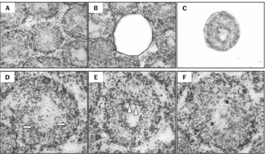
정자발생시기의 구별은
hematoxylin
에 의해 염색된 핵 에 의해서구별을하였다. I-V
단계는 버팀세포(sertoli cell)
깊숙이 정세관내의길쭉정자세포
(elongate spermatid)
를 볼수 있으며
, VII-VIII
단계는 정자방출을위해서 길쭉정자세포가 정세관 상피내강 표면에 배열되었다
. IX-XI
단계의정세관에는 길쭉정자세포가 존재하지 않으며
,
정자세포의핵 모양은 납작하며뚜렷한 응축이 나타나지 않았다
.
제VI
단계와XII
단계 정자세포는 발달의 진행이 다양하므로세포들의 모양을 근거로시기를 결정할 수가없었다
(Sluka
et al
., 2008).
각 단계별 정자발생과정 시료는real-time PCR
을 위하여각 단계별로32
개의 정세관이포함되도록LCM
을 하였으며,
시료는 각기 다른3
개의 고환에 대하여 각각
3
번 반복하여 채취하였다. LCM
은Arcturus XT Laser capture microdissection microscope (Molecular Devices, California, USA)
를 이용하였고,
각slide
는Prepstrip tissue preparation strip (Arcturus, USA)
을 이용하여 잔존물을 제거한 후 사용하였다
.
조직절편은20
배검경렌즈를 사용하여 관찰하였으며
, 30
µm
깊이의laser
를 사용하였고
power
는5 msec
간격으로20-30 mW
범위에서 사용하였다
.
세포는CapSure LCM macro caps (Arcturus, USA)
에 포획한 직후PicoPure RNA Isolation Kit (Arcturus, USA)
을 이용하여 시약설명서에 따라total RNA
를 추출하였다.
Real-time PCR을 통한 정량분석
위의방법으로부터 얻어진
total RNA
약1
µg
을cDNA synthesis kit (BIO-RAD, USA)
을 이용하여cDNA
를 합성한 후
real-time PCR
을 실시하였다. TaqMan Universal PCR Master Mix Kit (Applied Biosystems, USA)
으로 총25
µL
를 맞춘 후7500 Real-Time PCR System (Applied Biosystems, USA)
기계를 이용하여PCR
을 실시하였다.
SOD1
primers
와probe
는mouse
유전자 특이적으로TIB Mol-Bio Synthesis labor (Berlin, Germany)
로부터 제작구입하였으며
forward primer
는5'-CTT CTC GTC TTG CTC TCT CTG G-3'
였고reverse primer
는5'-TCC TGT AAAT TTG TCC TGA CAA CAC-3'
였으며JOE
가 표지된TaqMan probe
는5'-ACT GGT TCA CCG CTT GCC
TTC TGC
의 염기서열이었다. Mouse
SOD2에 대한primers
와probe
는Applied Biosystems (USA)
에서 판매되는
Assay on demand #Mm01313000
를 사용하였다. Mouse
SOD3primers
와probe
의 염기서열은 다음과 같다
. Forward primer: 5'-GGA GAT CTG GAT GGA
GCT AGG A-3'; reverse primer: 5'-CCC TGC AGA
TTG CAT GCA T-3'; FAM
이 표지된Taqman probe: 5'- AGG TGG ATGCTG CCG-3'.
β-actin (Assay on demand
#4352933E, Applied Biosystems, USA)
은 내부 표지인자로서 사용하였다
.
각 데이터(n=3)
는3
회 반복하여 실험하였으며
comparative Ct method
로mRNA
의 발현을 측 정하였다.
통계처리
결과의 통계분석은 표본수가 많지 않으므로
(n=3)
유의수준 P
<0.05
에서 비모수적 평균분석법인Kruskal-Wallis
법을 이용하였다
.
또한,
각 유전자의 발현정도는 각 단계별로 상관관계가 있는지를 편상관분석을 통해 검정하였다
(
P<0.05).
결 과
정자발생단계는 길쭉정자세포의 모양과 위치를 중심으 로 마우스 정자발생
12
단계 중VI
과XII
단계 외에10
단계에 대하여
I-V, VII-VIII
와IX-XI
의3
분류로 나누어 구별하였으며
, LCM
을 이용하여 각3
분류별 정세관을채취하여 SOD1, SOD2, SOD3에 대한
mRNA
유전자 발현양상을 비교하였다
(Figure 1).
세가지 SODsubtypes mRNA
는정자발생동안 관찰된 모든 단계의 정세관내에서 발현되 었으며 내부 표지유전자 β
-actin
에 대한comparative Ct
값으로 표현되었다
.
정자발생과정 중 SOD1mRNA
의 발현이 가장 높게 관찰되었으며
(282~556-fold),
SOD2mRNA
는3.5~5.2-fold,
SOD3mRNA
는1.28~1.70-fold
로 가장 낮게 발현되었다
(Figures 2-4).
SOD1mRNA
는 정자발생의I-V
단계에서 가장 높게 관찰되었고(556-fold), VII-VIII
단계에서는480-fold, IX-XI
단계는282-fold
로 정자가 성숙한 형태로 발달할수록 발현이 감소하였으며 유의 적인 차이를 보였다
(
P<0.05; Figure 2).
SOD2mRNA
는I-V
단계에서5.2-fold
였고, VII-VIII
단계에서는4.5-fold, IX-XI
단계는
3.5-fold
로 발생단계가 지날수록 감소하여 SOD1과 유사한 발현양상을 보였지만 유의적인 차이를 보이지 Figure 1.
Isolation of the specific seminiferous tubules using a laser capture microdissection (LCM) and classifications of the spermatogenic stages. Micrograph of intact testes section viewed by light microscopy prior to LCM (A), isolated tubule cross- section on the LCM cap following LCM (B), and remaining tissue section (C). Seminiferous tubules at the following stages of spermatogenesis were grouped: Arrows indicate compact elongated spermatid heads embedded within the seminiferous epithelium on stages I-V (D). Arrowheads indicate elongated spermatids adjacent to the tubule lumen prior to spermiation on stages VII-VIII (E). X indicates absence of elongate spermatids on stages IX-XI (F). Magnification: X100 (A-C) and X200 (D-F).
Figure 2.
Stage-specific expression of cytoplasmic Cu,Zn- superoxide dismutase (SOD1) mRNA during spermatogenesis.
Gene expression for SOD1 in cross-sectioned seminiferous
tubules defining the spermatogenic stages (I-V, VII-VIII and IX-
XI) excised by a laser capture microdissection was assessed
by real-time PCR (n=3). *Significantly different from stage I-V at
P<0.05.
않았다
(Figure 3).
그러나,
SOD3mRNA
는 정자발생VII- VIII
단계에서1.7-fold
로 가장 높게 발현되었으나, IX-XI
단 계는1.5-fold, I-V
단계에서는1.28-fold
의 순서로 감소되는 양상을 나타내었고 유의적인 차이는 보이지 않았다(Figure 4).
고 찰
Chromium, benzo[alpha]pyrene, delta-9-tetrahydrocannabinol, cypermethrin,
방사선 등 다양한 산화스트레스는 정세관에 손상을 주어 정자발생에 영향을 미치어
SOD
의 활성이나 유전자 발현 정도를 감소시킨다고 하였다
(Nonogaki
et al.
, 1992; Arafa
et al., 2009; Mandal and Das, 2009; Wang
et al., 2009; Chandra
et al., 2010).
이와같이
,
고환에서 산화스트레스에 대한SOD
의 발현변화와역할에 대한 보고는 다양하게 이루어지고있지만
,
정상적인 마우스 정자발생단계에서
SOD
의 각subtypes
별 발현 변화에 대한 연구는 거의 이루어지지 않고 있다.
이번 연구에서는 마우스 정자발생단계에서
ROS
에 대한 첫째 방어작용으로서 매우 중요하다고 알려진
SOD
의발현양상을
LCM
과real-time PCR
을 이용하여 각 단계별발현 정도를 비교하였다
.
정자발생과정에서LCM
을 이용하여 단계별 정세관의
total RNA
를 추출하여real-time PCR
을 통하여3
가지의 SOD유전자에 대한 발현양상을관찰한 결과
,
SOD1mRNA
가 가장 높은 발현을 나타냈으며
,
SOD3mRNA
의발현이 가장 낮게 관찰되었다.
SOD1은 전체
SODs
발현양 중 가장 많은 양을 차지하고 있었고
,
SOD2는다음 정도의 발현양상을나타냈으며,
SOD3는 가장 적은 양을 차지하고 있었다
.
이것은 정자의 발생과정 중
SOD
는 정자발생세포의 세포질내에 포함된SOD1
이 외부 및 내부요인의 산화스트레스에 대해서 주로 작용한다는 것을 시사한다
.
SOD1은
I-V
단계에서 가장 높은 발현을 보였으며, IX-XI
단계에서 발현이 감소하여 발생단계가 지날수록
mRNA
의 발현이 감소되었다
.
SOD1mRNA
는 주로 원형정자세포
(round spermatids)
와 길쭉정자세포에서 관찰되었으며(Gu and Hecht, 1997),
In situhybridization
후 간접적으로
laser densitometer
를 이용하여측정한Jow
등(1993)
의 보고에따르면랫드의 정자발생과정에서 SOD1
mRNA
의 발현은 정자배출
(spermiation; stages VI-VIII)
전에 정세관내에서 가장 높게 발현되었고
, LCM
을 이용한 본 실험결과와매우 유사하여
SOD1
은 정자발생과정에서 발생할수 있는
ROS
로부터 정자발생세포를 직접적으로 보호한다는 것을 알 수 있다
.
SOD2의 발현양상도 SOD1과 유사하게
I-V
단계에서가장 높은 발현을 보였으며
, IX-XI
단계에서 발현이 감소하여발생단계가 지날수록
mRNA
의 발현이 감소되었지만 유의적인 차이는보이지 않았다
.
이전연구에 따르면 SOD2는 원형정자세포에서 높은
mRNA
발현을 나타냈지만,
길쭉정자세포에서의 발현은 거의 관찰되지 않았다
(Gu and Hecht, 1996).
이러한 결과로부터SOD2
는 길쭉정자세포가 포함된
IX-XI
단계에서 가장 낮은 발현을 보인 것으로보이며 미토콘드리아의 활성이 관찰되는 원형정자세포에 서 주로 관찰되는 것으로 사료된다
.
SOD3
mRNA
는 다른 장기에 비하여 고환에서 높은 발현을 보였으며
,
버팀세포와 정조세포에서 발현이 관찰되었다는 보고는 있지만
(Mruk
et al., 1998),
정자발생단계에서의 연구는 이루어지지 않았다
.
본 연구결과 비록 발현정도는 낮았지만
,
SOD3는 정자의 형태학적 변화와 정자방출이 관찰되는
VII-VIII
단계에서 가장 높은 발현을 보였다
.
이것은SOD3
가 정자의 형태학적 변화를 겪으며방출되는 시기에나타날 수 있는 산화스트레스에 대하여 Figure 3.
Stage-specific expression of mitochondrial Mn-
superoxide dismutase (SOD2) mRNA during spermatogenesis.
Gene expression for SOD2 in cross-sectioned seminiferous tubules defining the spermatogenic stages (I-V, VII-VIII and IX- XI) excised by a laser capture microdissection was assessed by real-time PCR (n=3).
Figure 4.
Stage-specific expression of extracellular superoxide
dismutase (SOD3) genes during spermatogenesis. Gene
expression for SOD3 in cross-sectioned seminiferous tubules
defining the spermatogenic stages (I-V, VII-VIII and IX-XI)
excised by a laser capture microdissection was assessed by
real-time PCR (n=3).
중요한 역할을 할 수 있음을 시사한다.
본 연구결과로부터 정자발생과정에서 SOD의 subtypes 별 발현이 서로 다르게 조절되고 있음을 알 수 있으며, 본 연구결과가 정자발생단계에서 SOD의 역할을 연구하 는 데 있어서 기본적인 자료로 사용될 수 있다고 사료된다.
감사의 글
본 연구는 2008년 충북대학교 학술연구비로 수행하였 습니다.