Effects of N-Acetylcysteine on Nicotinamide Dinucleotide Phosphate Oxidase Activation and Antioxidant Status in Heart, Lung, Liver and Kidney in Streptozotocin-Induced Diabetic Rats
Shaoqing Lei,
1,2Yanan Liu,
2Huimin Liu,
2Hong Yu,
3Hui Wang,
1and Zhengyuan Xia
1,21Department of Pharmacology, School of Basic Medical Science, Wuhan University, Wuhan;
2Department of Anesthesiology, The University of Hong Kong, Hong Kong SAR;
3Department of Biochemistry and Molecular Biology, School of Basic Medical Science, Wuhan University, Wuhan, China.
Received: March 7, 2011 Revised: May 29, 2011 Accepted: May 31, 2011
Corresponding author: Dr. Zhengyuan Xia, Department of Pharmacology, School of Basic Medical Science, Wuhan University, Wuhan, Hubei Province, 430072, China.
Tel: 027-68758665, Fax: 027-68759222 E-mail: [email protected]
∙ The authors have no financial conflicts of interest.
© Copyright:
Yonsei University College of Medicine 2012 This is an Open Access article distributed under the terms of the Creative Commons Attribution Non- Commercial License (http://creativecommons.org/
licenses/by-nc/3.0) which permits unrestricted non- commercial use, distribution, and reproduction in any medium, provided the original work is properly cited.
Purpose: Hyperglycemia increases reactive oxygen species (ROS) and the result- ing oxidative stress plays a key role in the pathogenesis of diabetic complications.
Nicotinamide dinucleotide phosphate (NADPH) oxidase is one of the major sourc- es of ROS production in diabetes. We, therefore, examined the possibility that NADPH oxidase activation is increased in various tissues, and that the antioxidant N-acetylcysteine (NAC) may have tissue specific effects on NADPH oxidase and tissue antioxidant status in diabetes. Materials and Methods: Control (C) and streptozotocin-induced diabetic (D) rats were treated either with NAC (1.5 g/kg/
day) orally or placebo for 4 weeks. The plasma, heart, lung, liver, kidney were har- vested immediately and stored for biochemical or immunoblot analysis. Results:
levels of free 15-F2t-isoprostane were increased in plasma, heart, lung, liver and kidney tissues in diabetic rats, accompanied with significantly increased mem- brane translocation of the NADPH oxidase subunit p67phox in all tissues and in- creased expression of the membrane-bound subunit p22phox in heart, lung and kidney. The tissue antioxidant activity in lung, liver and kidney was decreased in diabetic rats, while it was increased in heart tissue. NAC reduced the expression of p22phox and p67phox, suppressed p67phox membrane translocation, and reduced free 15-F2t-isoprostane levels in all tissues. NAC increased antioxidant activity in liver and lung, but did not significantly affect antioxidant activity in heart and kid- ney. Conclusion: The current study shows that NAC inhibits NADPH oxidase ac- tivation in diabetes and attenuates tissue oxidative damage in all organs, even though its effects on antioxidant activity are tissue specific.
Key Words: Antioxidant status, diabetes, 15-F2t-isoprostane, N-acetylcysteine, NADPH oxidase
INTRODUCTION
Diabetes mellitus is a serious metabolic disease, and the number of people with di- abetes is rapidly increasing due to population growth, aging, urbanization, and in-
effectiveness of antioxidant therapies. During early diabe- tes, various tissues may enhance their antioxidant enzymes activity to withstand hyperglycemia-induced ROS produc- tion. However, little is known regarding the tissue specific changes of overall antioxidant capacity and the underlying mechanism in early diabetes.
We hypothesized that changes in the levels of both NADPH oxidase and SOD may be tissue specific, possibly having a significant impact on the total tissue antioxidant activity and on the effectiveness of antioxidant treatment in early diabetes. Therefore, this hypothesis was tested in an estab- lished rat model of STZ-induced type 1 diabetes. Since glu- tathione (GSH) depletion may play an important role in the development of diabetic complications23 and GSH deple- tion occurs in early diabetes,24 we also explored the treat- ment effects of N-acetylcystein, a GSH precursor that can enhance the GSH antioxidant defense.25
MATERIALS AND METHODS
Experiment design and induction of diabetes
Sprague-Dawley male rats aged 8 weeks and weighing 250 to 280 gram were purchased from the Animal Unit of Wu- han University, China, and allowed to adapt in their cages for three days before experiments. All rats had free access to standard chow and water. The experiments were per- formed in accordance with our institutional animal care guidelines which are in line with the use of Laboratory Ani- mals published by the US National Institutes of Health. The study was approved by Institutional Animal Care and Use Committee.
Diabetes was induced by a single tail vein injection (un- der halothane anaesthesia) of streptozotocin (Sigma, St.
Louis, MO, USA) (STZ, 60 mg/kg body weight, freshly dis- solved in 0.1 M citrate buffer, pH 4.5), while control rats (n=7) were injected equal volume citrate buffer alone. Three days following STZ injection, blood glucose levels were measured using a Glucose Analyzer (Beckman Instruments, Fullerton, CA, USA), and rats with hyperglycemia (set at plasma glucose ≥16.7 mM) were used for the experiments.
NAC, at the dose of 1.5 g/kg/day, had no effects on NADPH oxidase activity and SOD activity in non-diabetic control rats but had effects on these parameters in diabetic rats.16 In the current study, therefore, NAC was used only in diabetic rats. One week after the induction of diabetes, rats were randomly assigned into NAC-treated or untreated groups creasing prevalence of obesity.1,2 Hyperglycaemia-induced
oxidative stress, as a result of excessive production of reac- tive oxygen species (ROS), plays a key role in the patho- genesis of diabetes-related complications.3-5 Theoretically, suppression of oxidative stress using antioxidants should reverse adverse effects induced by hyperglycaemia in dia- betic patients. However, large randomized clinical trials have failed to provide convincing evidence for effective an- tioxidant therapy with classic antioxidants, such as vitamins E and C.6-8 Therefore, development of new therapeutic re- gimes is important for patients with diabetes.
Oxidative stress results from increased production of ROS or diminished antioxidant capacity in cells or tissues.
One of the major sources of ROS in tissues of diabetic pa- tients is nicotinamide adenine dinucleotide phosphate (NADPH) oxidase.9,10 NADPH oxidase contains two mem- brane-bound subunits gp91phox (Nox2) and p22phox and cytoplasmic subunits such as p47phox, p67phox and a low- molecular-weight G protein (rac 1 and rac 2).11 NADPH oxidase catalyzes the generation of superoxide anion using NADPH as the electron donor in cells.12 Superoxide is well known to be a mediator of inflammation and an inducer of apoptosis. Expression or activation of NADPH oxidase is increased in diabetes or high-glucose treated endothelial cells.13-15 We previously showed that long term treatment with the antioxidant N-acetylcysteine (NAC) could inhibit NADPH oxidase activation in the heart of diabetic rats, and it also abolished the compensatory increase in the antioxi- dant enzyme superoxide dismutase (SOD).16 It is, however, unknown whether or not short term NAC treatment may preserve the compensatory increase of endogenous SOD protein expression in diabetes, while maintaining its inhibi- tory effects on NADPH oxidase activation.
SOD, including cytosolic Cu/Zn-SOD, mitochondrial Mn-SOD and extracellular SOD,17 are enzymes involved in the scavenging of superoxide anions and play an important role in balancing ROS generation. Superoxide anions are mainly generated by the mitochondrial electron transport chain and are implicated in the progression of diabetes.18-20 Increased ROS production in diabetes can be prevented by the administration of SOD.21 A recent study showed that to- tal SOD activity and extracellular Cu/Zn-SOD activity were reduced, whereas Mn-SOD activity was increased in the heart of long-term diabetic rats.22 This indicates that indi- vidual endogenous tissue antioxidant enzymes and poten- tially the total tissue antioxidant activity may change differ- ently in diabetes. This complexity may potentially affect the
dant Status Assay Kit (Calbiochem, Darmstadt, Germany), which is dependent on the capacity in the sample to inhibit the oxidation of ABTSTM [2,2’-Azino-di-(3-ethylbenz-thiazoline sulphonate)] to ABTSTM•+ by metmyoglobin (a peroxidase).
The amount of ABTSTM•+ produced can be monitored by reading absorbance at 600 nm. Under the reaction conditions used, the antioxidants in the sample suppress absorbance at 600 nm by the degree proportional to their concentration. To- tal antioxidant activity was calculated and expressed as milli- mole per liter (mM) in plasma or millimole per gram protein in tissue samples (mmol/g protein).
Total superoxide dismutase activity assay
SOD activity was determined using a Superoxide Dismutase Assay Kit (Cayman chemical, Ann Arbor, MI, USA) fol- lowing the manufacturer’s instructions. Plasma samples were assayed immediately after being thawed. Tissue sec- tions (50 mg) were homogenized in 1 mL of cold 20 mM HEPES buffer, pH 7.2, containing 1 mM EGTA, 210 mM mannitol and 70 mM sucrose, and centrifuged at 1500×g for 5 min at 4°C, and the supernatants were collected for as- say. The assay utilizes a tetrazolium salt for detection of su- peroxide radicals generated by xanthine oxidase and hapo- xanthine. One unit of SOD is defined as the amount of enzyme needed to exhibit 50% dismutation of the superox- ide radical. SOD activity was calculated and expressed as units per milliliter (U/mL) in plasma or units per milligram protein (U/mg protein) in tissue preparations.
Tissue sample preparations and western blot analysis In order to characterize subcellular distribution of targeted proteins, tissue samples were processed to isolate membrane and cytosolic fractions as previously described.26 Briefly, 150 mg of heart, lung, kidney and liver tissue samples were pul- verized separately with mortar and pestle in liquid nitrogen, and then homogenized using a Polytron homogenizer in 1.5 mL of cold buffer A, which contained (in mM) 50 Tris-HCl (pH 7.5), 5 MgC12, 10 EGTA, 2 EDTA, 1 NaHCO3, 1 PMSF, 1 β-glycerophosphate, 1 NaF, 1 Na3VO4, and (in μg/mL) 20 leupeptin and 4 aprotinin. The homogenate was centrifuged at 1000×g for 10 min at 4°C. The supernatant was collected and used as total tissue extracts. The supernatant was then further centrifuged at 100000×g for 60 min at 4°C. The su- pernatant was removed as cytosol fractions. The pellets were resuspended in buffer B containing 50 mM Tris-HCl (pH 7.5), 150 mM NaCl, 5 mM EDTA, 1% Triton X-100, 10%
glycerol, 1 mM PMSF, 1 mM β-glycerophosphate, 60 mM (n=7, per group), and they were treated respectively with
the antioxidant NAC (1.5 g/kg/day, D+NAC) or an equal volume of 0.9% saline (D) delivered by oral gavage. Rats in the non-diabetic control group (C) were treated also with an equal volume of 0.9% saline delivered by oral gavage.
Treatments in all groups were continued for four weeks.
Plasma and tissue preparations
At the end of four weeks of treatment, the rats were anti-co- agulated with heparin (1000 IU/kg) and then anaesthetized with pentobarbital sodium (65 mg/kg body weight). Blood glucose level and body weight were measured by a Glucose Analyzer (Beckman Instruments, Fullerton, CA, USA) and a lab scale, respectively. Blood samples were collected in heparinized syringes by heart puncture after opening the thoracic cavity. The collected blood was immediately cen- trifuged at 3000×g, plasma separated, aliquoted into small microcentrifuge tubes, and stored at -80°C for further de- tection. Heart, kidney, liver and lung tissues were harvested immediately after sacrifice and frozen in liquid nitrogen and stored at -80°C for further experiments.
Inflammatory cytokines in plasma
Plasma levels of inflammatory cytokines tumor necrosis factor (TNF)-alpha and interleukin (IL)-6 were measured, using the commercially available rat ELISA kit (Bender Med, Vienna, Austria) according to the manufacturer’s in- structions.
Enzyme immunoassay (EIA) for free 15-F2t-isoprostane 15-F2t-isoprostane (15-F2t-IsoP), a specific marker of oxida- tive stress, was measured using an EIA kit (Cayman chemi- cal, Ann Arbor, MI, USA). Thus, plasma samples or homog- enized heart tissue (in PBS) were purified using Affinity Sorbent/Column (Cayman chemical, Ann Arbor, MI, USA) in the presence of 0.01% butylated hydroxytoluene and then processed for analysis of free 15-F2t-IsoP as described.26 The values of plasma or tissue 15-F2t-IsoP were expressed as pg/mL in plasma or pg/mg protein in tissue homogenates.
Total antioxidant activity determination
Tissue sections (50 mg) were homogenized in 1 mL of cold buffer (5 mM potassium phosphate, pH 7.4, containing 0.9% sodium chloride and 0.1% glucose) and were centri- fuged at 10000×g for 15 min at 4°C. The supernatant was collected for assay. Total antioxidant activity in plasma and tissue homogenate was measured using the Total Antioxi-
measurement in control rats.
Statistical analysis
The values are presented as mean±S.E.M. All biochemical assays were performed in duplicate and averaged before being analyzed. One-way or two-way analysis of variance was used for statistical analyses of data obtained within the same group and between groups, respectively. Tukey’s test was used for multiple comparisons of group means. Signifi- cance was defined as p<0.05.
RESULTS
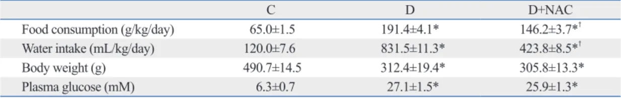
General characteristics and effects of NAC treatment Administration of STZ resulted in characteristic symptoms of diabetes including hyperglycemia, and reduced body weight gain along with increased food and fluid intake when compared to age-matched controls (Table 1). Food con- sumption, water intake and plasma glucose levels in the dia- betic group were much higher than those in the age-matched control group (p<0.01). NAC treatment significantly re- duced water intake and food consumption compared with the diabetic untreated group (p<0.05), but did not signifi- cantly affect glucose levels. Body weight in the diabetic group was lower than that of the control group (p<0.05), and NAC treatment did not have significant effects on body weight gain (Table 1).
Octyl β-glucopyranoside, 1 mM NaF, 0.1 mM Na3VO4, and (in μg/mL) 20 leupeptin and 4 aprotinin. The suspensions were homogenized again, held on ice for 30 min, and cen- trifuged at 100000×g for 60 min at 4°C. The supernatant was collected as membrane fractions. The proteins from each fraction (50-100 μg) were resolved via SDS-PAGE and subsequently transferred to PVDF membrane for immunob- lot analysis. The membranes were blocked in 5% non fat milk for 2 h at room temperature, washed three times with TBS-T buffer each for 5 min, and then incubated overnight at 4°C with primary antibodies raised against Cu/Zn-SOD (rabbit antiserum), Mn-SOD (rabbit polyclonal IgG) (1 : 1000, Millipore Corporation), p22phox and p67phox (rab- bit polyclonal antibody, 1 : 500, Santa Cruz Biotechnolo- gy), respectively. After washing three times with TBS-T for 30 min, the blot was incubated for 1 h at room temperature with anti-rabbit IgG, HRP-linked antibody (1 : 10000, Cell Signaling). After washing the membranes, the blot was de- veloped with enhanced chemiluminescence reagent (GE Healthcare) and then exposed to X-ray film. The same blot was stripped and reblotted with antibodies to ward GAPDH (1 : 2000, Cell Signaling) in total tissue extracts and cytosol fractions, or Na, K-ATPase (1 : 2000, Cell Signaling) in membrane fractions as an internal control. The intensity of the bands was quantified using image analysis software (Bio-Rad, Hercules, CA, USA), and the results were nor- malized to corresponding internal controls to correct for loading. Data are presented as percent change relative to the
Table 1. General Characteristics of Rats at the End of the Study
C D D+NAC
Food consumption (g/kg/day) 65.0±1.5 191.4±4.1* 146.2±3.7*†
Water intake (mL/kg/day) 120.0±7.6 831.5±11.3* 423.8±8.5*†
Body weight (g) 490.7±14.5 312.4±19.4* 305.8±13.3*
Plasma glucose (mM) 6.3±0.7 27.1±1.5* 25.9±1.3*
Control (C) or streptozotocin-induced diabetic rats were either untreated (D) or treated with the antioxidant N-acetylcysteine (1.5 g/kg/
day, D+NAC) by oral gavage for four weeks.
All values are expressed as means±S.E.M., n=7.
*p<0.05 compared with control group.
†p<0.05 compared with diabetic group.
Table 2. Inflammatory Cytokines at the End of the Study
C D D+NAC
Plasma TNF-alpha (pg/mL) 18.5±1.1 41.2±5.5* 20.3±2.5†
Plasma IL-6 (pg/mL) 42.7±2.7 58.0±2.4* 44.7±4.2†
TNF, tumor necrosis factor; IL, interleukin.
Control (C) or streptozotocin-induced diabetic rats were either untreated (D) or treated with the antioxidant N-acetylcysteine (1.5 g/kg/
day, D+NAC) by oral gavage for four weeks.
All values are expressed as means±S.E.M., n=7.
*p<0.05 compared with control group.
†p<0.05 compared with diabetic group.
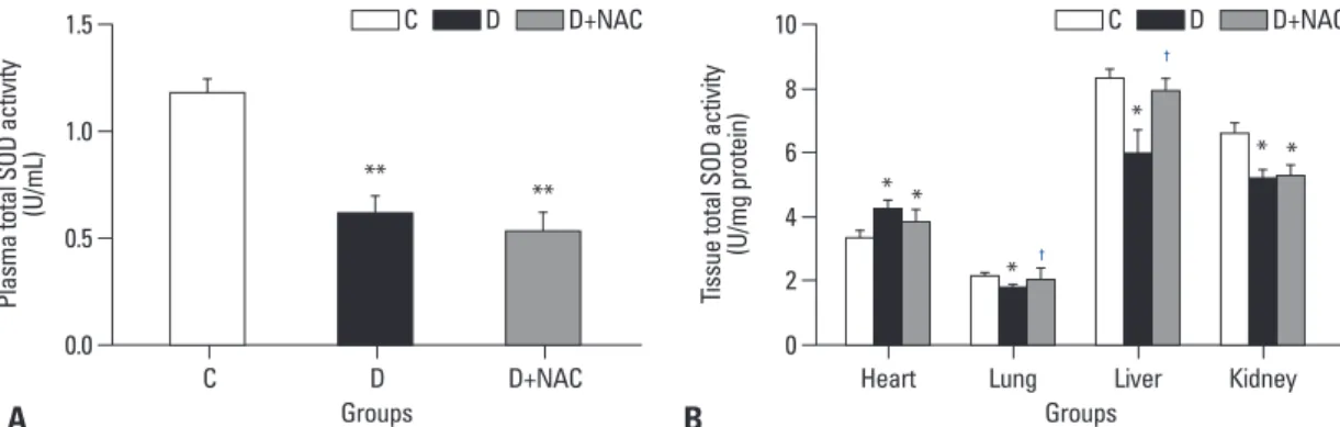
pacity in tissues or in the plasma. Plasma total antioxidant activity in diabetic rats was higher than that of the control group (p<0.01) (Fig. 2A). NAC treatment significantly at- tenuated this increase in plasma total antioxidant levels in diabetic rats (p<0.05, D+NAC vs. D) (Fig. 2A). Changes in tissue total antioxidant activity and the effects of NAC treat- ment were tissue-specific. The total antioxidant activity in the heart in the diabetic group was higher than that in the control group (p<0.05) and was unaffected by NAC treat- ment (Fig. 2B). In contrast, however, total antioxidant ac- tivity in the lung, liver and kidney tissues was significantly decreased in the diabetic group as compared with control group (p<0.05) (Fig. 2B). NAC treatment elevated total an- tioxidant activity in the lung and liver (p<0.05, D+NAC vs.
D), but not in the kidney.
Total SOD activity in plasma and various tissues The amount of SOD present in the cellular and extracellular environments is crucial for prevention of ROS mediated oxidative cellular damage. Plasma total SOD activity in the diabetic rats was much lower than that in the control group Inflammatory disorder
Diabetes is associated with disorders of many inflammatory cytokines.27 As shown in Table 2, the plasma levels of TNF- alpha and IL-6 in diabetic rats were increased significantly as compared to control rats (p<0.05), which were prevented by treatment with NAC.
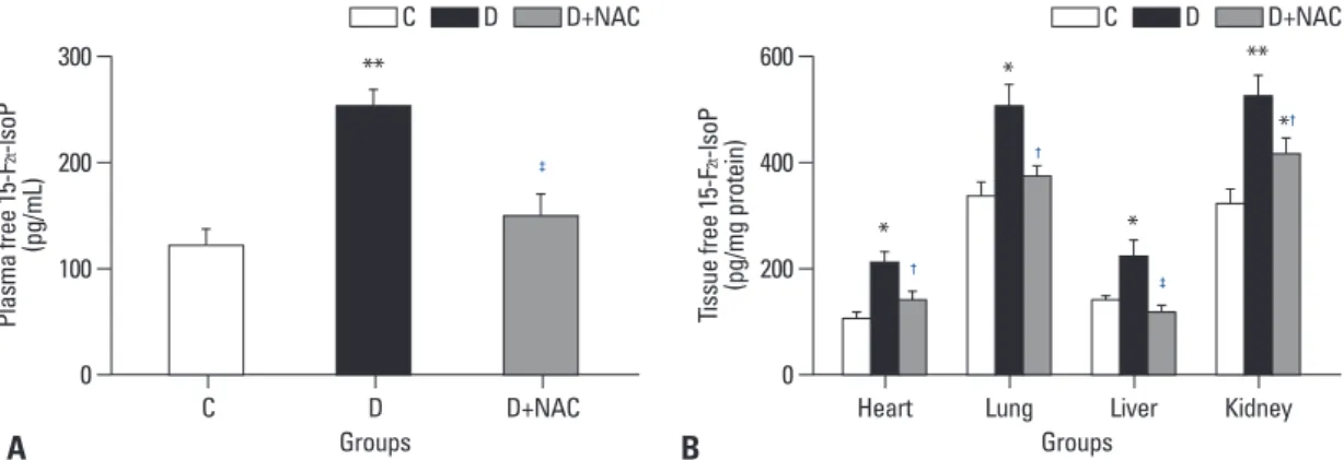
Oxidative stress marker: free 15-F2t-IsoP levels in plasma and tissue
Plasma (Fig. 1A) and tissue (Fig. 1B) levels of free 15-F2t- IsoP in the diabetic group were much higher than those in the control group (all p<0.05 or p<0.01). NAC treatment sig- nificantly reduced free 15-F2t-IsoP concentrations in the plasma and prevented the significant increase of 15-F2t-IsoP in the heart, lung and liver tissues seen in the diabetic group (all p<0.05, D+NAC vs. D). NAC significantly reduced, but did not normalize 15-F2t-IsoP levels in the kidney (p<0.05, D+NAC vs. D; p<0.05, D+NAC vs. C) (Fig. 1B).
Total antioxidant activity in plasma and various tissues Total antioxidant activity represents the total antioxidant ca-
Fig. 2. Effects of N-acetylcysteine treatment on the level of total antioxidant activity in plasma (A) and various tissues (B). Control (C) or STZ-induced diabetic rats were either untreated (D) or treated with the antioxidant N-acetylcysteine (1.5 g/kg/day, D+NAC) by oral ga- vage for four weeks. Results are expressed as means±S.E.M., n=7, *p<0.05, **p<0.01 vs. C; †p<0.05 vs. D. STZ, streptozotocin.
Fig. 1. Effects of N-acetylcysteine treatment on the level of free 15-F2t-isoprostane in plasma (A) and various tissues (B). Control (C) or STZ-induced diabetic rats were either untreated (D) or treated with the antioxidant N-acetylcysteine (1.5 g/kg/day, D+NAC) by oral ga- vage for four weeks. Results are expressed as means±S.E.M., n=7, *p<0.05, **p<0.01 vs. C; †p<0.05, ‡p<0.01 vs. D. STZ, streptozotocin.
A
A
B
B
0
0.0
0
0 100
0.5
200
1 200
1.0
400
2 300
1.5
600
3
Plasma free 15-F2t-lsoP (pg/mL)Plasma total antioxidant activity (mM) Tissue free 15-F2t-lsoP (pg/mg protein)Tissue total antioxidant activity (mmol/g protein)
Groups
Groups
Groups
Groups C
C
Heart
Heart
Lung
Lung
Liver
Liver
Kidney
Kidney D
D
D+NAC
D+NAC
**
**
*†
*
*
*
**
*
**
**
*
†
*
†
†
‡
†
*†
* C D D+NAC
C D D+NAC C D D+NAC
C D D+NAC
‡
significantly in diabetic rats as compared to age-matched controls (all p<0.05) (Fig. 3B), and NAC treatment pre- vented the decrease in total SOD activity in the liver, but not in the kidney.
Protein expression of Cu/Zn-SOD and Mn-SOD In the heart and lung tissues, the levels of Cu/Zn-SOD pro- tein in the diabetic group increased significantly compared with controls (all p<0.05, D vs. C) (Fig. 4A). NAC attenu- ated this increase in Cu/Zn-SOD protein expression in the heart (p< 0.05, D+NAC vs. D), but did not significantly af- fect Cu/Zn-SOD protein expression in the lung (Fig. 4A).
In contrast, however, Cu/Zn-SOD protein expression in liv- er and kidney tissues in diabetic rats was lower than that of (p<0.01) (Fig. 3A), NAC did not have significant effects on
plasma total SOD activity. In the control rats, tissue total SOD activities in the heart and lung were much lower than those in the liver and kidney (Fig. 3B), suggesting that SOD might not be the predominant endogenous antioxidant enzyme in the heart and lung in rats under physiological conditions. In the heart, total SOD activity was slightly in- creased in diabetic rats as compared to control rats (p<0.05, D vs. C), and this compensatory increase in SOD activity was not significantly affected by NAC treatment. While to- tal SOD activity in the lung, was slightly decreased in dia- betic rats as compared to control rats, NAC prevented the reduction of SOD activity in the lung. Furthermore, total SOD activities both in the liver and kidney were decreased
Fig. 4. Western blots analysis of Cu/Zn-SOD and Mn-SOD protein expression in various tissues. Control (C) or STZ-induced diabetic rats were either untreated (D) or treated with the antioxidant N-acetylcysteine (1.5 g/kg/day, D+NAC) by oral gavage for four weeks. (A) (top) Representative Western blot showing Cu/Zn-SOD expression with GAPDH as a loading control in total tissue extracts; (bottom, graph) Cu/Zn-SOD densitometric values were normalized to their corresponding GAPDH densitometric values and expressed as percent change relative to the measurement in control rats. (B) (top) Representative Western blot showing Mn-SOD expression with GAPDH as a loading control in total tissue extracts; (bottom, graph) Mn-SOD densitometric values were normalized to their corresponding GAPDH densitometric values and expressed as percent change relative to the measurement in control rats. All the results are expressed as means±S.E.M., n=7, *p<0.05, **p<0.01 vs. C; †p<0.05 vs. D. SOD, superoxide dismutase; STZ, streptozotocin.
A B
Fig. 3. Effects of N-acetylcysteine treatment on the level of total SOD activity in plasma (A) and various tissues (B). Control (C) or STZ- induced diabetic rats were either untreated (D) or treated with the antioxidant N-acetylcysteine (1.5 g/kg/day, D+NAC) by oral gavage for four weeks. Results are expressed as means±S.E.M., n=7 per group, *p<0.05, **p<0.01 vs. C; †p<0.05 vs. D. SOD, superoxide dismutase;
STZ, streptozotocin.
A B
0.0 0
0.5 4
2
1.0 6
8
1.5 10
Plasma total SOD activity (U/mL) Tissue total SOD activity (U/mg protein)
Groups Groups
C D D+NAC Heart Lung Liver Kidney
** ** *
*
*
*
*
†
†
* C D D+NAC C D D+NAC
00 0
100
100 100
50
50 50
150 150 200 150 200 250
250 200
Cu/Zn-SOD expression (% of control)Cu/Zn-SOD expression (% of control) Mn-SOD expression (% of control)
Groups
Groups Groups
Heart
Heart LungLung LiverLiver KidneyKidney Heart Lung Liver Kidney
**
**
**
** *
*
**
**
*
*†
*†
††
*
*†
*†
**† C D D+NAC
C D D+NAC C D D+NAC
Heart
Heart Heart
Cn/Zn-SOD GAPDH Cn/Zn-SOD GAPDH
Mn-SOD GAPDH
Lung
Lung LiverLiver KidneyKidney Lung Liver Kidney
C D D+NAC
C D D+NAC C D D+NACC D D+NAC C D D+NACC D D+NAC C D D+NACC D D+NAC C D D+NAC C D D+NAC C D D+NAC C D D+NAC
†
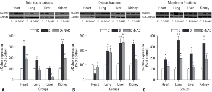
in various tissues. As shown in Fig. 5A, p22phox protein levels in the heart, lung and kidney of the diabetic group were significantly higher than those of the control group, and NAC prevented the increase of p22phox protein ex- pression in the lung, but did not normalize their increase in the heart and kidney (all p<0.05, D+NAC vs. D) (Fig. 5A).
While there was no significant difference in p22phox pro- tein expression in the liver between control and diabetic rats, 4 weeks of NAC treatment decreased its expression signifi- cantly (p<0.05, D+NAC vs. D or C) (Fig. 5A).
The p67phox protein expression was significantly in- creased both in the cytosol and membrane fractions in the lung, liver and kidney tissues of diabetic rats relative to control rats (Fig. 5B and C), reflecting increased p67phox activation as well as increased total protein levels which were in line with the increases of total p67phox protein ex- pression in these tissues (data not shown). However, p67phox protein expression in the heart was significantly increased in the membrane fractions while markedly de- creased in the cytosol fractions, reflecting increased p67phox activation. NAC prevented the increase of p67phox membrane translocation in the heart and liver and significantly attenuated the increase of p67phox membrane translocation in the lung and kidney tissues (all p<0.05, D+NAC vs. D) (Fig. 5C).
age-matched control rats (p<0.05 in liver, p<0.01 in kidney) (Fig. 4A). NAC prevented the decrease of Cu/Zn-SOD pro- tein in the liver and attenuated its reduction in the kidney (all p<0.05, D+NAC vs. D) (Fig. 4A).
The changes of Mn-SOD protein expression in various tissues were different from those of Cu/Zn-SOD. There was no significant difference in Mn-SOD protein expression in the heart tissue among control, diabetic and NAC-treated diabetic groups (Fig. 4B). However, Mn-SOD protein ex- pression in both the lung and the kidney decreased signifi- cantly in the diabetic group as compared to the control group, and NAC prevented the reduction of Mn-SOD pro- tein in the lung (p<0.05, D+NAC vs. D) but further de- creased its level in the kidney (p<0.05, D+NAC vs. D) (Fig.
4B). It is of an interest to note that Mn-SOD protein expres- sion in the liver increased significantly in diabetic rats as compared control rats, but this increase was not affected by NAC treatment (Fig. 4B).
Expression of NADPH oxidase subunit: p22phox and p67phox
NADPH oxidase has been shown to be a major source of ROS production,10,28 and its subunits p22phox and p67phox are increased in the myocardium of STZ-induced diabetic rats.16 In the present study, we detected p22phox and p67phox
Fig. 5. Western blots analysis of NADPH oxidase subunits p22phox and p67phox protein expression in various tissues. Control (C) or STZ-induced diabetic rats were either untreated (D) or treated with the antioxidant N-acetylcysteine (1.5 g/kg/day, D+NAC) by oral gavage for four weeks. (A) (top) Representative Western blot showing p22phox expression with GAPDH as a loading control in total tissue extracts; (bottom, graph) p22phox densitometric values were nor- malized to their corresponding GAPDH densitometric values and expressed as percent change relative to the measurement in control rats. (B) (top) Representative Western blot showing p67phox expression with GAPDH as a loading control in cytosol fractions; (bottom, graph) p67phox densitometric values were normalized to their corresponding GAPDH densitometric values and expressed as percent change relative to the measurement in control rats. (C)(top) Representative Western blot showing p67phox expression with Na, K-ATPase as a loading control in membrane fractions; (bottom, graph) p67phox densito- metric values were normalized to their corresponding Na, K-ATPase densitometric values and expressed as percent change relative to the measurement in control rats. All the results are expressed as means±S.E.M., n=7, *p<0.05, **p<0.01 vs. C; †p<0.05 vs. D. NADPH, nicotinamide dinucleotide phosphate; STZ, streptozotocin.
A B C
A
0 100 200 300 400
p22phox expression (% of control)
Groups
Heart Lung Liver Kidney
**
*
†
*† *†
**
*†
C D D+NAC C D D+NAC
Heart
p22phox GAPDH
Lung Liver Kidney
C D D+NAC C D D+NAC C D D+NAC C D D+NAC
0 0
100 100
200
200 300
300 400
p67phox expression (% of control) p67phox expression (% of control)
Groups Groups
Heart Lung Liver Kidney Heart Lung Liver Kidney
*
*
*
**
*
*†
† †
† *†
* **
**
†
**
*
C D D+NAC
Heart Heart
p67phox GAPDH
p67phox Na,K-ATPase
Lung Liver Kidney Lung Liver Kidney
C D D+NAC C D D+NAC C D D+NAC C D D+NAC C D D+NAC C D D+NAC C D D+NAC C D D+NAC
Total tissue extracts Cytosol fractions Membrane fractions
15-F2t-isoprostane was increased despite these increases in systemic and heart tissue antioxidant activity. This indicates that up-regulation of antioxidant defenses is not sufficient to withstand hyperglycemia-induced increase in ROS pro- duction. It is also worth to note that in the control rats, the tissue total SOD activities in the heart and lung were much lower than those in the liver and kidney, which may suggest that SOD might not be the predominant endogenous anti- oxidant enzyme in the heart and lung in rats under physio- logical conditions. Other endogenous antioxidant enzymes such as glutathione peroxidase also play important roles in maintaining tissue antioxidant activity in these tissues un- der physiological conditions as well as in diabetes.36 Indeed, glutathione peroxidase and catalase are increased in the heart and lung of diabetic rats.36 This may explain the dis- crepancy between total antioxidant activity and SOD activi- ty in plasma observed in the current study.
NADPH oxidase, a transmembrane enzyme located in intracellular organelles, is one of the major sources of ROS in diabetes.9,10 Recent studies show that the NADPH oxi- dase p22phox and gp91phox subunits were over-expressed in rat aorta,37 and that the p22phox, p47phox and p67phox subunits were increased in testes in STZ-induced diabetic rats.28 Our recent study shows that p22phox and p67phox, but not the p47phox, are robustly enhanced in the diabetic myocardium, and that NAC can prevent their increase.16 Therefore, in the current study, we specifically examined the changes of p22phox and p67phox in various tissues in diabetic rats, and found that p22phox and p67phox subunits were over-expressed or activated in various tissues in early diabetic rats, and that NAC treatment prevented or attenuat- ed these changes in diabetes. NAC mediated reduction of p67phox translocation to membrane in various tissues was seen in the current study. This finding is in keeping with a previous study showing that NAC can inhibit NADPH oxi- dase activation.10 Thus, inhibition of NADPH oxidase over- expression and activation and the subsequent ROS produc- tion may represent a mechanism by which NAC reduces oxidative stress in diabetes in addition to its ROS scaveng- ing property.
Mitochondrial dysfunction is another major intracellular source of ROS through the mitochondrial respiratory chain (MRC).10 About 4% of oxygen consumed in the mitochon- dria is converted to ROS.38 Impaired activities of enzymes in the MRC and mitochondrial DNA mutations were detected in patients with diabetes and in animal models of diabetes.39 Mitochondrial derived superoxide anion is viewed as a sin-
DISCUSSION
We have shown in the current study that the level of 15-F2t- IsoP, a biomarker of oxidative stress,29,30 was elevated both in plasma and various tissues of type 1 diabetic rats. In con- trast to reductions in total antioxidant activity in the lung, liver and kidney tissues, total antioxidant activity in the heart was elevated in the diabetic rats accompanied by an increase in heart tissue total SOD activity which indicated a significant up-regulation of self-defense mechanisms in the heart. The variations in SOD activity and protein expression in diabetes were tissue-specific. NAC treatment up-regulat- ed Mn-SOD protein expression in diabetic rats in the lung tissue, but unexpectedly reduced Mn-SOD protein expres- sion in the kidney. The underlying mechanisms governing the tissue-specific effects of NAC on SOD protein expres- sion are not clear, nevertheless, it might be due to tissue-spe- cific effectiveness of NAC in reducing the production of su- peroxide.16 Consequently, the organism may regulate the levels of SOD activation or protein expression on a basis of needs. It should be noted that NAC uniformly reduced the increase of p22phox protein expression and p67phox activa- tion in all tissues studied, most likely inhibiting the activa- tion of NADPH oxidase, a major source of ROS production in diabetes. This result is keeping with findings of a previ- ous study which showed that NAC can inhibit NADPH ox- idase activity in osteoblast precursor cells.31 Thus, our pres- ent studies together with others suggest that NAC can break vicious cycle of ROS-induced ROS production via NADPH oxidase activation in diabetes.
Increased oxidative stress can be the result of several dia- betes-induced abnormalities, including auto-oxidation of glu- cose, the formation of advanced glycation end products, and impairment of defense system.32 The impaired defense system is always associated with reduced antioxidant capac- ity, abnormal activity or expression of antioxidant enzymes in diabetes.33-35 Among various antioxidant enzymes, SOD plays an important role in balancing ROS generation and the overall tissue antioxidant capacity. In our study, total an- tioxidant activity and SOD activity in the lung, liver and kidney tissues and total SOD activity in plasma were com- promised in diabetic rats. It should be noted total antioxi- dant activity both in the plasma and heart tissues and total SOD activity in heart tissues were increased in diabetic rats, reflecting increased systemic antioxidant capacity overall and in the heart in particular. However, heart tissue level of
ACKNOWLEDGEMENTS
This is supported by grant 30872447 from the National Natural Science Foundation of China (NSFC) and General Research Fund grants 782910M from Research Grants Council of Hong Kong.
REFERENCES
1. Yang W, Lu J, Weng J, Jia W, Ji L, Xiao J, et al. Prevalence of dia- betes among men and women in China. N Engl J Med 2010;362:
1090-101.
2. Hakim FA, Pflueger A. Role of oxidative stress in diabetic kidney disease. Med Sci Monit 2010;16:RA37-48.
3. Baynes JW, Thorpe SR. Role of oxidative stress in diabetic com- plications: a new perspective on an old paradigm. Diabetes 1999;
48:1-9.
4. Ceriello A, Testa R. Antioxidant anti-inflammatory treatment in type 2 diabetes. Diabetes Care 2009;32 Suppl 2:S232-6.
5. Brownlee M. Biochemistry and molecular cell biology of diabetic complications. Nature 2001;414:813-20.
6. Robinson I, de Serna DG, Gutierrez A, Schade DS. Vitamin E in humans: an explanation of clinical trial failure. Endocr Pract 2006;12:576-82.
7. Hasnain BI, Mooradian AD. Recent trials of antioxidant therapy:
what should we be telling our patients? Cleve Clin J Med 2004;
71:327-34.
8. Ristow M, Zarse K, Oberbach A, Klöting N, Birringer M, Kiehntopf M, et al. Antioxidants prevent health-promoting effects of physical exercise in humans. Proc Natl Acad Sci U S A 2009;106:8665-70.
9. Gao L, Mann GE. Vascular NAD(P)H oxidase activation in diabe- tes: a double-edged sword in redox signalling. Cardiovasc Res 2009;82:9-20.
10. Shen GX. Oxidative stress and diabetic cardiovascular disorders:
roles of mitochondria and NADPH oxidase. Can J Physiol Phar- macol 2010;88:241-8.
11. Lyle AN, Griendling KK. Modulation of vascular smooth muscle signaling by reactive oxygen species. Physiology (Bethesda) 2006;21:269-80.
12. Griendling KK, Sorescu D, Ushio-Fukai M. NAD(P)H oxidase:
role in cardiovascular biology and disease. Circ Res 2000;86:494- 13. Liu S, Ma X, Gong M, Shi L, Lincoln T, Wang S. Glucose down-501.
regulation of cGMP-dependent protein kinase I expression in vas- cular smooth muscle cells involves NAD(P)H oxidase-derived re- active oxygen species. Free Radic Biol Med 2007;42:852-63.
14. Li L, Renier G. Activation of nicotinamide adenine dinucleotide phosphate (reduced form) oxidase by advanced glycation end products links oxidative stress to altered retinal vascular endothe- lial growth factor expression. Metabolism 2006;55:1516-23.
15. Picchi A, Gao X, Belmadani S, Potter BJ, Focardi M, Chilian WM, et al. Tumor necrosis factor-alpha induces endothelial dys- function in the prediabetic metabolic syndrome. Circ Res 2006;99:
69-77.
16. Guo Z, Xia Z, Jiang J, McNeill JH. Downregulation of NADPH
gle unifying mechanism for diabetic complications.18,40 Over-expression of Mn-SOD, the major scavenger of mito- chondrial superoxide anion, prevents high glucose-induced oxidative stress, cell apoptosis and mitochondrial DNA damage in retinal endothelial cells,19,20 and protects the reti- na from diabetes-induced oxidative stress in mice.41 In our present study, Mn-SOD expression in diabetic lung and kid- ney tissues decreased significantly, whereas Mn-SOD ex- pression in diabetic liver tissue increased but was unchanged in heart tissue. This suggests that the change in mitochondrial SOD in response to hyperglycemia-induced oxidative stress is tissue-specific in early diabetes. NAC increased Mn-SOD in the lung, however, it did not increase it in the liver and further reduced its protein expression in the kidney, which might be the reason of why NAC could not bring down kid- ney tissue 15-F2t-IsoP content to a level comparable to that in the control rats.
Inflammation has been widely recognized as a key pro- cess of the progression of diabetes.42,43 Some inflammatory factors, such as TNF-alpha and IL-6, are elevated in patients with type 2 diabetes,44 and correlate with the incidence of diabetic macrovascular complications, especially athero- sclerosis development.45 Our previous studies showed that long term (8 weeks) treatment with NAC attenuated myo- cardial levels of IL-6 protein expression and reduced plas- ma level of TNF-alpha in STZ induced diabetic rats.16,46 In the present study, we found that plasma levels of TNF-al- pha and IL-6 were significantly increased in STZ-induced diabetic rats early at 4 weeks after the establishment of dia- betes, and that NAC treatment for 4 weeks decreased the secretion of TNF-alpha and IL-6 cytokines, which reflects a suppression of inflammatory responses. Similar to our study, NAC has been demonstrated to have anti-inflammatory property both in vitro in lipopolysaccharide-activated mac- rophages47 and in vivo in humans.48
In summary, the current study shows that oxidative stress occurs at an early stage of diabetes, and changes in the ma- jor pro-oxidant enzyme NADPH oxidase and the antioxi- dant enzyme SOD are tissue-specific in STZ-induced type 1 diabetic rats. Antioxidant NAC can confer protection against oxidative stress by suppressing NADPH oxidase activation and restoring or enhancing SOD enzyme activity and/or protein expression. The effects of NAC are tissue-specific, being more effective in the heart, lung and liver and less ef- fective in the kidney. The current findings may have poten- tial clinical implications in developing therapies for the treatment of diabetic complications.
chem J 2011;433:393-402.
32. Wold LE, Ceylan-Isik AF, Ren J. Oxidative stress and stress sig- naling: menace of diabetic cardiomyopathy. Acta Pharmacol Sin 2005;26:908-17.
33. Du D, Shi YH, Le GW. Oxidative stress induced by high-glucose diet in liver of C57BL/6J mice and its underlying mechanism.
Mol Biol Rep 2010;37:3833-9.
34. Ahmed FN, Naqvi FN, Shafiq F. Lipid peroxidation and serum antioxidant enzymes in patients with type 2 diabetes mellitus. Ann N Y Acad Sci 2006;1084:481-9.
35. Ramakrishna V, Jailkhani R. Oxidative stress in non-insulin-de- pendent diabetes mellitus (NIDDM) patients. Acta Diabetol 2008;45:41-6.
36. Hünkar T, Aktan F, Ceylan A, Karasu C; Antioxidants in Diabetes- Induced Complications (ADIC) Study Group. Effects of cod liver oil on tissue antioxidant pathways in normal and streptozotocin- diabetic rats. Cell Biochem Funct 2002;20:297-302.
37. Olukman M, Orhan CE, Celenk FG, Ulker S. Apocynin restores endothelial dysfunction in streptozotocin diabetic rats through regulation of nitric oxide synthase and NADPH oxidase expres- sions. J Diabetes Complications 2010;24:415-23.
38. Lenaz G, Bovina C, D’Aurelio M, Fato R, Formiggini G, Genova ML, et al. Role of mitochondria in oxidative stress and aging. Ann N Y Acad Sci 2002;959:199-213.
39. Fosslien E. Mitochondrial medicine--molecular pathology of defec- tive oxidative phosphorylation. Ann Clin Lab Sci 2001;31:25-67.
40. Sack MN. Type 2 diabetes, mitochondrial biology and the heart. J Mol Cell Cardiol 2009;46:842-9.
41. Kowluru RA, Kowluru V, Xiong Y, Ho YS. Overexpression of mitochondrial superoxide dismutase in mice protects the retina from diabetes-induced oxidative stress. Free Radic Biol Med 2006;41:1191-6.
42. Hotamisligil GS. Endoplasmic reticulum stress and the inflamma- tory basis of metabolic disease. Cell 2010;140:900-17.
43. Casas-Agustench P, Bulló M, Salas-Salvadó J. Nuts, inflammation and insulin resistance. Asia Pac J Clin Nutr 2010;19:124-30.
44. Navarro-Gonzalez J, Mora-Fernandez C, Gomez-Chinchon M, Muros M, Herrera H, Garcia J. Serum and gene expression profile of tumor necrosis factor-alpha and interleukin-6 in hypertensive diabetic patients: effect of amlodipine administration. Int J Immu- nopathol Pharmacol 2010;23:51-9.
45. Wu W, Wang M, Sun Z, Wang X, Miao J, Zheng Z. The predictive value of TNF-alpha and IL-6 and the incidence of macrovascular complications in patients with type 2 diabetes. Acta Diabetol 2010. [Epub ahead of print]
46. Tsai GY, Cui JZ, Syed H, Xia Z, Ozerdem U, McNeill JH, et al.
Effect of N-acetylcysteine on the early expression of inflammatory markers in the retina and plasma of diabetic rats. Clin Experiment Ophthalmol 2009;37:223-31.
47. Palacio JR, Markert UR, Martínez P. Anti-inflammatory properties of N-acetylcysteine on lipopolysaccharide-activated macrophages.
Inflamm Res 2011;60:695-704.
48. Nascimento MM, Suliman ME, Silva M, Chinaglia T, Marchioro J, Hayashi SY, et al. Effect of oral N-acetylcysteine treatment on plasma inflammatory and oxidative stress markers in peritoneal dialysis patients: a placebo-controlled study. Perit Dial Int 2010;
30:336-42.
oxidase, antioxidant enzymes, and inflammatory markers in the heart of streptozotocin-induced diabetic rats by N-acetyl-L-cyste- ine. Am J Physiol Heart Circ Physiol 2007;292:H1728-36.
17. Sandström J, Nilsson P, Karlsson K, Marklund SL. 10-fold increase in human plasma extracellular superoxide dismutase content caused by a mutation in heparin-binding domain. J Biol Chem 1994;269:19163-6.
18. Brownlee M. The pathobiology of diabetic complications: a unify- ing mechanism. Diabetes 2005;54:1615-25.
19. Kowluru RA, Atasi L, Ho YS. Role of mitochondrial superoxide dismutase in the development of diabetic retinopathy. Invest Oph- thalmol Vis Sci 2006;47:1594-9.
20. Madsen-Bouterse SA, Zhong Q, Mohammad G, Ho YS, Kowluru RA. Oxidative damage of mitochondrial DNA in diabetes and its protection by manganese superoxide dismutase. Free Radic Res 2010;44:313-21.
21. Reis JS, Bosco AA, Veloso CA, Mattos RT, Purish S, Nogueira- Machado JA. Oxidizing and reducing responses in type 1 diabetic patients determined up to 5 years after the clinical onset of the dis- ease. Acta Diabetol 2008;45:221-4.
22. Ivanović-Matić S, Mihailović M, Dinić S, Martinović V, Bogojević D, Grigorov I, et al. The absence of cardiomyopathy is accompa- nied by increased activities of CAT, MnSOD and GST in long- term diabetes in rats. J Physiol Sci 2010;60:259-66.
23. Ghosh S, Pulinilkunnil T, Yuen G, Kewalramani G, An D, Qi D, et al. Cardiomyocyte apoptosis induced by short-term diabetes re- quires mitochondrial GSH depletion. Am J Physiol Heart Circ Physiol 2005;289:H768-76.
24. Yue KK, Chung WS, Leung AW, Cheng CH. Redox changes pre- cede the occurrence of oxidative stress in eyes and aorta, but not in kidneys of diabetic rats. Life Sci 2003;73:2557-70.
25. Aruoma OI, Halliwell B, Hoey BM, Butler J. The antioxidant ac- tion of N-acetylcysteine: its reaction with hydrogen peroxide, hy- droxyl radical, superoxide, and hypochlorous acid. Free Radic Biol Med 1989;6:593-7.
26. Xia Z, Kuo KH, Nagareddy PR, Wang F, Guo Z, Guo T, et al. N- acetylcysteine attenuates PKCbeta2 overexpression and myocar- dial hypertrophy in streptozotocin-induced diabetic rats. Cardio- vasc Res 2007;73:770-82.
27. Meng X, Tancharoen S, Kawahara KI, Nawa Y, Taniguchi S, Hashiguchi T, et al. 1,5-Anhydroglucitol attenuates cytokine re- lease and protects mice with type 2 diabetes from inflammatory reactions. Int J Immunopathol Pharmacol 2010;23:105-19.
28. Xu M, Dai DZ, Zhang Q, Cheng YS, Dai Y. Upregulated NADPH oxidase contributes to diabetic testicular complication and is re- lieved by strontium fructose 1,6-diphosphate. Exp Clin Endocrinol Diabetes 2010;118:459-65.
29. Wu HC, Wang Q, Yang HI, Ahsan H, Tsai WY, Wang LY, et al.
Urinary 15-F2t-isoprostane, aflatoxin B1 exposure and hepatitis B virus infection and hepatocellular carcinoma in Taiwan. Carcino- genesis 2008;29:971-6.
30. Faure P, Polge C, Monneret D, Favier A, Halimi S. Plasma 15-F2t isoprostane concentrations are increased during acute fructose loading in type 2 diabetes. Diabetes Metab 2008;34:148-54.
31. Mandal CC, Ganapathy S, Gorin Y, Mahadev K, Block K, Ab- boud HE, et al. Reactive oxygen species derived from Nox4 medi- ate BMP2 gene transcription and osteoblast differentiation. Bio-