Original Article
Laser Peripheral Iridotomy with Iridoplasty in Primary Angle Closure Suspect: Anterior Chamber Analysis by Pentacam
Jong Rak Lee, Jin Young Choi, Yeon-Deok Kim, Jaewan Choi
HanGil Eye Hospital, Incheon, Korea
Purpose: To compare conventional laser peripheral iridotomy (LPI) and LPI combined with laser peripheral irido- plasty in eyes with primary angle closure suspect (PACS) by assessment of anterior chamber dimensional changes using a Pentacam.
Methods: Forty-eight eyes of 24 subjects with bilateral PACS were recruited consecutively. Each eye was randomly allocated to treatment with conventional LPI, argon LPI only, or LPI plus iridoplasty, which consisted of simulta- neous argon LPI and peripheral iridoplasty. Anterior chamber measurements were performed on each eye using a Pentacam, both before and after treatment. Mean anterior chamber depth (ACD), anterior chamber volume (ACV), and anterior chamber angle were measured, and topographic ACD analysis was performed. Results were compared between the two treatment groups.
Results: After treatment with either conventional LPI or LPI plus iridoplasty, the mean ACD and ACV increased significantly. Topographic ACD analysis revealed that the mid-to-peripheral ACD increase was significantly greater in the LPI plus iridoplasty group than in eyes treated with conventional LPI. Intraocular pressure changes and post-LPI complications did not differ between the groups.
Conclusions: Compared with conventional LPI, our study showed that LPI plus iridoplasty improved the mid-to-pe- ripheral ACD increase. This procedure may have a role as an adjunct for reducing angle closure by simulta- neously eliminating pupillary and non-pupillary block components.
Key Words:Laser peripheral iridoplasty, Laser peripheral iridotomy, Pentacam, Primary angle closure suspect
ⓒ2011 The Korean Ophthalmological Society
This is an Open Access article distributed under the terms of the Creative Commons Attribution Non-Commercial License (http://creativecommons.org/licenses /by-nc/3.0/) which permits unrestricted non-commercial use, distribution, and reproduction in any medium, provided the original work is properly cited.
Received: August 3, 2010 Accepted: November 9, 2010
Corresponding Author: Jaewan Choi, MD, PhD. HanGil Eye Hospital,
#543-36 Bupyeong-dong, Bupyeong-gu, Incheon 403-010, Korea. Tel:
82-32-503-3322, Fax: 82-32-503-0801, E-mail: [email protected]
* This article was presented at the 100th Korean Ophthalmological Society Annual Meeting, November 1, 2008, Goyang, Korea.
The guidelines of the European Glaucoma Society (EGS) [1] state that if gonioscopic measurements indicate the ante- rior chamber angle (ACA) is in appositional contact between the iris and the posterior trabecular meshwork over at least 180°, the eye should be designated primary angle closure suspect (PACS). If peripheral anterior synechiae (PAS) are present, primary angle closure (PAC) is diagnosed. Further, if glaucomatous optic neuropathy (GON) and a correspond- ing visual field defect are evident in eyes with PAC, the con- dition is termed primary angle closure glaucoma (PACG).
Acute PACG, if left untreated, can cause devastating blind-
ness in a very short time. In some Asian countries, PACG is the most prevalent form of glaucoma, ranging from 0.12% in Singapore [2] to 2.5% in Myanmar [3]. In a population-based study [4,5], 22% of PACS patients progressed to PAC, and 28.5% of PAC patients developed PACG within 5 years if no treatment was given.
Laser peripheral iridotomy (LPI) has been the standard therapeutic modality for treatment of PACS. LPI can elimi- nate the pupillary block component and may widen the ACA by equilibrating the pressure between the anterior and poste- rior chambers. In a questionnaire-based survey conducted in Singapore [6], 84.9% of responding ophthalmologists re- ported that they routinely perform LPI on asymptomatic PACS patients to prevent acute angle closure. Another study found that only 9.3% of PAC patients developed PACG in the 5 years after LPI treatment and suggested that LPI appeared to alter the natural course of angle closure [7].
LPI can increase the limbal anterior chamber depth (ACD) in PACS eyes. Nevertheless, some eyes show residual angle
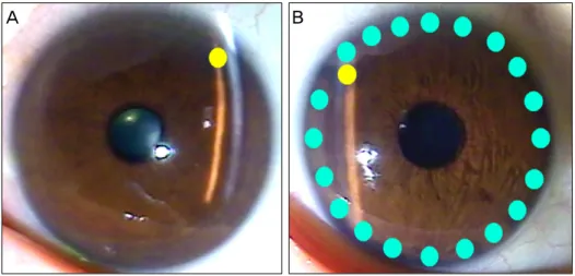
A B
Fig. 1. Conventional laser periph- eral iridotomy (LPI) and the LPI combined with iridoplasty technique.
(A) Conventional LPI refers to LPI alone. (B) The LPI combined with iridoplasty approach consists of LPI followed by laser peripheral irido- plasty administered in the same session. Yellow circle: LPI site, cy- anine-blue circle: laser peripheral iridoplasty site; about 20 burn spots are placed alongside the limbus.
closure even after LPI [8]. In other words, the etiology of PACS eye development may be a combination of both pupil- lary and non-pupillary block components. Laser peripheral iridoplasty applies a thermal energy that contracts the periph- eral iris away from the trabecular meshwork, thus providing amelioration of any appositional angle closure caused by a mechanism other than pupillary block. Thus, we devised a modified LPI technique in which standard LPI was combined with laser peripheral iridoplasty to prevent pupillary block and to reduce PAS formation by widening the closure angle.
The Pentacam (Oculus Inc., Wetzlar, Germany) is a rotating Scheimpflug camera that provides 50 cross-sectional images within a few seconds and has the advantage of generating both a three-dimensional model of the ACD as well as two-dimensional single scans. In the present study, we com- pared conventional LPI with LPI plus iridoplasty in PACS eyes with respect to the resulting anterior chamber dimen- sional changes visualized using a Pentacam.
Materials and Methods
This study adhered to the tenets of the Declaration of Helsinki. Patients were consecutively and prospectively en- rolled from the HanGil Eye Hospital, Incheon, Republic of Korea. After written informed consent was obtained, each patient underwent slit-lamp, optic disc, and fundus examina- tions; intraocular pressure (IOP) measurement by Goldmann applanation tonometry (GAT); and gonioscopy employing a Sussman four-mirror goniolens.
Only subjects in whom both eyes were PACS were eligible for inclusion in the study. Based on the EGS definition de- scribed above, a PACS eye showed an appositional contact between the iris and the posterior trabecular meshwork ex- tending over at least 180° on gonioscopic examination and an IOP ≤ 21 mmHg by GAT. Patients were excluded if they met any of the following criteria: 1) IOP > 21 mmHg by GAT; 2) PAS, defined as abnormal adhesion of the iris at any angle that was at least half a clock-hour in width and was located in the anterior trabecular meshwork or higher; 3) GON such as neuroretinal rim notching and/or thinning and/or disc hemor-
rhage and an associated retinal nerve fiber layer defect; 4) visual field defects indicative of GON; 5) a previous episode of acute angle closure attack; or 6) secondary angle closure.
Conventional LPI was performed on one randomly se- lected eye by a single glaucoma specialist (JC) using an ar- gon laser. One week later, the complementary eye was treat- ed with LPI combined with laser peripheral iridoplasty. Prior to each laser procedure, one drop of pilocarpine 2% and one drop of brimonidine tartrate 0.15% were instilled into the eye.
Conventional LPI used an argon laser to first create a con- traction burn; the laser was activated at a setting of 200 to 250 mW with a spot size of 500 μm for 0.6 seconds. In addition, a penetrating burn was achieved using the argon laser at a set- ting of 1,000 mW with a spot size of 50 μm for 0.05 seconds.
In LPI plus iridoplasty, the conventional procedure (described above) was followed by peripheral iridoplasty in the same session, during which the argon laser was used at a setting of 200 to 250 mW for 0.6 seconds, which created burn spots on the peripheral iris alongside the limbus. Each patient re- ceived about 20 burn spots over 360 degrees. After each pro- cedure, prednisolone acetate 1% was instilled 4 times daily for 1 week (Fig. 1).
Before and 1 week after treatment with either the conven- tional or the LPI plus iridoplasty technique, anterior chamber volume (ACV), ACD, and ACA were analyzed using the Pentacam. To permit detailed data analysis, we devised novel advanced ACD topographic parameters based on the simple ACD output data from the Pentacam. Thus, 1) “Central ACD” was the average of ACD values taken at nine topo- graphic points within 2 mm from the center of the eye; 2)
“Mid1 ACD” was the average of ACD measurements at 12 topographic points within 2-4 mm from the center of the eye;
3) “Mid2 ACD” was the average of ACD values taken at 16 topographic points within 4 to 6 mm from the center of the eye; and 4) “Mid3 ACD” was the average of ACD measure- ments at 20 topographic points within 6 to 8 mm from the center of the eye (Fig. 2).
Changes in the basic anterior chamber parameters and ad- vanced topographic ACD parameters, and IOP after LPI,
Mid1 ACD: mean ACD (2-4 mm)
Central ACD: mean ACD (-2 mm)
Mid2 ACD: mean ACD (4-6 mm)
Mid3 ACD: mean ACD (6-8 mm)
Fig. 2. Advanced anterior chamber depth (ACD) topographic parameters derived from routine ACD data from the Pentacam output. Central ACD is the average of ACD values at nine topographic points within 2 mm from the center of the eye; Mid1 ACD is the average of ACD measurements at 12 topographic points within 2 to 4 mm from the eye center; Mid2 ACD is the average of ACD values at 16 topographic points within 4 to 6 mm from the center of the eye; Mid3 ACD is the average of ACD measurements at 20 topographic points within 6 to 8 mm from the eye center.
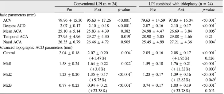
Table 1. Descriptive statistics for basic and advanced topographic anterior chamber depth parameters measured in the two study groups
Conventional LPI (n = 24) LPI combined with iridoplasty (n = 24)
Pre Post p-value Pre Post p-value
Basic parameters (mm)
ACV 79.96 ± 15.30 95.63 ± 17.26 <0.001* 79.63 ± 14.59 97.83 ± 16.04 <0.001* Deepest ACD 2.07 ± 0.17 2.10 ± 0.18 <0.001* 2.07 ± 0.16 2.10 ± 0.17 <0.001*
Mean ACA 25.10 ± 5.14 25.83 ± 4.39 0.382 24.98 ± 4.47 26.69 ± 3.84 0.005*
Temporal ACA 27.95 ± 4.96 29.27 ± 4.30 0.019* 28.98 ± 5.05 29.88 ± 4.66 0.21 Nasal ACA 26.35 ± 6.79 26.46 ± 4.72 0.905 25.45 ± 4.99 27.21 ± 4.36 0.004* Advanced topographic ACD parameters (mm)
Central 2.04 ± 0.18 2.07 ± 0.20 0.004* 2.05 ± 0.16 2.08 ± 0.17 <0.001*
(+1.47%) (+1.95%) 0.526
Mid1 1.58 ± 0.24 1.64 ± 0.22 0.022* 1.59 ± 0.18 1.76 ± 0.21 <0.001*
(+3.8%) (+11.32%) 0.003†
Mid2 1.23 ± 0.20 1.35 ± 0.17 <0.001* 1.23 ± 0.17 1.39 ± 0.16 <0.001*
(+9.75%) (+12.82%) 0.049†
Mid3 0.77 ± 0.23 0.94 ± 0.21 <0.001* 0.74 ± 0.17 1.00 ± 0.19 <0.001*
(+23.38%) (+33.78%) 0.202
Data are means ± standard deviations.
LPI = laser peripheral iridotomy; ACV = anterior chamber volume; ACD = anterior chamber depth; ACA = anterior chamber angle.
*p < 0.05, paired t-test comparing pre- and post-LPI parameters in the conventional LPI and LPI combined with iridoplasty groups, respectively.
†p < 0.05, unpaired t-test between the conventional LPI and LPI combined with iridoplasty groups with respect to increases in the levels of advanced topographic parameters after LPI.
were compared in each group using the paired t-test.
Additionally, any increases in the values of the advanced topographic ACD parameters and complications after LPI were compared between the two groups employing the un- paired t-test. Transient elevation of IOP (an IOP spike) was defined as an IOP increase above 5 mmHg from baseline within 1 week of a laser procedure. In all analyses, a differ- ence with a probability p-value < 0.05 was considered to be statistically significant.
Results
Forty-eight eyes of 24 patients were enrolled in the study;
mean patient age was 59.1 ± 9.0 years (range, 40 to 74 years).
Four patients were male and 20 were female. The basic pa- rameters of ACV and deepest ACD and all of the advanced topographic ACD parameters (Central ACD, Mid1 ACD, Mid2 ACD, and Mid3 ACD) increased significantly in both the conventional and LPI plus iridoplasty groups. The extent of increase in the advanced topographic ACD parameters
Table 2. Intraocular pressure changes following conventional LPI and LPI combined with iridoplasty Intraocular pressure (mmHg)
Pre-LPI 1 hr 1 day 1 wk 1 mon 3 mon
Conventional LPI 15.4 ± 3.3 18.9 ± 4.6 14.0 ± 2.8 14.5 ± 3.0 14.6 ± 3.6 14.2 ± 2.6 LPI combined with iridoplasty 14.7 ± 2.7 18.0 ± 6.1 13.5 ± 3.1 13.8 ± 3.3 13.4 ± 3.2 13.9 ± 3.7 No between-group statistical significance was apparent at any intraocular pressure measurement time point.
LPI = laser peripheral iridotomy.
Table 3. Complications after intervention in the conven- tional LPI and LPI combined with iridoplasty groups
Conventional
LPI LPI combined with iridoplasty
Transient IOP spike (≥5 mmHg) 8 8
Hyphema 1 1
Persistent uveitis 1 wk post-LPI 2 1
Transient atonic pupil 0 1
Numbers refer to the number of eyes.
No statistically significant difference in complication rate or nature was found between the conventional LPI and LPI combined with iridoplasty groups.
LPI = laser peripheral iridotomy; IOP = intraocular pressure.
was compared between the groups, and an increase in the Central ACD of was found approximately 2.0% for eyes treated by LPI plus iridoplasty and approximately 1.5% for eyes that received conventional LPI; these values were not significantly different (p = 0.526). However, the increase in the Mid1 ACD was approximately 11.3% for eyes treated with LPI plus iridoplasty and 3.8% for eyes that received conventional LPI (p = 0.003). The increase in the Mid2 ACD was approximately 12.8% for eyes treated with LPI plus iri- doplasty and 9.8% for eyes that received conventional LPI (p
= 0.049). Although the increase in the Mid3 ACD was ap- proximately 33.8% for eyes that received LPI plus iridoplasty and 23.4% for eyes treated with conventional LPI, the difference was not statistically significant (p = 0.202). In brief, changes in the Mid1 ACD and Mid2 ACD, namely the mid-peripheral ACD, were significantly greater for eyes treated with LPI plus iridoplasty than in those treated by conventional LPI, whereas the central and far-peripheral mid3 ACD values did not differ significantly between the two groups (Table 1).
The baseline IOP did not significantly differ between eyes treated by the conventional and LPI plus iridoplasty techniques. Post-procedure IOP elevation was not significant in either group, and also did not significantly differ between the two groups during the 3 months after treatment (Table 2).
Post-procedural complications were similar in frequency and severity in the two groups. IOP spikes occurred in eight eyes from both groups. Hyphema occurred in one eye treated with conventional LPI, whereas post-laser uveitis persisted in two eyes for 1 week after conventional LPI treatment. In eyes receiving the LPI plus iridoplasty procedure, hyphema was evident in one eye, persistent post-laser uveitis was evi- dent in one eye, and a transient atonic pupil occurred in one
eye over the 3-month follow-up period (Table 3).
Discussion
Previously, Kumar et al. [9] reported that approximately 30% of PACS eyes had plateau iris prior to LPI, and this con- dition persisted in 75% of such eyes after LPI. Another study, in an Asian cohort, found that 30% of PACG eyes previously treated with LPI had plateau iris [10]. This implies that a mechanism other than pupillary block may play a significant role in the occurrence and progression of angle closure. One non-pupillary block mechanism seen in PACS, PAC, and PACG eyes with plateau iris is bunching of the peripheral iris resulting in closure of the anterior chamber angle despite a patient iridotomy. The suggested mechanism of this effect is an anteriorly dislocated and/or large ciliary body serves as a contact between the iris and the angle [11,12]. In such eyes, laser peripheral iridoplasty may help to eliminate apposi- tional angle closure and widen the ACA, because iridoplasty pulls the peripheral iris away from the angle [13,14].General indications for laser peripheral iridoplasty include the fol- lowing: acute and chronic angle closure, phacomorphic glau- coma, and nanophthalmos. Further, the technique may be used as an adjunctive method to deepen the anterior chamber, facilitating selective laser trabeculoplasty [13]. One histo- pathologic study suggested that laser peripheral iridoplasty not only induced short-term thermal shrinkage of stromal collagen, but the contraction effect may persist long-term mediated by fibroblast-like cells [15].
In the present work, we investigated the efficacy and safety of LPI combined with iridoplasty, which consisted of simul- taneous argon LPI and peripheral iridoplasty. We measured anterior chamber dimensional changes and IOP and assessed post-laser complications after application of two different la- ser procedures to PACS eyes. We found that the mid-periph- eral ACD was significantly increased in eyes treated with LPI plus iridoplasty, without any additional complications, compared to eyes that received conventional LPI. As pre- viously mentioned by van Herick et al. [16], far-peripheral ACD is widely regarded as one of the important risk factors for angle closure. However, increases in the far-peripheral ACD and ACA did not differ significantly between the two groups. This may be attributable to the rotating Scheimpflug camera, Pentacam. Boker et al. [17] previously reported that the Pentacam was unable to directly visualize the ACA, i.e., on Pentacam images, it is difficult to identify the most pe-
ripheral part of the iris and the base of the ACA. Because the Pentacam assumes and automatically localizes the apex of the ACA, the ACA and adjacent far-peripheral ACD meas- urements can be incorrect and unreliable.
Gonioscopy is the gold standard modality for assessment of ACA. However, this method is relatively subjective, and accuracy increases with the experience of the ophthalmologist.
Further, it is difficult to quantitatively describe the AC morphology.
Previous studies have found that ultrasound biomicroscopy may offer a detailed view of anatomical structures in the ACA [11,18]. However, this technique requires that patients are maintained in a supine position, with an ocular cup, saline bath, and probe contacting the eyeball. We analyzed ante- rior chamber dimensions in PACS eyes using a noncontact rotating Scheimpflug camera, the Pentacam, and estimated novel parameters representing the ACD at different loca- tions within the eye. Kurita et al. found that the Pentacam was potentially useful for examining both PACS and PAC eyes, except for those with plateau iris, to calculate the ACV and ACD, but not ACA [19]. In addition, they found the Pentacam measurement of anterior chamber dimensions was reliable [20].
The present study has some limitations. First, the data re- flect only changes in ACD, and not the detailed variations in angle structure, thus, the number of subjects in this study with plateau iris was not determined. Second, we have not shown that the LPI plus iridoplasty technique is better than conventional LPI in preventing the progression of PACS eyes to PACG. A long-term follow-up study is needed to ex- plore whether the LPI plus iridoplasty technique is superior to the conventional LPI approach in this regard. Third, our relatively small sample size may limit data interpretation.
Fourth, no study has optimized either the laser beam width or spot number when laser peripheral iridoplasty is used to treat PACS eyes. Although we found no significant difference in the extent of complications between the two groups during the short-term follow-up, laser peripheral iridoplasty can cause permanent iris burn scars which can affect pupil size or pupil constriction.
The present study is the first to explore ACD topographic changes after use of different laser techniques. Both of the methods increased the anterior chamber volume and depth in PACS eyes. However, the LPI plus iridoplasty technique was more effective in elevating the mid-peripheral ACD than was conventional LPI, while the complication rate did not differ between the two groups. In conclusion, the LPI plus irido- plasty technique may be effective and safe for inhibiting the development of unwanted anterior chamber dimensional changes in PACS eyes. Further work is needed to determine if this combined laser technique prevents the progression of PACS eyes to eyes with PAC or PACG.
Conflict of Interest
No potential conflict of interest relevant to this article was
reported.
References
1. European Glaucoma Society. Terminology and guidelines for glaucoma. 3rd ed. Savona: Dogma; 2008.
2. Shen SY, Wong TY, Foster PJ, et al. The prevalence and types of glaucoma in malay people: the Singapore Malay Eye Study.
Invest Ophthalmol Vis Sci 2008;49:3846-51.
3. Casson RJ, Newland HS, Muecke J, et al. Prevalence of glau- coma in rural Myanmar: the Meiktila Eye Study. Br J Ophthalmol 2007;91:710-4.
4. Thomas R, George R, Parikh R, et al. Five year risk of pro- gression of primary angle closure suspects to primary angle closure: a population based study. Br J Ophthalmol 2003;87:
450-4.
5. Thomas R, Parikh R, Muliyil J, Kumar RS. Five-year risk of progression of primary angle closure to primary angle closure glaucoma: a population-based study. Acta Ophthalmol Scand 2003;81:480-5.
6. Ang MH, Baskaran M, Kumar RS, et al. National survey of ophthalmologists in Singapore for the assessment and man- agement of asymptomatic angle closure. J Glaucoma 2008;
17:1-4.
7. Pandav SS, Kaushik S, Jain R, et al. Laser peripheral iridot- omy across the spectrum of primary angle closure. Can J Ophthalmol 2007;42:233-7.
8. He M, Friedman DS, Ge J, et al. Laser peripheral iridotomy in primary angle-closure suspects: biometric and gonioscopic outcomes: the Liwan Eye Study. Ophthalmology 2007;114:
494-500.
9. Kumar RS, Baskaran M, Chew PT, et al. Prevalence of plateau iris in primary angle closure suspects an ultrasound biomicro- scopy study. Ophthalmology 2008;115:430-4.
10. Kumar RS, Tantisevi V, Wong MH, et al. Plateau iris in Asian subjects with primary angle closure glaucoma. Arch Ophthalmol 2009;127:1269-72.
11. Pavlin CJ, Ritch R, Foster FS. Ultrasound biomicroscopy in plateau iris syndrome. Am J Ophthalmol 1992;113:390-5.
12. Ritch R. Plateau iris is caused by abnormally positioned ciliary processes. J Glaucoma 1992;1:23-6.
13. Ritch R, Tham CC, Lam DS. Argon laser peripheral irido- plasty (ALPI): an update. Surv Ophthalmol 2007;52:279-88.
14. Ritch R, Tham CC, Lam DS. Long-term success of argon laser peripheral iridoplasty in the management of plateau iris syndrome. Ophthalmology 2004;111:104-8.
15. Sassani JW, Ritch R, McCormick S, et al. Histopathology of argon laser peripheral iridoplasty. Ophthalmic Surg 1993;24:
740-5.
16. Van Herick W, Shaffer RN, Schwartz A. Estimation of width of angle of anterior chamber. Incidence and significance of the narrow angle. Am J Ophthalmol 1969;68:626-9.
17. Boker T, Sheqem J, Rauwolf M, Wegener A. Anterior chamber angle biometry: a comparison of Scheimpflug photography and ultrasound biomicroscopy. Ophthalmic Res 1995;27 Suppl 1:104-9.
18. Pavlin CJ, Harasiewicz K, Foster FS. Ultrasound biomicro- scopy of anterior segment structures in normal and glaucoma- tous eyes. Am J Ophthalmol 1992;113:381-9.
19. Kurita N, Mayama C, Tomidokoro A, et al. Potential of the pen- tacam in screening for primary angle closure and primary an- gle closure suspect. J Glaucoma 2009;18:506-12.
20. Rabsilber TM, Khoramnia R, Auffarth GU. Anterior chamber measurements using Pentacam rotating Scheimpflug camera. J Cataract Refract Surg 2006;32:456-9.