https://doi.org/10.12750/JET.2017.32.1.25
Endoplasmic Reticulum (ER) Stress and Apoptosis in Parthenogenetic Porcine Embryos following Different Combination of Activation Methods
*Hye-Bin Park1, Yeo-Reum Park1, Hwa-Yeon Lee1, Hyo-Kyung Bae1, Seunghyung Lee2, Choon-Keun Park2, Boo-Keun Yang2, Hee-Tae Cheong1†
1College of Veterinary Medicine and Institute of Veterinary Science,
and 2College of Animal Life Sciences, Kangwon National University, Chuncheon 24375, Korea
ABSTRACT
This study was conducted to investigate the effect of activation method on the endoplasmic reticulum (ER) stress induction, apoptosis and in vitro development of porcine parthenogenetic embryos. Porcine in vitro matured oocytes were activated by four activation methods; 1) electric stimulus (ES) (E), 2) ES+10 μM Ca-ionophore (A23187) treatment (EC), 3) ES+2 mM 6-dimethylaminopurine (6-DMAP) treatment (ED), or 4) ES+A23187 and 6-DMAP treatments (ECD). Parthenogenetic embryos were sampled to analyze x-box binding protein 1 (Xbp1) mRNA, ER stress-associated genes and apoptosis genes at 3 h after ES and the 1-cell and blastocyst stages. In the EC group, the band intensity of spliced Xbp1 (Xbp1s) mRNA was higher than those of the other groups at the 3 h and 1-cell stage, and higher than that of the E group at the blastocyst stage. Four ER stress-associated genes were expressed at the highest level in the EC group and weakly expressed in the ED group at 3 h after activation. However, most of the genes were highly expressed at the 1-cell and blastocyst stages with some variation in the EC and ECD groups.
Expression of Bcl-2-associated X protein (Bax) and caspase-3 mRNA was significantly higher in the EC group than in the other groups at all development stages. The developmental rates to the blastocyst stage were higher in the ED and ECD groups than in the E and EC groups. These results suggest that the intracellular ER stress of parthenogenetic porcine embryos is affected by the activation method and subsequently lead to the apoptosis of embryos.
(Key words: Parthenogenesis, endoplasmic reticulum stress, apoptosis, pig)
* This study was supported by 2015 Research Grant from Kangwon National University (No. 520150285)
†Corresponding author address:
Phone: +82-33-250-8659 E-mail: [email protected]
INTRODUCTION
The endoplasmic reticulum (ER) is a cell organelle with various functions such as protein folding, transport of synthesized proteins, lipid biosynthesis, and calcium storage.
Various physiological and pathological conditions, as well as various pharmacological agents may interfere with proper ER function. The disruption of ER homeostasis affects the proper protein folding of cells, causing accumulation of unfolded or misfolded protein in ER lumen. If the accumulation of unfolded or misfolded protein exceeds the folding capacity of the ER, which condition is called ER stress, the cells activates a defense mechanism called the ER stress response or unfolded protein response (UPR) to reduce the accumulation of unfolded/misfolded protein (Mori, 2000; Boyce and Yuan, 2006; Malhotra and Kaufman, 2007).
Under ER stress conditions, the UPR signaling sensor/
pathway, the inositol-requiring enzyme 1ɑ (IRE1ɑ), the PKR-like ER kinase (PERK) and the activating transcription factor 6 (ATF6) are activated in the ER luminal domains (Boyce and Yuan, 2006; Yoshida, 2007). Thus, ER stress in cells can be estimated by detecting UPR signaling sensors (Schröder and Kaufman, 2005; Samali et al., 2010). Excessive ER stress can cause cellular damage such as apoptosis (Yoshida, 2007). In somatic cell nuclear transfer (SCNT) procedure, several physical and chemical stresses caused by manipulation can cause ER stress in the SCNT embryos.
Activation of reconstituted oocytes is one of the essential steps in the SCNT procedure, and various activation methods have been used in animal SCNTs.
This study was conducted to examine the effect of activation method on the induction of ER stress, and
subsequent apoptosis and in vitro development of porcine parthenogenetic embryos.
MATERIALS and METHODS
Chemicals
All chemicals and reagents were purchased from Sigma-Aldrich Chemical Co. (St Louis, Mo, USA) unless otherwise indicated.
Collection and in vitro maturation of oocytes
Porcine ovaries were obtain from a local slaughterhouse and cumulus-oocyte complexes (COCs) were collected by aspirating antral follicles (3~6 mm diameter) using a 10 ml syringe with an 18-gauge needle. COCs were cultured for 42-44 h in in vitro maturation (IVM) medium at 39°C, 5%
CO2 in air. IVM medium was tissue culture medium 199 (TCM199; Gibco, Grand Island, NY, USA) supplemented with 0.1% polyvinyl alcohol (PVA), 3.05 mM D-glucose, 0.91 mM NA-pyruvate, 75 μg/ml penicillin G, 50 μg/ml streptomycin, 0.57 mM cysteine, 10 μg/ml epidermal growth factor (EGF), 0.5 μg/ml luteinizing hormone (LH), and 0.5 μg/ml follicle- stimulating hormone (FSH).
Parthenogenetic activation and in vitro culture
Cumulus cells of matured oocytes were removed by vortexing with PBS containing 0.1% (w/v) hyaluronidase and 0.1% (w/v) PVA for 3 min. The oocytes were activated by four activation methods; 1) electric stimulus (ES) with the same conditions as our fusion condition for the SCNT experiment (E), 2) ES followed by additional treatment with 10 μM Ca-ionophore (A23187) for 5 min (EC), 3) ES followed by treatment with 2 mM 6-dimethylaminopurine (6‐
DMAP) in PZM 3 for 3 h (ED), or 4) ES followed by treatments with 10 μM A23187 for 5 min and then 2 mM 6‐
DMAP for 3 h (ECD). For ES, oocytes were placed between two wire electrodes (1 mm apart) covered with 0.3 M mannitol solution, supplemented with 0.1 mM MgCl2, 0.05 mM CaCl2 and 0.5 mM HEPES (Duchefa Biochemie, Haarlem, Netherlands), and 2 pulses of direct-current (DC) of 1.25 kV/cm were applied for each 30 μsec using a BTX Electro Cell Manipulator 200 (BTX, San Diego, CA, USA).
After activation, parthenogenetic embryos were cultured in
vitro in PZM-3 medium (Yoshioka et al., 2002) for 6 days at 39°C, 5% CO2 in air. To analyze ER stress and apoptosis, parthenogenetic embryos were sampled at 3 h, 20 h and 6 days after ES.
Isolation of mRNA and cDNA synthesis
The mRNAs of parthenogenetic embryos at 3 h, 24 h (1-cell stage) and 6 days (blastocyst) after ES were isolated according to the manufacturer’s protocol using the Dynabeads mRNA Direct kit (Invitrogen, Carlsbad, CA, USA). In brief, each embryo group was washed with 0.3% PVP-PBS and sampled with 10 μl RNAlater® solution (Invitrogen) at low temperature before use and stored at -70°C. After thawing, 10 μl of Dynabeads oligo (dT) was added to each sample at room temperature to hybridize oligo (dT) and the poly (A) tail of the mRNA for 2 min. Samples were lysed in 200 μl of lysis/binding buffer for 2 min at room temperature and beads were separated from binding buffer using a Dynal magnetic bar. Separated poly (A) mRNAs and beads were washed with Buffer A and B and poly (A) mRNA was eluted at 63°C with 6.5 μl of Tris-HCl buffer. The cDNA was synthesized according to the manufacturer’s protocol using Rever Tra Ace® qPCR RT Master Mix (Toyobo, Osaka, Japan). A total 6 μl of the isolated mRNA was denatured at 65°C for 5 min.
To remove genomic DNA, 2 μl of 4x DN Master Mix was added to the RNA template and incubated at 4°C for 5 min, then 2 μl of 5x DN Master Mix was added and incubated at 37°C for 20 min for reverse transcription. The products were incubated at 50°C for 5 min and the reaction was terminated at 98°C for 5 min. The synthetic DNA was used as a template for reverse transcription-polymerase chain reaction (RT-PCR) and real-time quantitative RT-PCR (RT-qPCR).
RT-PCR and RT-qPCR
Expression of x-box binding protein 1 (Xbp1) mRNA, a key transcription factor in the ER stress response, was detected by RT-PCR analysis, and expressions of ER stress-associated genes C/EBP homologous protein (CHOP), binding protein (BiP), activating transcription factor 4 (ATF4) and glucose- regulated protein 94 (GRP94), and apoptotic genes were analyzed by RT-qPCR. For RT-PCR analysis, the cDNA template was amplified according to the manufacturer’s protocol using AccuPower® Taq PCR PreMix (Bioneer, Daejeon, Korea). The initial denaturation was carried out at
Genes Primer Sequence (5’-3’) Length (bp)
GenBank Acc. No.
Annealing Temp (ºC)
Xbp1 F-GGCAGAGACCAAGGGGAATG
R-GGGTCGACTTCTGGGAGCTG 263 FJ213449.1 60
CHOP F-AAGACCCAGGAAACGGAAAC
R-TCCAGGAAAGGTCAGCAGTA 261 NM_001144845.1 58
BiP F-ACCAATGACCAAAATCT
R-GTGACTTTCCAGCCAAA 246 J03214.1 58
ATF4 F-TGAGCCCTGACTCCTATCTG
R-TCCAGCTCTTTACATTCGCC 277 NM_001123078.1 58
GRP94 F-CTGCTGAAGGGGAAGTTACC
R-ATCATCTGAGTCCACAACGC 197 Y09136.1 58
Bax F-ACTGGACAGTAACATGGAGC
R-GTCCCAAAGTAGGAGAGGAG 294 XM003127290.3 55
Caspases-3 F-GAGGCAGACTTCTTGTATGC
R-CATGGACACAATACATGGAA 237 NM_214131 60
GAPDH F-GGGCATGAACCATGAGAA
R-AAGCAGGGATGATGTTCT 230 AF017079 58
Table 1. All primer sequences used for RT-PCR.
72°C for 1 min, and then 34 cycles of amplification were performed. Each cycle consisted of denaturation at 95°C for 30 sec, annealing at 58°C for 30 sec, and extension at 72°C for 50 sec. The reaction was terminated by final extension step at 72°C for 5 min. The PCR products were resolved by electrophoresis on a 2% agarose gel (Amresco, Cleveland, OH, USA) stained with ethidium bromide (Bioneer). Target bands were identified by UV irradiation using an UV transilluminator (Bio-Rad, Berkeley, CA, USA). RT-qPCR was performed with the power SYBR Green PCR master Mix (TOPrealTM qPCR 2X PreMIX; SYBR Green with high ROX, Enzynomics, Daejeon, Korea) in a StepOne Plus instrument (Applied Biosystems, Foster City, CA, USA). The comparative CT method (∆∆CT method) was used to quantify the mRNA level of target genes. Glyceraldehyde 3-phosphate dehydro- genase (GAPDH) was used as an internal control for the normalization of target gene expression. The Primer sequences of each gene were shown in Table 1.
Statistical analysis
Each activation method was replicated at least 5 times. Data were analyzed by the analysis of variance (ANOVA) and Duncan’s multiple-range test using the Statistical Analysis System software package (SAS Institute, Cary, NC, USA)
RESULTS
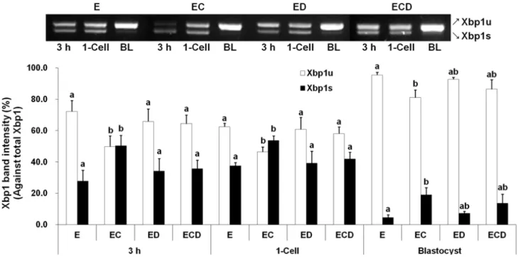
Spliced Xbp1 mRNA expression in parthenogenetic embryos The band intensity of Xbp1s mRNA in the EC group was higher (P<0.05) than those of other groups at 3 h and the 1-cell stage, and higher than that of E group at the blastocyst stage (Fig. 1). Xbp1s mRNA was weakly detected at the blastocyst stage.
ER stress-associated gene expression in parthenogenetic embryos Expression of ER stress-associated genes such as CHOP, BiP, ATF4 and GRP94 was highest in EC group at 3 h, and weak in the ED group at 3 h and the 1-cell stage (Fig. 2).
However, most of the genes were highly expressed in the EC and ECD groups at the 1-cell and blastocyst stages with some variation.
Apoptosis in parthenogenetic embryos
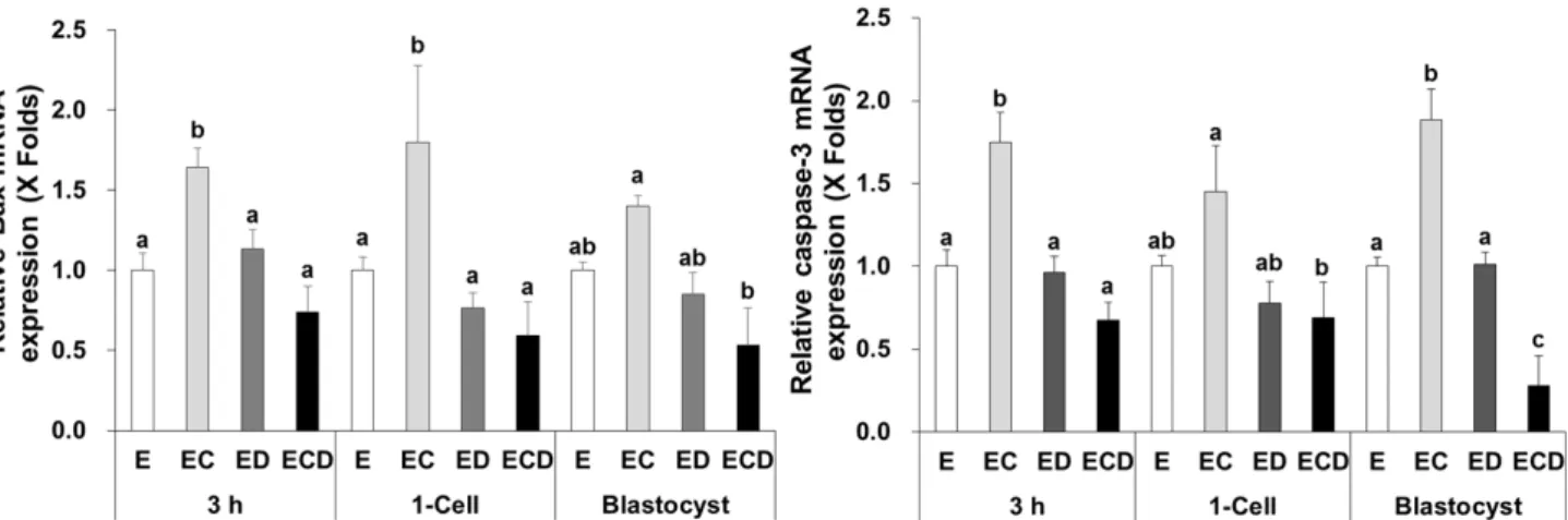
Expressions of Bcl-2-associated X protein (Bax) and caspase-3 mRNAs, which are known as apoptosis-related genes, were significantly higher (P<0.05) in the EC group than other three groups at 3 h and the 1-cell and blastocyst stages, except the expressions of Bax mRNA in the blastocyst stage and caspase-3 mRNA in the 1-cell stage (Fig. 3).
Fig. 1. Xbp1 mRNA expression in porcine parthenogenetic embryos. Spliced Xbp1(Xbp1s) and unspliced Xbp1 (Xbp1u) mRNAs were detected by RT-PCR at 3 h after electric stimulation and the 1-cell and blastocyst stages, and band intensity was measured densitometrically. E, electric stimulus (ES); EC, ES+10 μM Ca-ionophore (A23187) treatment; ED, ES+2 mM 6-dimethylaminopurine (6‐DMAP) treatment; ECD, ES+10 μM A23187 and 2 mM 6‐DMAP treatments; BL, blastocyst. Data were presented as means ± SEM of five replicates. a,bValues were significantly different from each group within the same stage (P<0.05).
Fig. 2. ER stress-associated gene expression in porcine parthenogenetic embryos. ER stress-associated gene expression was quantified by RT-qPCR at 3 h after electric stimulus and the 1-cell and blastocyst stages. E, electric stimulus (ES); EC, ES+10 μM Ca-ionophore (A23187) treatment; ED, ES+2 mM 6-dimethylaminopurine (6‐DMAP) treatment; ECD, ES+10 μM A23187 and 2 mM 6‐DMAP treatments.
Data were presented as means ± SEM of five replicates. a-dValuesweresignificantlydifferentfromeachgroupinthesamegeneandstage(P<0.05).
Treatments No. of embryos cultured % of embryos developed to
≥2-Cell Blastocyst
E 277 88.8±1.7 26.1±3.9a
EC 279 88.9±2.0 25.5±1.0a
ED 276 91.9±1.5 32.1±3.8b
ECD 290 91.5±1.6 34.6±2.2b
E, electric stimulus (ES); EC, ES+10 μM Ca-ionophore (A23187) treatment; ED, ES+2 mM 6-dimethylaminopurine (6-DMAP) treatment;
ECD, ES+10 μM A23187 and 2 mM 6-DMAP treatments. Data were presented as means ± SEM of seven replicates. a,bValues in the same column were significantly different (P<0.05).
Table 2. Development of parthenogenetic embryos following various activation methods
Fig. 3. Expression of apoptotic genes in parthenogenetic embryos. Bax and caspase-3 mRNA expressions were quantified by RT-qPCR at 3 h after electric stimulus and the 1-cell and blastocyst stages. E, electric stimulus (ES); EC, ES+10 μM Ca-ionophore (A23187) treatment; ED, ES+2 mM 6-imethylaminopurine (6‐DMAP) treatment; ECD, ES+10 μM A23187 and 2 mM 6‐DMAP treatments. Data were presented as means ± SEM of five replicates. a-cValues were significantly different from each group in the same gene and stage (P<0.05).
Development of parthenogenetic embryos
The cleavage rate of parthenogenetic embryos was not influenced (88.8±1.7 to 91.5±1.6%) by activation method, whereas the developmental rate to the blastocyst stage was higher (P<0.05) in ED (32.1±3.8) and ECD (34.6±2.2%) groups than those of E (26.1±3.9%) and EC (25.5±1.0) groups (Table 2).
DISCUSSION
When the ER stress occurs, the unfolded and misfolded proteins in the ER lumen bind BiP, and activates UPR.
Generally, BiP binds to IRE1ɑ, PERK and ATF6 and held inactivation. By accumulation of unfolded/misfolded protein in
the ER, BiP is dissociated from IRE1, PERK and ATF6 and UPR is activated (Yoshida, 2007). Activated IRE1ɑ acts as an endoribonuclease that splits the 26 base pair nucleotide intron from Xbp1 mRNA, leading to subsequent frame shift (Schröder and Kaufman, 2005; Hetz and Glimcher, 2009).
Xbp1s mRNA is translated into a stable and active UPR transcription factor. Thus, measuring Xbp1s is a reliable method for determining IRE1αactivation. PERK is activated through oligomerization and phosphorylation, and the α -subunit of eukaryotic translational initiation factor 2 (elf2α) promotes the translation of ATF4 (Fels and Koumenis, 2006), which stimulates the expression of CHOP (Nishitoh, 2012).
Activated ATF6 is translocated to Golgi (Yoshida, 2007), then regulates the expression of GRP78, GRP94, XBP1, and CHOP (Yoshida et al., 2001; Adachi et al., 2008; Parmar and
Schröder, 2012).
Various chemicals such as tunicamycin, thapsigargin, and dithiothreitol evoke ER stress. Tunicanycin is an antibiotic that inhibits N-glycosylation and is often essential for protein folding (Dorner et al., 1990). Ca2+ metabolic disruptors such as Ca-ionophore (A23187) and Ca2+ pump inhibitor thapsigargin are ER stressors (Dornor et al., 1990; Price et al., 1992). Ca2+metabolic disturbance by chemicals invokes ER stress because high concentration of Ca2+ ion is retained in the ER and ER chaperones require Ca2+ ions. Dithiothreitol and 2-mercaptoethanol also cause ER stress by disrupting disulfide bonds (Brostrom et al., 1995; Fernandez et al., 1996). Hypoxia is also an ER stressor that can inhibit N-glycosylation through a decrease in glucose levels (Ikeda et al., 1997).
Various expression levels of Xbp1s mRNA were detected stage-dependently in porcine parthenogenetic embryos (Zhang et al., 2012), suggesting that ER stress-derived Xbp1 splicing modulates early embryonic genome activation. Although a certain level of ER stress may be essential for early embryonic development, excessive ER stress causes apoptotic cell death (Yoshida, 2007). In this study, A23187 treatment (e.g. EC group) increased Xbp1s mRNA expression, while 6-DMAP treatment prevented Xbp1 splicing by A23187 at the early embryonic stage. Xbp1s mRNA was also increased by treatment with A23187. The expression of ER stress-associated genes such as CHOP, ATF4, BiP, and GRP94 tended to be increased by A23187 treatment and suppressed by 6-DMAP treatment at the early embryonic stage. However, most of the genes were highly expressed in the EC and ECD groups at the 1-cell and blastocyst stages with some variation (see Fig. 2), which was slightly different from the Xbp1s mRNA expression pattern. ER stress induction in the embryos at the early developmental stage may be affected by the activation agents, but ER stress induction at the blastocyst stage may be influenced by the culture environment as well as by the activation method.
ER stress-induced apoptosis was associated with Caspase-3 (Groenendyk and Michalak, 2005) and Bcl-2 family proteins (Szegezdi et al., 2006). Prolonged UPR induces the expressions of Caspase-3 and Bcl-2 family proteins and increases apoptosis (Tabas and Ron, 2001; Gorman et al., 2012). In this study, the expression of Bax and Caspase-3 mRNA was increased by additional A23187 treatment, suggesting that A23187-induced ER stress induces apoptosis of
the parthenogenetic embryos. A23187 increased cytosolic free calcium concentration and induced apoptosis in cells (Caron-Leslie et al., 1994). Additional chemical activation after ES has been reported to increase the frequency of apoptosis in porcine SCNT embryos (Im et al., 2006). On the other hand, the increased apoptotic activity by A23187 treatment was reduced by 6-DMAP treatment in ECD group at all stages, which was not exactly the same as the result of ER stress induction. In the ER stress result of this study, most of the ER stress-associated genes were strongly expressed at the 1-cell stage and especially at the blastocyst stage of ECD group. Our results suggested that the pro-apoptotic activity of A23187 could be reduced by 6-DMAP treatment regardless of the induction of ER stress, suggesting that increased activation by ECD treatment in view of the high blastocyst formation rate (see Table 1).
In this study, the in vitro development of parthenogenetic embryos was affected by the activation method. The blastocyst formation rate, however, was not directly affected by the ER stress because the blastocyst formation rate was not lower in the EC group than in the E group and significantly higher in the ECD group with high ER stress. In the EC group, ER stress-induced apoptosis was not recovered at all stages, unlike the ECD group, which may be a cause of low blastocyst development. A more valid reason for the difference in parthenogenetic development may be the difference in activating ability of the activation agent or method. A variety of activation methods can affect the ability of activation and in vitro development of the oocyte (Im et al., 2006).
These results suggest that the intracellular ER stress in the parthenogenetic porcine embryos is affected by the activation method and subsequently induces apoptosis of the embryo. ER stress and apoptosis can be highly induced by A23187 treatment. However, it is unclear whether the developmental capacity of parthenogenetic porcine embryos is affected by ER stress or by the activating ability of the activation method.
REFERENCES
Adachi Y, Yamamoto K, Okada T, Yoshida H, Harada A and Mori K. 2008. ATF6 is a transcription factor specializing in the regulation of quality control proteins in the endoplasmic reticulum. Cell Struct. Funct. 33:75-89.
Boyce M and Yuan J. 2006. Cellular response to endoplasmic reticulum stress: a matter of life or death. Cell Death Differ. 13:363-373.
Brostrom MA, Prostko CR, Gmitter D and Brostrom CO.
1995. Independent signaling of grp78 gene transcription and phosphorylation of eukaryotic initiator factor 2 alpha by the stressed endoplasmic reticulum. J. Biol. Chem.
270:4127-4132.
Caron-Leslie LM, Evans RB and Cidlowski JA. 1994. Bcl-2 inhibits glucocorticoid-induced apoptosis but only partially blocks calcium ionophore or cyclohecimide-regulated apoptosis in S49 cells. FASEB J. 8:639-645.
Dorner AJ, Wasley LC, Raney P, Haugejorden S, Green M and Kaufman RJ. 1990. The stress response in Chinese hamster ovary cells. Regulation of ERp72 and protein disulfide isomerase expression and secretion. J. Biol.
Chem. 26:22029-22034.
Fels DR and Koumenis C. 2006. The PERK/eIF2alpha/ATF4 module of the UPR in hypoxia resistance and tumor growth. Cancer Biol. Ther. 5:723-728.
Fernandez F, Jannatipour M, Hellman U, Rokeach LA and Parodi AJ. 1996. A new stress protein: synthesis of Schizosaccharomyces pombe UDP-Glc: glycoprotein glucosyltransferase mRNA is induced by stress conditions but the enzyme is not essential for cell viability. EMBO J.
15:705-713.
Gorman AM, Healy SJ, Jäger R and Samali A. 2012. Stress management at the ER: regulators of ER stress-induced apoptosis. Pharmacol. Ther. 134:306-316.
Groenendyk J and Michalak M. 2005. Endoplasmic reticulum quality control and apoptosis. Acta Biochim. Polonica 52:381-395.
Hetz C and Glimcher LH. 2009. Fine-tuning of the unfolded protein response: Assembling the IRE1alpha interactome.
Molecular Cell 35:551-556.
Ikeda J, Kaneda S, Kuwabara K, Ogawa S, Kobayashi T, Matsumoto M, Yura T and Yanagi H. 1997. Cloning and expression of cDNA encoding the human 150 kDa oxygen-regulated protein, ORP150. Biochem. Biophys. Res.
Commun. 230:94-99.
Im GS, Seo JS, Hwang IS, Kim DH, Kim SW, Yang BC, Yang BS, Lai L and Prather RS. 2006. Development and apoptosis of pre-implantation porcine nuclear transfer embryo activated with different combination of chemicals.
Mol. Reprod. Dev. 73:1094-1101.
Malhotra JD and Kaufman RJ. 2007. The endoplasmic reticulum and the unfolded protein response. Semin. Cell Dev. Biol. 18:716-731.
Mori K. 2000. Tripartite management of unfolded proteins in the endoplasmic reticulum. Cell 101:451-454.
Nishitoh H. 2012. CHOP is a multifunctional transcription factor in the ER stress response. J. Biochem. 151:217-219.
Parmar VM and Schröder M. 2012. Sensing endoplasmic reticulum stress. Adv. Exp. Med. Biol. 738:153-168.
Price BD, Mannheim-Rodman LA and Calderwood SK. 1992.
Brefeldin A, thapsigargin, and AIF4- stimulate the accumulation of GRP78 mRNA in a cycloheximide dependent manner, whilst induction by hypoxia is independent of protein synthesis. J. Cell Physiol. 152:545-552.
Samali A, FitzGerald U, Deegan S and Gupta S. 2010. Methods for monitoring endoplasmic reticulum stress and the unfolded protein response. Int. J. Cell Biol. 2010 ID 830307.
Schröder M and Kaufman R. 2005. ER stress and the unfolded protein response. Mutat. Res. 569:29-63.
Szegezdi E, Logue SE, Gorman AM and Samali A. 2006.
Mediators of endoplasmic reticulum stress-induced apoptosis. EMBO Rep. 7:880-885.
Tabas I and Ron D. 2001. Integrating the mechanisms of apoptosis induced by endoplasmic reticulum stress. Nature Cell Biol. 13:184-190.
Yoshida H. 2007. ER stress and diseases. FEBS J.
274:630-658.
Yoshida H, Matsui T, Yamamoto A, Okada T and Mori K.
2001. XBP1 mRNA is induced by ATF6 and spliced by IRE1 in response to ER stress to produce a highly active transcription factor. Cell 107:881-891.
Yoshioka K, Suzuki C, Tanaka A, Anas IM and Iwamura S.
2002. Birth of piglets derived from porcine zygotes cultured in a chemically defined medium. Biol. Reprod. 66:112-119.
Zhang JY, Diao YF, Oqani RK, Han RX and Jin DI. 2012.
Effect of endoplasmic reticulum stress on porcine oocyte maturation and parthenogenetic embryonic development in vitro. BMC Mol. Biol. 86:128.
Received November 18 2016, Revised December 06, 2016, Accepted March 18, 2017