Expression of Leukemia-Associated Antigen, JL1, in Bone Marrow and Thymus
Young Kee Shin,*
†Eun Young Choi,*
Seok Hyung Kim,* Junho Chung,
‡Doo Hyun Chung,* Weon Seo Park,
§Kyeong Cheon Jung,
¶Heung Sik Kim,
储Seonyang Park,** Hee Jin Kim,
††Myoung Hee Park,
††Chang Ki Min,
‡‡Chun Choo Kim,
‡‡and Seong Hoe Park*
From the Departments of Pathology,* Internal Medicine,**
Clinical Pathology,
††and Biochemistry,
‡Seoul National University College of Medicine, Seoul; DiNonA Inc.,
†Suwon; the Department of Pathology,
§Kangwon National University College of Medicine, Chunchon; the Department of Pathology,
¶Hallym University College of Medicine, Chunchon; the Catholic HSCT Center,
‡‡St. Mary’s Hospital, The Catholic University of Korea, Seoul; and the Department of Pediatrics,
储Keimyung University College of Medicine, Taegu, Korea
The identification of immunophenotypic markers with restricted expression has long been a critical issue in diagnostic and therapeutic advances for acute leukemias. We previously developed a monoclonal antibody against a new thymocyte surface antigen , JL1 , and showed that JL1 is expressed in the majority of acute leukemia cases. In this study , using multipa- rameter flow cytometric analyses , we found that JL1 was uniquely expressed in subpopulations of normal bone marrow (BM) cells , implying the association of JL1 with the differentiation and maturation process.
Although CD34
ⴙCD10
ⴙlymphoid precursors and some of maturing myeloid cells express JL1 , neither CD34
ⴙCD38
ⴚ/lonor CD34
ⴙAC133
ⴙnoncommitted pluripotent stem cells do. As for the myeloid precur- sors , CD34
ⴙCD33
ⴙcells do not express JL1. During lymphopoiesis , JL1 on the earliest lymphoid precur- sors disappear in the CD20
ⴙsIgM
ⴙstage of B-cell development or after CD1a down-regulation in thy- mocytes. Despite the highly restricted expression of JL1 in normal BM cells , most of the leukemias express JL1 irrespective of their immunophenotypes. These results indicate that JL1 is not only a novel differen- tiation antigen of hematopoietic cells , but also a leu- kemia-associated antigen. Therefore , we suggest that JL1 be a candidate molecule in acute leukemia for the diagnosis and immunotherapy that spares the normal BM stem cells. (Am J Pathol 2001, 158:1473–1480)
Leukemia-specific phenotypes, identified by monoclonal antibodies (mAbs) recognizing various cell surface anti- gens, have long played essential roles in the diagnosis and classification of leukemia. Because the antigens found on certain leukemic cells are also expressed on the surfaces of the normal counterparts in a developmental stage-dependent manner, they can be used as markers for lineage and differentiation. For instance, differentia- tion antigens such as CD34, CD10 (CALLA), CD13, CD19, CD7, CD20, CD33, and CD13 that are expressed on specific subsets of normal hematopoietic cells have been used as diagnostic markers for leukemic cells. There- fore, the characterization of molecules on the surface of hematopoietic cells is critical for the diagnosis of leukemia as well as for the understanding of hematopoiesis.
Acute leukemia still remains a therapeutic challenge in medical practice even in the age of high cure rates for pediatric leukemia with the advent of intensification of chemotherapy along with hematopoietic stem cell res- cue.
1It is therefore not surprising that alternative strate- gies, possessing distinct action mechanisms to comple- ment currently used treatment approaches, are needed and immunotherapy has been an appealing candi- date.
2–5In the past trials, mAb-based immunotherapy targeting leukocyte-specific antigens such as CD33, CD45, and CDw52 has been evaluated for the treatment of leukemia.
6 –16Although immunotherapy might be an attractive approach for the treatment of patients with leukemia, all these antibody-based immunotherapies are dependent on limited antigenic properties themselves.
As a consequence, it is worth trying to develop mAb(s) that recognize(s) specific antigen(s) with the restricted expression profile.
We previously reported a mAb against a novel human thymocyte differentiation antigen, designated as JL1,
17which is not expressed on mature T cells. We further showed that the anti-JL1 mAb broadly recognizes various types of acute leukemias of myeloid and B cell origins as well as T cell lineage.
18This strong co-relationship be- tween JL1 positivity and the diagnosis of leukemia prompted us to investigate the expression pattern of JL1 antigen in normal leukocytes during hematopoiesis in
Supported by the 1999 BK21 project for Medicine, Dentistry, and Phar- macy, and the 99 ⬘ DiNonA Inc. R&D Project, Suwon, Korea.
Accepted for publication December 21, 2000.
Address reprint requests to Seong Hoe Park, Dept. of Pathology, Seoul National University College of Medicine, 28 Yongon-dong, Chongno-gu, Seoul 110-799, Korea. E-mail: [email protected].
1473
detail. Although JL1 antigen was initially reported not to be expressed in the majority of normal unfractionated bone marrow (BM) cells, if not all, there remained a possibility that a small proportion of BM cells do express JL1, because of the high heterogeneity of mononuclear cells (MNCs) in normal BM.
18In the present study, to dissect the expression patterns of JL-1 antigen on the leukocytes of different lineage and maturation stages in BM, cord blood (CB), and thymus, we fractionated the MNCs using lineage-specific markers and re-evaluated JL1 expression on their surfaces. We found that JL1 molecules are expressed on some of the precursor cells of lymphoid and myelomonocytic lineages but not on pluripotent stem cells. As most of the leukemias express JL1 antigen on the cell surfaces, anti-JL1 mAb may have the implications for the immunotherapeutic potential for the treatment of leukemia.
Materials and Methods Cell Preparation
CB cells (n ⫽ 7) were obtained at Keimyung University Hospital, and normal (n ⫽ 3) or G-CSF-primed (n ⫽ 10) BM cells were aspirated from the posterior iliac crests of healthy adult and pediatric transplantation donors with written consents following guidelines approved by the Institutional Review Board for Human Research, Seoul National University Hospital or Catholic University Hospi- tal in conformity with the Helsinki protocols. Recombinant human G-CSF (Filgrastim; Amgen, Thousand Oaks, CA) was administered subcutaneously at a dosage of 10
g/kg/day for 3 consecutive days. Both BM aspirates and CB were drawn into 10-ml syringes containing 100 U of preservative-free heparin and then diluted with RPMI 1640 (Life Technologies, Inc., Grand Island, NY) supple- mented with 100 U/ml penicillin/streptomycin (Life Tech- nologies, Inc.). MNCs were isolated by density-gradient centrifugation on Ficoll-Hypaque (1077 g/cm
3; Pharma- cia, Uppsala, Sweden). Normal thymic specimens (n ⫽ 5) were obtained from children ( ⬍3 years old) undergoing corrective cardiac surgery. Thymocyte single-cell sus- pensions were prepared, washed, and used for immuno- fluorescence staining.
Antibodies
The murine mAbs, anti-JL1, were produced and purified as previously described.
17,19Biotinylation of anti-JL1 mAb was performed with biotin hydrazine (Pierce, Rock- ford, IL) according to the manufacturer’s protocols.
20,21The directly labeled mAbs were purchased as follows:
CD1a (YG19-FITC), CD4 (YG22-PE), CD8 (DN17-FITC), CD10 (D4-3-1-PE), CD16 (GRM-1-PE), CD19 (SJ25-C1- FITC, -PE), CD20 (B-Ly1-PE), CD33 (WM53-PE), CD45 (AP4-FITC), CD56 (ERIC-1-PE), and CD71 (RVS10-PE) from DiNonA Inc. (Suwon, Korea); CD3 (UCHT1-FITC, -PE) from Southern Biotechnology Associates, Inc. (Bir- mingham, AL); CD34 (HPCA2-FITC, -PE) from Becton Dickinson Immunocytometry Systems (BDIS, San Jose,
CA); CD23 (M-L233-PE), CD38 (HIT2-PE), IgD (IA6-2- PE), IgM (G20 –127-PE) from PharMingen (San Diego, CA); AC133 (AC133/2-PE) from Miltenyl Biotec (Bergisch Gladbach, Germany). As for the control experiments, cells were incubated with fluorochrome-labeled isotype- matched controls (IgG1-FITC, -PE; Ig2a-FITC, -PE, bio- tinylated IgG1). For biotinylated antibodies, streptavidin- cychrome (PharMingen) were used as third fluorochrome for the analysis. Especially, JL1
locells were well visual- ized, because cychrome-conjugated streptavidin has ex- cellent quantum efficiency. All stained cells were ana- lyzed using FACScalibur (BDIS) flow cytometry.
Immunofluorescence Staining and Flow Cytometric Analysis
Three-color flow cytometric analysis was used for anti- JL1 mAb screening. The 10
6cells were first incubated with biotinylated mAbs in phosphate-buffered saline (PBS) containing 1% bovine serum albumin and 0.1%
sodium azide for 30 minutes at 4°C. These cells were then washed with PBS and stained with cychrome-con- jugated streptavidin. To minimize nonspecific staining in three-color fluorescence-activated cell sorting (FACS) analysis, this step was followed by incubation with unla- beled irrelevant-murine IgG1 (MOPC21; Sigma, St. Louis, MO) or mouse serum (DAKO, Carpinteria, CA). After indirect staining, relevant fluorochrome-labeled mAbs were used. Before analysis, the cell suspension was passed through a 30- m nylon mesh (Swiss Silk Bolting Mfg. Co., Zurich, Switzerland). Flow cytometric analysis was performed on a FACScalibur (BDIS) equipped with an argon laser tuned with 488 nm. Forward light scatter- ing, orthogonal light scattering, and fluorescence signals (FL-1-FITC, FL-2-PE or -Red613, FL-3-cychrome) were stored in list mode data files. Results were analyzed for at least 10,000 cells (10,000 to 30,000 cells) per test using the CellQuest software program (BDIS).
Immunophenotyping of Leukemia and Statistical Analysis
To analyze JL-1 expression in leukemic cells from the
clinical material, BM specimens from the consecutive
patients diagnosed as leukemia in the Seoul National
University Hospital were used. The classification of the
acute leukemia was made according to the French-Amer-
ican-British classification. MNCs were isolated by Ficoll-
Hypaque density gradient centrifugation. The immuno-
phenotype of the leukemic cells was determined by the
same method described previously.
18According to the
standard criteria, samples were considered positive
when the percentage of stained cells exceeded that of
the control by at least 20%. Expression of JL1 or CD34
antigen in leukemia cases was given as a percentage of
positive cases. For the statistical analysis of correlation,
the expressions of JL1 and CD34 antigens were consid-
ered as discontinuous variables. All chi-square P values
indicated were two-tailed and reported as statistically
significant if less than 0.05. The processing and statistical analysis of the data were performed with the software SPSS V6.1.2. (SPSS, INC., Chicago, IL).
Results
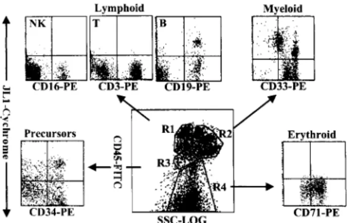
JL1 Expression of Lineage-Committed Leukocytes in BM
JL1 expression in fresh BM MNCs was investigated using multiparameter FACS analysis with a panel of conjugated mAbs directed against leukocyte surface antigens. Al- though we previously reported that JL1 antigen was not detectable in unfractionated BM cells,
18we have repro- ducibly observed a minimal subpopulation of JL1-ex- pressing cells in subsequent experiments. To confirm the presence of JL1
⫹subpopulations in BM cells, we first performed FACS analysis with a use of CD45 intensity side-scatter gating and painting and identified JL1
⫹cells in various leukocyte lineages.
22–25As shown in Figure 1, BM MNCs were able to be separated into four distinct clusters such as lymphoid lineages (R1), myelomono- cytic lineages (R2), early precursors (R3), and nucleated
red blood cells (R4). The types of corresponding leuko- cytes in each of the gated regions were confirmed by staining with the respective lineage markers (data not shown). JL1
⫹cells were detected in CD19
⫹B cells and CD34
⫹early precursors, whereas CD3
⫹T cells, CD16
⫹NK cells, and nucleated erythrocytes did not express JL1 antigen. The expression pattern of JL1 in the myelomono- cytic region was heterogeneous and divided into three subgroups (Figure 3, see below). These results were also observed in CB MNCs (data not shown).
JL1 Expression in CD34 ⫹ Cells of Human BM and CB
The proportion of JL1
⫹cells showed a great interdonor variation (5 to 50%) in the early precursor compartment, and was remarkably reduced in CB MNCs (1 to 10%) (data not shown). We correlated JL1 reactivity with the expression of CD34, along with those of AC133, CD38, CD10, CD33, and CD71 to allow identification of noncom- mitted and various lineage-committed progenitor cells (Figure 2). Pluripotent stem cells were defined as AC133
⫹CD34
⫹or CD38
⫺/dimCD34
⫹cells in BM or CB,
26 –30and they did not express JL1 molecules at all. In addition, JL1 was not expressed on CD34
⫹CD33
⫹cells that contain virtually all colony-forming cells such as pro- genitor cells capable of forming granulocytes, erythro- cytes, monocytes, megakaryocytes (CFU-GEMM), CFU- GM, and burst-forming unit erythrocytes (Figure 2B).
CD34
⫹CD71
brighterythroid-committed progenitor cells were also JL1-negative (Figure 2B). In contrast, as shown in Figure 2B, the expression of JL1 antigen was observed on the majority of lymphoid-committed CD10
⫹CD34
⫹precursors.
The expression profile of the JL1 molecule in CD34
⫹CB cells was almost same as that in BM cells. However, less CD34
⫹cells were JL1-positive as compared with that in BM cells (data not shown), and these results are directly attributed to the fact that there are less AC133
⫺CD34
⫹cells in CB MNCs. Therefore, these data indicate that JL1 expression is restricted to only lymphoid precursor cells among the CD34
⫹BM or CB MNCs.
Figure 2. The expression of JL1 molecules on CD34
⫹BM progenitor cells.
BM cells were labeled with biotinylated JL1 mAb and SA-cychrome, followed by staining with CD34-FITC in combination with CD38-PE, AC133-PE, CD10- PE, CD33-PE, or CD71-PE, respectively. CD34
⫹cells were gated (R1; A) and JL1 expression in R1 gate was compared on each of the progenitor cells (B).
A square indicates CD71
brightcell population.
Figure 3. Three-color flow cytometric analysis of JL1 expression in myeloid population of G-CSF-treated BM MNCs. MNCs obtained by density-gradient separation were labeled with biotinylated JL1 mAb and SA-cychrome, fol- lowed by staining with CD45-FITC and CD33-PE. Each lineage cell cluster was painted on the CD45 versus side-scatter plot and JL1 expression was compared. Dashed lines represent isotype control levels for each popula- tion. R5, promonocytes and monocytes; R6, promyelocytes, myelocytes, and metamyelocytes; R7, band forms.
Figure 1. Flow cytometric analysis of JL1 expression of BM leukocytes.
MNCs obtained by density-gradient separation were labeled with biotinyl- ated JL1 mAb and SA-cychrome, followed by staining with CD45-FITC in combination with CD3-PE, CD19-PE, CD16-PE, CD33-PE, CD71-PE, or CD34- PE, respectively. Normal BM cells were divided into four normal populations by CD45-side scatter analysis and the expressions of JL1 as well as lineage- specific markers were measured on the corresponding regions. R1, lympho- cytes and lymphoblasts; R2, myelomonocytic lineage; R3, early precursors;
R4, erythroid cells including nucleated red blood cells.
JL1 Expression and Myeloid Differentiation in Human BM
Because the cells in the myelomonocytic lineage showed a heterogeneous pattern of JL1 expression, BM cells of
myeloid-lineage were enriched by G-CSF administra- tion
31and they were subdivided on the basis of CD33 and CD45 expression intensities.
32,33CD33 positivity is strong on promonocytes and monocytes (R5 in Figure 3);
median on promyelocytes, myelocytes, and metamyelo- cytes (R6); and dim on band forms (R7). CD33
medpro- myelocytes, myelocytes, and metamyelocytes showed none or only dim expression of JL1. Most remarkably, the CD33
dimband forms expressed JL1 at high levels, whereas mature granulocytes were completely JL1-neg- ative (data not shown). JL1 was also expressed on pro- portions of CD33
brightpromonocytes, whereas peripheral monocytes did not express JL1 antigen (data not shown).
JL1 Expression and B-Cell Differentiation in Human BM
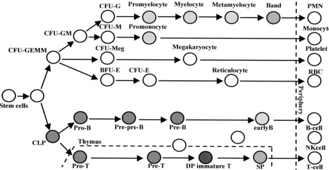
The coordinate expression of the markers such as CD34, CD19, CD20, and CD45RA has been exploited to char- acterize the developmental pathway of B lymphopoiesis in BM, and the development of B cell precursors could be divided into three major stages.
34,35To investigate the relationship between the expression of JL1 molecule and B cell development, cells of lymphoid and precursor-rich regions (R1 and R3 gate in Figure 1) were only analyzed, and mAbs against CD19 and surface-IgM (sIgM) were included in the staining protocols.
As shown in Figure 4, at least three major stages of B cell differentiation could be identified. The earliest B- lineage cells are characterized by their expression of CD34 and CD19, and these CD34
⫹CD19
⫹cells showed a high level of JL1 expression. The next stage B cell pre- cursors, characterized by the loss of CD34 expression, weak CD10 expression, and intermediate CD45RA density (CD45RA
int) on their surfaces, can be subdivided into two subsets, CD20
⫺/dimsIgM
⫺cells and CD20
highsIgM
⫹cells
Figure 4. Three-color cytometric analysis of the JL1 expression of B-lineage cells in BM MNCs. BM cells were labeled with biotinylated JL1 mAb and SA-cychrome, followed by staining with CD34-FITC and CD19-PE, or CD19- FITC in the combination with CD20-PE and IgM-PE, respectively. Dashed lines represent isotype control levels for each of the colored populations.
Figure 5. Three-color flow cytometric analysis of the JL1 expression in human thymocytes. Thymocytes were labeled with biotinylated JL1 mAb and SA-cychrome,
followed by staining with CD34-FITC and CD3-PE (A), or CD8-FITC and CD4-PE (B), or CD1a-FITC and CD3-PE (C). Dashed lines represent isotype control
levels for each of the colored populations.
according to the expression patterns of CD20 and sIgM.
The most mature B cell precursors are CD20
highsIgM
⫹cells with CD19
⫹CD10
⫺immunophenotype. Although JL1 antigen was detected on CD20
⫺/dimor sIgM
⫺lym- phoblasts, it was no longer observed on CD20
highor sIgM
⫹cells. Therefore, these results revealed that JL1 antigen appeared from the earliest CD34
⫹CD10
⫹lym- phoid precursor cells and disappeared in the CD20
highsIgM
⫹stage of B cell development.
JL1 Expression and T Cell Differentiation in Human Thymus
Because T cell development in human thymus proceeds along the sequential acquisition and loss of the antigens such as CD34, CD1a, CD3, CD4, and CD8, their expres- sion pattern has been used to assess the differentiation pathway of T lymphocytes (Figure 5).
36When JL1 ex- pression was analyzed for T-cell differentiation stage, the expression of JL1 was detected from the earliest CD34
⫹CD1a
⫺CD3
⫺thymic precursor cells to mature sin- gle-positive medullary thymocytes through the double- positive CD4
⫹CD8
⫹thymocytes (Figure 5, A and B).
Most remarkably, the JL1 expression disappeared after CD1a down-regulation in single-positive CD3
highCD4
⫹or CD3
highCD8
⫹thymocytes (Figure 5C). CD1a
highCD4
⫹CD8
⫹CD3
⫺/lowimmature cortical thymocytes dis- played the highest JL1 density on their surfaces among all leukocytes (Figure 5B). In addition, a subpopulation of CD3
⫺CD4
⫺CD8
⫺thymocytes did not express JL1 anti- gen and this subset included virtually all of the CD56
⫹NK cells (data not shown).
JL1 Expression in Leukemia
As summarized in Table 1, JL1 antigen was expressed on more than 20% of blast cells in 181 (87.0%) of 208 leukemia cases and was effective for the detection of
leukemias regardless of the phenotype. However, in sIg
⫹non-T-ALL, JL1 positivities were significantly lower than the other subtypes, which is in accordance with the JL1 expression pattern in BM. JL1 positivity was slightly higher than CD34 positivity (76.4%) in all types of leuke- mia, and this difference is attributed to higher positivity of JL1 antigen in non-T-ALL (P ⫽ 0.027). No statistically significant co-relationship was observed between ex- pression of JL1 and that of CD34 in leukemia. Most of the JL1-positive leukemias, including AML, however, ex- pressed CD34 molecules. Furthermore, either JL1 or CD34 was expressed in most of leukemia cases tested, indicating that flow cytometric analysis using both anti- JL1 and anti-CD34 mAbs might be able to detect almost all types of leukemia.
Discussion
JL1 was discovered during the development of mAbs recognizing human thymocyte-specific antigens.
17JL1 is detectable on cortical thymocytes but not on peripheral lymphocytes. In earlier studies using unfractionated hu- man BM, subsets of JL1-positive cells were not readily detected.
18In the present study, we re-examined the level of the expression of JL1 antigen on human leuko- cytes in BM, CB, and thymus using a multiparameter flow cytometric analysis, to reveal the level of its expression in CD34
⫹and other lineage-committed precursor cells. Es- pecially, immunostaining was performed in a biotin- streptavidin complex cychrome system to increase the sensitivity of the detection of JL1 antigen. In this study, we provide the evidence that JL1 antigen is a novel differentiation antigen and its expression is restricted to certain subsets of lymphoid and myeloid lineages and not expressed on pluripotent stem cells in BM and CB.
Currently available antigen to identify the pluripotent and progenitor cells in human BM is CD34.
37The CD34
⫹population in BM, however, represents heterogeneous Table 1. FACS Profiles of Anti-CD34 and Anti-JL1 mAb Immunofluorescence in Leukemia
Type of leukemia Percent of JL1
⫹cases Percent of CD34
⫹cases Percent of CD34
⫹JL1
⫹cases
AML 90.1 (82*/91
†) 83.5 (76/91) 76.9 (67/91)
M0 33.3 (1/3) 100.0 (3/3) 33.3 (1/3)
M1, M2 97.4 (38/39) 92.3 (36/39) 89.7 (35/39)
M3 100.0 (7/7) 42.9 (3/7) 42.9 (3/7)
M4, M5 84.4 (27/32) 81.3 (26/32) 68.8 (22/32)
Others
‡90.0 (9/10) 80.0 (8/10) 70.0 (7/10)
Non-T-ALL 80.9 (55/68) 64.7 (44/68) 61.8 (42/68)
CD10
⫹90.6 (48/53) 73.6 (39/53) 67.9 (36/53)
CD10
⫺83.3 (5/6) 66.7 (4/6) 66.7 (4/6)
sIg
⫹22.2 (2/9) 11.1 (1/9) 11.1 (1/9)
T-ALL 87.0 (20/23) 70.0 (16/23) 60.9 (14/23)
Acute biphenotypic leukemia 100.0 (13/13) 84.6 (11/13) 84.6 (11/13)
CML in blast crisis 84.6 (11/13) 92.3 (12/13) 84.6 (11/13)
Total 87.0 (181/208) 76.4 (159/208) 71.2 (148/208)
*No. of positive cases.
†
No. of cases tested.
‡