약학회 지 제 51 권 제 2 호 108-사14 (2007)
Yakhak Hoeji
Vol. 51, No. 2MLSb
계 항생물질 유도 내성 세균에서M
vitro로L선발된 지속성 내성형erm(A)
와erm(C)
의 분자적 특성 규명윤은정 ■ 진 성 혜 * • 최 응 칠 ^ • 심 미 자 *
서 울대 학교 약학대 학 ■ 종합 약학 연구소, *서울시 럽대학교 생명과학과 (Received February 15, 2007; Revised March 14, 2007)
Molecular Analysis of Spontaneous Mutations in
erm{A)
andermiC)
SelectedIn vitro
as a Constitutive MLSg Resistant StaphylococciEun-Jeong Yoon, Sung-Hye Jin*, Eung-Chil Choi* and Mi-Ja Shim*'^
College of Pharmacy and Research Institute of Pharmaceutical Sciences, Seoul National University, Seoul 151-742, Korea
^'Department of Life Science, University of Seoul, Seoul 130-743, Korea
Abstract - The predominant Macrolides-Lincosamide-Streptogramin B (MLSp^) antibiotics resistance genes in staphy
lococci are
erm(A)
anderm{C).
There is the phenomenon that the ratio of constitutively MLS^ antibiotics resistance (cMLS) inerm{A)
is much higher than inerm(C).
Thus, we confirmed that the difference of the mutation ratio betweenerm{A)
anderm{C)
makes the phenomenon. We examined 8 staphylococci carrying inducibly expressed (iMLS)erm{A)
orerm{C)
genes.After overnight incubation in the presence of the non-inducer MLS^ antibiotics, spontaneous mutants constitutively expressed MLSg resistance were selected. Against our expectation, the mutation ratio of
erm(A)
was lower thanerm{C).
Therefore, possibilities of other factors determining the ratio of cMLS phenotype might be concerned. All the mutants showed sequence alterations in translational attenuator and all the alterations seemed to give rise to change the second structure of mRNA to express constitutively. For
erm{k),
4 different types of sequence deletions ranging from 72 bp to 122 bp and 3 different types of duplications ranging 24 bp to 93 bp were detected. Also, there were 9 different types of duplications ranging 15bp to 154bp in
erm{Q).
Keywords □ MLSr resistance,
Staphylococcus aureus,
coagulase-negative staphylococci,erm{A), erm(C)
포도상구균(staphylococci)은인간의 점막이나피부에 정상균 총으로존재하는그람앙성구5이다. 그중
Staphybcoccus aureus
는coagulase효소룰가지고있어다ft 종과구별되는포도상구
균으로, 유임.하게병원성으로분류되어있는종이다."'^
S, aureus
에의학감염은능가진과같온피부감염이 기장훈하며, 균혈증, 심내막염, 수막염, 폐렴, 화농성 관절염, 골수염, 식중독등매우
다양하다_2) Coagulase 효소를가지고있지않은포도상구균을
coagulase-negative staphylococci(CNS)로통청하는데, 파거에는 임상적 의의를두지앉는경우가많았지만면역력이 저하된환 자에게 심내막염이나복막염을일으키는원인균으로밝혀져그
타]- 논문에 관한 문의는 저자에게로 (선화) 02-880-7874 (팩스) 02-872-1795 (E-mail) ecchoi(f/ snu.ac.kr
(선화) 02-2210-2490 (팩스) 02-2210-2490 (E-mail) mjshim^f uos.ac.kr
중요성이 점차증대되고있다.=»
이들
S.
awreMs나CNS 감염증에1차치료제는3-lactam계항 생물질이나, 이계열의항생물질은앨러지의위험이있고, 내성 율도메우높아제한적으로만쓰이고있다/*""I 이에 2차치료제로추천되는약물이 quinolone계항생물질 및마크로러의드-린
코사마이드-스트렘토그라민 B{Macrolide-Lincosamide-Strepto- gramin B, MLS,,) 계열의 항생물질이다. 그러나이들약물에대 한내성균도증가하고있는추 세 이 다MLSb 계열항생물질에 대한내성의유형은, 그표현형에따라유출형내성형, 유도내
성형, 지속성내성형의 세가지로나누어 생각할수있다.®* MLSb
계열항생물질에대한내성의 가장흔한형태인유도내성형과 지속성내성형의경우, 그원인유전자가erythromycin resistance methylase(mM) 유견자이며,
erm
유전자의모형은유도제인항 생제에 의해다른항생제의 내성여부를결정하게 되는반면, inverted repeat(IR) site의일부에번이가일어나게되면지속성자잘적 지속성 내성 번이주의 분석 109
내성형을보이게된다.3' 임 상 균주들의내성표현형에대한 지속적인관찰결과에따르면본래
erm
유전자의내성형인유도성내성형보다그번이형인지속성내성형을보이는 MLSb 계
열항생물질 내성균주의비율이 절대적으로높은것이확인되
고있 다 MLSb 계열항생물질에내성율이높아지고있
다는것자체도주목해야할문제이지만, 지속성내성형을보이 는포도상구균의 경우에마크로라미드계열항생물질의발견형 인케토타이드계열항생물질에조차내성을보이므로유도성 내 성형을보이는
erm
유전자가지속성 내성형으로번이되지않도 록관리하는것또한중요하다.Lim 등의 2002년발표에따르면, 우리나라에서임상분리된
S.
awrews의경우 enw(A)의비율이 mw(C)에버해 매우 높으며
MLSb 계열항생물질에내성인균주의 대부분이지속성내성형
인반면, CNS의경우 em(C)의비율이 em(A)에비해높고
S.
에비해유도성내성형의비율이높은것으로나타났다
이를통해 mw(A)와 em(C)의자발적번이율차이가이와같은
현상을유발한것이리라는예상을하게되었다. 이에본연구에
서는 em(A)와 erw(C)의자발적 번이율에 어떤차의가있으며,
자발적변이주의분자적특성이어떠한지를확민해보기로하였다.
실험 방법
균주의 선발
균주는 1999년과 2001년에서울과대견의 3차의료기관에서 환자로부터 분러한 coagulase-negative staphylococci(CNS)와 2001년에식약청에서 정상인에게서분리한
S. aureus
를이용하 였다. 1차로 Clinical Laboratory Standards Institute(CLSI,USA)의기준에 의거한디스크실험을통해 MLSg 계열항생물
질에유도내성표현형을가지는균주률선 발 하 였 다 선 발 된균주률 대상으로 Bioneer 사(Daejeon, Korea)에서 합성한 primer(Table I)듭을 이용해 multiplex Polymerase Chain
Reaction(PCR)을설시하여, 어떤내성유전자를가지고 있는지
확인하였다.^® 이를통해, MLSb 계열항생물질에 유도내성형
의표현형을보이면서 그분자적 원인이 mw(A)이거나
erm{C)
임이확인된 8개의세균균주률선발하였다. 이들대상균주를
대상으로 CLSI의기준에따라항생물질의 최소억제농도를측
정하고실험을진행하였다.
항생물질
14환마크로라이드계 항생물질인 erythromycin(Sigma Chem
ical Co., MO, USA), 15환마크로e}이드계항생물질인 josamycin (ICN Biomedical, CA, USA), 린코사마이드계 항생물질인 clindamycin(Sigma Chemical Co., Mo, USA), 스트렘토그라민 B 계항생물질인 quinuprisitine(Handok Co., Seoul, Korea), 케토 라이드계항생물질인 telithromycin(Handok Co., Seoul, Korea) 을사용하였다.
자발적 변어율의 확인 및둘연변이체 선발
대상균주를 Mueller Hinton(MH) broth(Difco, MD, USA)에 서진탕배양하여, 그균액을 4배최소저지농도의 항생물질이 포함된 MH agar(Difco, MD, USA)에도말하였다. 이를 37°C에 서 18시간동안배양공1여 생겨난돌연변이체의 집탁을관찰하였 다. 항생물질을포함하지 않은 MH agar 배지에같은균액을도 말-5}여 생균수률 하고, 돌연변이체 집락의수와비교하식 번 이율을계산하였다. 생겨난돌연변이체가자발적 변이에의해지 속성내성형을 획득하였는지률확인하였다. 이를위해, CLSI에
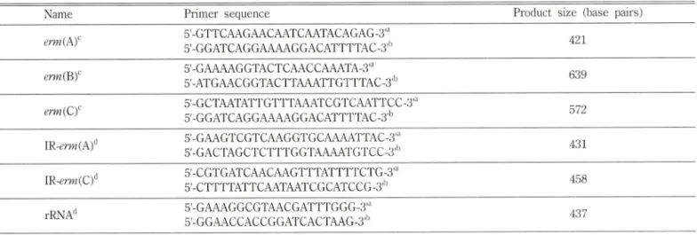
Table I - Primers used in PCR
Name Primer sequence Product size (base pairs)
erm{PCf 5-GTTCAAGAACAATCAATACAGAG-3'"
5'-GGATCAGGAAAAGGACATTTTAC-3''^ 421
5'-GAAAAGGTACTCAACCAAATA-3'"
5'-ATGAACGGTACTTAAATTGTTTAC-3'•그 639
erm{Cf 5'-GCTAATATTGTTTAAATCGTCAATTCC-3'"
5'-GGATCAGGAAAAGGACATTTTAC-3'^ 572
\R-erm{k)^ 5'-GAAGTCGTCAAGGTGCAAAATTAC-3'"
5'-GACTAGCTCTTTGGTAAAATGTCC-3'^ 431
\R-^rm{Cf 5'-CGTGATCAACAAGTTTATTTTCTG-3'"
5'-CTTTTATTCAATAATCGCATCCG-3'*" 458
rRNA'* 5'-GAAAGGCGTAACGATTTGGG-3'"
5'-GGAACCACCGGATCACTAAG-3'*^ 437
단Forward primer.
'^Reverse primer.
^Primer set for multiplex PCR.
선Primer set for PCR and sequencing analysis.
Vol. 51, No. 2, 2007
110 운T은정 • 진성혜 • 최응칠 • 심머자
의거한 agar dilution 법으로최소저지농도률견정하고, 디스크 법으로지속성 내성형을보이고었는지률보았다. 내성을유도 하기 견후의 생장곡선을비교하의내성유도와관계없이지속
적으로 MLSii 계열 항생제 전반에 내성임을확인하고, pulse-
field gel electrophoresis(PFGE) 설험을실시하여돌연번이체가 모형균주에서발생한것임을확인하였다.
변이위처의결정
Genomic DNA extraction kit인 G spin*보(INtRON, Gueonggi,
Korea)을이용하여 모형 균주및지속성 내성형 돌연변이체의
total DNA률 얻었다. 이 total DNA를주형으로하여, Table I의 IR~mw(A)와 IR-mw(C) primer률 이용하여 Peltier Thermo cycler(MJ Research, MA, USA)에서 PCR 하였다. 최초에 95X 에서 5분간 denaturing을하고, 이어 97X에서 10초, primer별
적합 annealing 온도에서 3Q초, 74®C에서 45초간 elongation하는 반응을 25회반복한후 74T에서 10분간마무러 elongation을 하였다. 이렇게 얻어진 DNA단편을INtRON사외 PCR quick spin™으로정제하고 Bionics사(Seoul, Korea)에 sequencing 분 석을의뢰하여번이위처롤결정하였다.
실험 결과 및 고 찰
최소저지능도및디스크실험을통해분명한유노내성형균 주임을확인하여 선발된실험대상모형균주듭은 14환마크로 라이드계항생물질인 erythromycin에의해내성이유도되어 16 환마크로라어드계인 josamycin, 린코사마이드계인 clindamycin, 스트렘토그라민 B 계인 quinupristine, 케토라이드계인 telithro- mycin에내성을가지게되는것으로보였다(Table II와 Fig. 1).
Table II - The characters of parental isolates and their mutants
Erythromycin Josamycin Clindamycin Quinupristine Telithromycin
p/t mutants P/t mutants p/t mutants p/t mutants p/t mutants
mw(A)
S. aureus NSTA191 >64 >64 8 >64 8 >64 4 >64 0 . 1 2 >64
NSTA819 >64 >64 8 >64 8 >64 4 >64 0 . 1 2 >64
CNS 1999-122 >64 >64 4 >64 0.25 >64 0.25 >64 <0.06 >64
1999-147 >64 >64 4 >64 0.5 >64 0.5 >64 0 . 1 2 >64
ermiO
S. aureus NSTA534 >64 >64 16 >64 16 >64 8 >64 0.25 >64
NSTA777 >64 >64 16 >64 16 >64 8 >64 0.25 >64
CNS KY46 >64 >64 8 >64 8 >64 0.5 >64 0 . 1 2 >64
KY50 >64 >64 8 >64 8 >64 0.5 >64 0 . 1 2 >64
A parental isolate (iMLS type) A selected spontaneous mutant (cMLS type)
Fig. 1 - Phenotypes of a parental isolate and its selected spontaneous mutant. Disks marked E contains erythromycin 15 lag/disc, C contains clindamycin 2 ^ig^disc, J contains josamycin 15 fig/disc, T contains telithromycin 0.2 jag/disc and Q contains quinupristin 2 f^g/disc. We could find D-shaped zone of inhibition around the disks of C, J, T and Q at the panel of the parental isolate. However there was no zone of inhibition around the discs of all MLSj^ antibiotics.
자팔적 지속성 내성 번이주의 분석 111
이는, 내성을유도히는항생물질인 erythromycin을처리한뒤에 다른항생물질이 포함된배지에서 배양한균의성장곡선으로도 확인되었다. 이에, non-inducer인 4종의항생제률이용하식자발 적지속성내성형돌연번이체률선발하였다. 더불어 생균수를측
정하여균액에 돌어있는생7스에서생겨난돌연번이체의 비율을 계산SK자 Table III에나타내었다. 돌연변이체 선발실험은 3회 에걸쳐실시히식
: 1
평균처룰구하였다. 언어진돌연변이체에대해서는 triple-disc test 상에서 저지환이 생기지 않옴을확인
Table III - The ratio of spontaneous mutation
Selected by
Josamycin Clindamycin Quinupristine Telithromycin
ermiA)
S. aiiretis
NSTA191 NSTA8194.5x10■피 1.1x10■효**
3.6x10■브* 3.7x10-^^^
4.5x10■오다
신 Q-13a 2.7x10■조요 9.7x10■그
CNS 1999-122
1999-147
8.1x10■으
<10■며
5.1x10■소스*
1.2x10•으 <10■오^
8.1x10'^^^
3.9xl0'^^
erm{C)
S. aureus
NSTA534 NSTA7771.1
X
10■은4.4x10'은
4.9x10'^
4.3x10■은
3.4x10'^
3.6x10*^
4.6x10'^
2.2x10■완
CNS KY46
KY50
1.2x10'^
1.7x10■구
5.7x10-^
7.
IX
10■은 4.2x102.6x10■■으오조조 3.0x101.1x10■■은구^No mutants were selected and the number of viable cells were near 1(P CFU/ml.
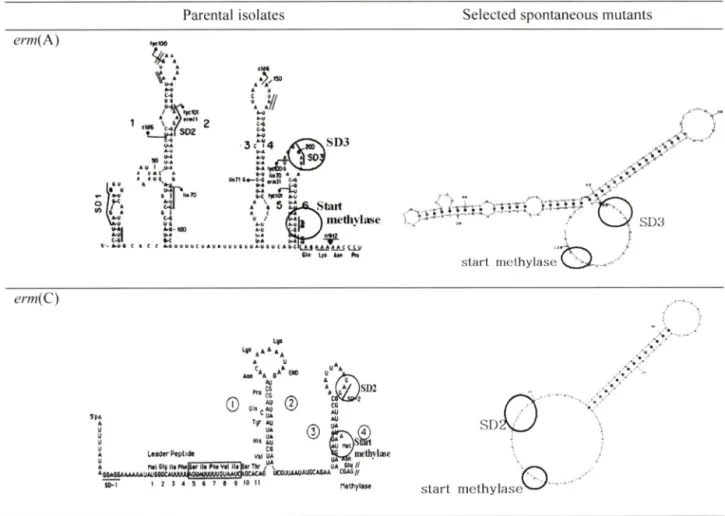
Parental ^rm(A)
S O I O R F o f th e SE>2 O R F o *«N e 15-s b p « p 1 id « 19- M p « p t id *
116 bp deetion (5412-5297) --니
S 0 1 O R F o f ttM 3 D 3 ORf o f th e
p«p1i<5« 19-m p * p t h te
122 bp deletion (5417-5296)
1 2 3
—~i-m
S O I oORFof th« *
hbY'
1S-4B
72 bp deletion (5417-5346)
ttW*"
1 9-M p « p ik i«
-THZ
S O I ... •• ■ -
O R F o f t t w S D 2 O A F o tfth * I Sh i* p « p t M « 19-m p * p fit f*
95 bp deletion (5437-5343)
1 2 3
니■HTTTT"'
S 0 1 O R F o f t h i *
19-M p * p t t d *
24 bp duplication (5297-5274)
1 2
-> r
5 0 1 O R F o f t t w 3 0 2 1 S ^ s p * p t k f «
> "h
3 0 O R F o f ttw
19- M p ^ p t f d *
25 bp duplication (5297-5273)
1 2
'■ . h사 ;
S O I O R F o f t h e 3 D 2 0 « F 19-m 1S-«B p « p t M «
93 bp duplication (5341-5249) 1
— H * —■
s o i O R F o f t h e 3 D 2 C M F o f l t a 1S-«» p « p t t d « 1 9*M p * p ( W t
S 0 3 5* • n d o f
5 6
S D 3 5- • n d o f • r m (A )
S 0 3 5' A nd o f •rr» (A )
- IJ T i
—t H_
S 0 3 o f
Parental erm(C)
S D 1 O R F o f tfve 1 9 a a p e p t i d e
57 bp deletion (2816-2872)
1 2
S O I O R F o f t h e 1
58 bp deletion (2800-2857)
I peptide
2
f - r
—t
S O I O R F o f t
60 bp deletion (2798-2857) I p « p t i d «
2
— *
S D 1 O R F o f t h e 1 9 a a p e p t i d «
68 bp deletion (2801-2868)
S D 1 O R F o f t h e 1 9 • « p « p t i 6 e
71 bp deletion (2798-2868)
S O I O R F o f t h e 1 9 a a p e p t i d e
120 bp deletion (2749-2868)
1 2
S O I O R F o f t h e 1 9 a a p « p t i d e
25 bp deletion (2816-2840)
1 2
S O I O A F o f t f w 1 9 • « p e p t i d e
15 bp deletion (2763-2777)
903 y vnd
o f
Fig. 2 - Detected mutations in the translational attenuator region of
erm
genes.—P"-f•그 ~ '
S O I O R F o f t h e 1 9 a a p e p t i d e
154 bp deletion (2664-2817)
S O I O R F o f m e « a p « p t i d e
S D 2 S ' «MKi o f • m r C
S D 2 5 ' «0<1 o f o r m C
S 0 2 5 ' » n d o f Trr/C.
S 0 2 S ' »1><1 o f 9rrvG
으* 스 ____
!
H-
S 0 2 5 ' * n d o f «rmC
■ H i
S 0 3 S ' » n d o f • r m C
3 4
■ "F-hLt
S 0 3 5 ' * n d o f • m i C
Vol. 51, No. 2, 2007
Tn554 (GI: 43726) 5412-5297 5417-5296 5417-5346 5437-5343 5297-5274 5297-5273 5341-5249
S. aureus
3 5 3
CNS 6
erm{k)
Deletion Deletion Deletion Deletion Duplication Duplication Duplication
erm{C)
Deletion 57
in pE194 (GI: 49489772)
2816-2872 3
Deletion 58 2800-2857 2 1
Deletion 60 2798-2857 1 -
Deletion 6 8 2801-2868 1 -
Deletion 71 2798-2868 4 -
Deletion 1 2 0 2749-2868 1 -
Deletion 25 2816-2840 3 -
Deletion 15 2763-2777 2 -
Deletion 154 2664-2817 - 4
112 윤은•정 • 진성혜 • 최응칠 • 심미자
하였고, 최소저지 농도와성장곡선으로지속성내성화되었움을 재확인하였다. 그번이율은 10■쳐1서 정도로매우낮은편 이었다. 번이율은
erm
유전자의중류나선발항생물질에따라조 금씩 달랐으나, 균종별차이는크지 않았다. 선발항생물질로 quinupristine 및 telithromycin을쓴 경우의번이율이 전반적으 로가장낮아, mw(A)를가지는S.
flwrews와 CNSS] 경우, 번이주률하나도얻지못한경우도있었다. em(C)보다는
ermiA
)를가지는균주의 번이율이더낮았다. mK(A)는지속성내성형번
이주를하나노얻지못하식 보다도낮은변이율을보인경
우부터 8.1XKT®의 번이율을 보이는 경우까지 있었다. 반면,
mw(C)의경우가장낮게는 4.2x10■겨서높게는 1.7x10'M지 선반적으로 m«(A)에비해높은번이율을보이는것을관찰할 수있었다. mw(A)와
ermiO
유전자에서보여진이와같은처<>1 는임상균주에서의 내성표현형과유전자형의비율을통해 에 측한바와는상반되는결과였다. 이룰통해, 임상에서분리되는균주가 MLS„ 계열항생물질에대해지속성내성화되는원인에
는자발적 번이이외에다른요인이 작용하고있으리라예측된 다. 실제 MLS[;계열항생물질에유도내성형인
S. aureus
감염 증을치료할때 non-inducer인 clindamycin 등도처료실패경 험이 있어임상적으로는이들을사용하지 앉을것을권하고있 다. 이와같이낮은번이율에도불구하고 non-inducer 시'§■시치 료실패와같은결과가나타나는것은, 인체내에다른인자가 유도내성형균주의지속성내성화에관여하고있다는뜻어라해 석할수있겠다.수차례의 실험을통해안정적으로일정한양의돌연번이체률
언을수 있었던 clindamycin으로선발한균주둘을이후의 실험
에이용하였다. 이둘돌연변이체돌은모형균주와달러내성유
도없이도 MLSij 계열항생물질견반에대해높은최소저지농
도를나타내었다(Table II). MLSb 계열항생물질에대헤지속성 내성형을 가지게된균주들은
erm
유전자의 translational 흑은 transcriptional attenuation을결 정 히IR site의번이에의한경 우가일반적이다. 따라서이부분을증폭하여 얻어낸 DNA 단편 을 sequencing analysis로확인하였다. 더불어 MLSg 계열항생 물질의 결합부위도증폭히M 분석하였다. 이때시용한 primer는 Table I의 IR-mM(A)와 IR-erm(C), 그리고 rRNA set였다. MLS,, 계열항생물질의결합부위인 ribosomal RNA에는아무•런번이노 일어나지않았옴을확인하였고, 반면에 IR site에서는 밍^ > 번 이를 관찰할 수 있었다(Table IV). em(A)의 IR site에서는 deletion 및 duplication의번이가관찰되었다. Deletion 타입의 경우는 72 bp에서 122 bp까지 4가지종류의유전자조각상실을 확인하였으며, duplication 타입의 경우는 24bp, 25bp 또는 93bp의유전자조각이 반복되는 3가지의 번이를확인하였다. em(C)의 IR site에서는 15 bp에서 154 bp의유전자가상실되는 9가지타입의 deletion 변이가관찰되었다. 이들번이된 IR site sequence의 mRNA 2차구조를에상해본결과/®•조구* Fig. 3에서 보여지듯구조가변형되어erm
유전자의 시작코돈앞에위치한 ribosome 결합부위에 ribosome의결합이용이하도록노출됨으 로써, 별다른유도없이유전자가발현되는구조률만드는것으 로보였다.결 론
Non-inducer인 MLSg 계열 항생물질에 의한 유도 내성형
staphylococci의지속성내성형자발적 변이율은낮은편이었으
며, 그들의 번이 원인은 inverted repeat site의 번이에 의한
mRNA의 2차구조의 번화때문인것을확인할수있었다.
Table IV - Detected mutations in the translational attenuator region of erm genes
Mutation type Structural alteration (base pairs) Positions of alterations No. of mutants
16 2272
95 24 25 93
erm(C)
A m i III Plw|S«rll» ^mV«I lltB tr Tfcr ,
* M»66A*AAAAIMUM0CAIAWW0UMWAI0UMW>(K*C*<S
■ ^ iT I 2 3 4 S 6 7 a 9 JO II
*« .*
methylase
CSAS//«u//
n«triylM« start methylase^
> y
Fig. 3 - Representative estimated secondary structures of mRNA in the translational attenuator region of erm (A) and efjn(C).
감사의 말씀
이 논문은 서울시럽대학교 2005년 학술연구조성비에 으1하여 연구되었습니다. 이에 서울시럽대 학교에 감사하는 바입니다.
참고문헌
1) Thouverez, M., Muller, A., Hocquet, D., Talon, D. and Bertrand, X. : Relationship between molecular epidemiology and antibiotic susceptibility of methicillin-resistant Staphylococcus aureus (MRSA) in a French teaching hospital. Journal of Medical Microbiology 52(Pt 9), 801 (2003).
2) Kim, H. B., Jang, H. C., Nam, H. J., Lee, Y. S., Kim, B. S., Park, W B., Lee, K. D., Choi, Y. J., Park, S. W, Oh, M. D., Kim, E.
C. and Choe, K. W : In vitro activities of 28 antimicrobial agents against Staphylococcus aureus isolates from tertiary- care hospitals in Korea: A nationwide survey. Antimicrobial Agents and Chemotherapy 48(4), 1124 (2004).
3) Lampson, B. C. and Parisi, J. T. : Naturally occurring
Staphylococcus epidermidis plasmid expressing constitutive
macrolide lincosamide streptogramin B resistance contains a deleted attenuator. Journal of Bacteriology166(2), 479 (1986).
4) F'elmingham, D., Reinert, R. R., Hirakata, Y. and Rodloff, A. ; Increasing prevalence of antimicrobial resistance among isolates of Streptococcus pneumoniae from the PROTEKT surveillance study, and compatative in vitro activity of the ketolide, telithromycin. J. Antimicrob. Chemother. 50 Suppl
SI, 25 (2002).
5) Uh, Y., Jang, I. H., Hwang, G. Y., Lee, M. K., Yoon, K. J. and Kim, H. Y.: Antimicrobial susceptibility patterns and macrolide resistance genes of beta-hemolytic streptococci in Korea.
Antimicrob Agents Chemother. 48(7), 2716 (2004).
6) Shobha, K. L., Rao, R S. and Thomas, J. : Survey of
Staphylococcus isolates among hospital personnel, environment and their antibiogram with special emphasis on methicillin resistance. Indian J. Med. Microbiol. 23(3), 186 (2005).
7) Lim, J. A., Kwon, A. R., Kim, S. K., Chong, Y., Lee, K.
and Choi, E. C. : Prevalence of resistance to macrolide, lincosamide and streptogramin antibiotics in Gram-positive cocci isolated in a Korean hospital. J. Antimicrob, Chemother.
49(3), 489 (2002).
Vol. 51 No. 2, 2007
V*
SD3
s ta r t m e t h y la s e'
erm{A)
자발적 지속성 내성 변이주의 분석 113
Parental isolates Selected spontaneous mutants
114 윤^ • 진성혜 • 최옹철 • 심미자
8) Fiebelkorn, K. R., Crawford, S. A., McElmeel, M. L. and Jorgensen, J. H. : Practical disk diffusion method for detection of inducible clindamycin resistance in Staphylococcus aureus
and coagulase-negative Staphylococci. Journal of Clinical Microbiology 41(10), 4740 (2003).
9) Schmitz, E J., Petridou, J., Milatovic, D., Verhoef, J., Fluit, A. C.
and Schwarz, S. : In vitro activity of new ketolides against macrolide susceptible and resistant Staphylococcus aureus
isolates with defined resistance gene status. Journal of Antimicrobial Chemotherapy 49(3), 580 (2002).
10) Schmitz, E J., Petridou, J., Jagusch, H., Astfalk, N., Scheuring, S. and Schwarz, S. : Molecular characterization of ketolide- resistant t?m(A)-carrying Staphylococcus aureus isolates selected in vitro by telithromycin, ABT-773, quinupristin and clindamycin. Journal of Antimicrobial Chemotherapy 49(4), 611 (2002).
11) Schmitz, E J., Petridou, J., Astfalk, N., Scheuring, S., Kohrer, K., Verhoef, J,, Fluit, A. C. and Schwarz, S. : Structural alterations in the translational attenuator of constitutively expressed ^rm(A) genes in Staphylococcus aureiis. Antimicrobial Agents and Chemotherapy 45(5), 1603 (2001).
12) Werckenthin, C., Schwarz, S. and Westh, H. : Structural alterations in the translational attenuator of constitutively expressed ermC genes. Antimicrobial Agents and Chemotherapy
43(7), 1681 (1999).
13) Lina, G., Quaglia, A., Reverdy, M. E., Leclercq, R., Vandenesch, K and Etienne, J . : Distribution of genes encoding resistance to Macrolides, Lincosamides, and Streptogramins among Staphylococci. Antimicrobial Agents and Chemotherapy
43(5), 1062 (1999).
14) Luthje, R and Schwarz, S .: Molecular analysis of constitutively expressed erm{C) genes selected in vitro in the presence of the non-inducers pirlimycin, spiramycin and tylosin. Journal of Antimicrobial Chemotherapy 59(1), 97 (2007).
15) Khan, S. A., Nawaz, M. S., Khan, A. A. and Cerniglia, C. E, : Simultaneous detection of erythromycin resistant methylase genes erm A and ermC from Staphylococcus spp. by multiplex- PCR. Molecular and Cellular Probes 13(5), 381 (1999).
16) Sandler, P and Weisblum, B .: Erythromycin induced ribosome stall in the ermk leaderra barricade to 5'-to-3' nucleolytic cleavage of the ermh transcript. Joimial of Bacteriology,
171(12), 6680 (1989).
17) Zuker, M. : Mfold web server for nucleic acid folding and hybridization prediction. Nucleic Acids Research 31(13), 3406 (2003).
18) Clinical and Laboratory Stansdards Institute (CLSI). Per
formance standards for antimicrobial susceptibility testing:
15* Informational Supplement. Document M100-S15. CLSI, Wayne, PA (2005).