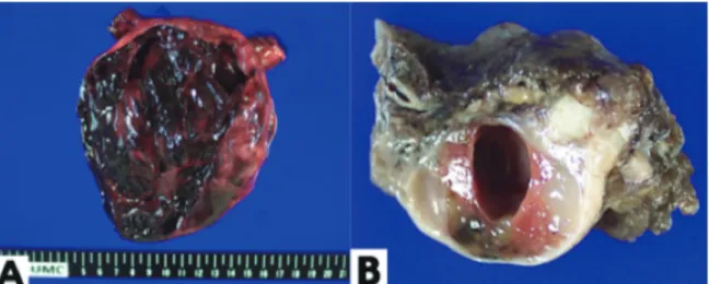
Malignant Struma Ovarii Detected Incidentally Due to Contralateral Ovarian Torsion
6
0
0
전체 글
(2)
(3)
(4)
(5)
(6)
수치

관련 문서
