rates, comparing with standard shunt placement.
MATERIALS AND METHODS Patient population and study design
A retrospective analysis was performed on 72 patients who underwent first VP shunt surgery at our institution within four years from October 2007 to September 2010. All adult and pe- diatric patients with hydrocephalus of any origin participated in this study. Patients with slit ventricles and those with revision cases were excluded to minimize selection bias.
Each patient had a precise chart review with multiple vari- ables including age, causes of hydrocephalus, the number of shunt revisions, interval time between the first shunt operation and revision, and reason for shunt failure. All patients had pre- operative and postoperative brain imaging to reveal the out- comes of shunt surgery.
All patients were divided into 2 groups according to the use INTRODUCTION
Proximal catheter obstruction is known to be the most com- mon cause of malfunctioning of ventriculoperitoneal (VP) shunt followed by infection and disconnection14,17). The optimal position of the catheter tip was focused to reduce potential oc- clusion of proximal catheter by ventricular parenchyma or cho- roidal tissues1,4,12). Many studies including Tuli et al.22) have shown that a ventricular catheter tip surrounded by cerebrospi- nal fluid (CSF) could decrease the risk of shunt failure25).
Accurate placement of ventricular catheter is related with both proper insertion trajectory and proper catheter tip positioning.
So recently, many studies have validated accuracy in VP shunt with the development of frameless neuronavigation in the field of neurosurgery2,25). However, it has not clearly proven that the role of the catheter position using navigation system on failure rates. The authors conducted a retrospective study of navigated shunt placement about real impact on clinical shunt failure
Effect of Electromagnetic Navigated
Ventriculoperitoneal Shunt Placement on Failure Rates
Nayoung Jung, M.D., Dongwon Kim, M.D., Ph.D.
Department of Neurosurgery, Dongsan Medical Center, Keimyung University College of Medicine, Daegu, Korea
Objective : To evaluate the effect of electromagnetic (EM) navigation system on ventriculoperitoneal (VP) shunt failure rate through comparing the result of standard shunt placement.
Methods : All patients undergoing VP shunt from October 2007 to September 2010 were included in this retrospective study. The first group re- ceived shunt surgery using EM navigation. The second group had catheters inserted using manual method with anatomical landmark. The relation- ship between proximal catheter position and shunt revision rate was evaluated using postoperative computed tomography by a 3-point scale. 1) Grade I; optimal position free-floating in cerebrospinal fluid, 2) Grade II; touching choroid or ventricular wall, 3) Grade III; tip within parenchyma.
Results : A total of 72 patients were participated, 27 with EM navigated shunts and 45 with standard shunts. Grade I was found in 25 patients from group 1 and 32 patients from group 2. Only 2 patients without use of navigation belonged to grade III. Proximal obstruction took place 7% in grade I, 15% in grade II and 100% in grade III. Shunt revision occurred in 11% of group 1 and 31% of group 2. Compared in terms of proximal catheter position, there was growing trend of revision rate according to increase of grade on each group. Although infection rate was similar between both groups, the result had no statistical meaning (p=0.905, chi-square test).
Conclusion : The use of EM navigation in routine shunt surgery can eliminate poor shunt placement resulting in a dramatic reduction in failure rates.
Key Words : Hydrocephalus · Neuronavigation · Ventriculoperitoneal shunt · Revision.
Clinical Article
•Received : August 3, 2012 •Revised : December 27, 2012 •Accepted : February 25, 2013
•Address for reprints : Dongwon Kim, M.D., Ph.D.
Department of Neurosurgery, Dongsan Medical Center, Keimyung University College of Medicine, 56 Dalseong-ro, Jung-gu, Daegu 700-712, Korea Tel : +82-53-250-7333, Fax : +82-53-250-7356, E-mail : [email protected]
•This is an Open Access article distributed under the terms of the Creative Commons Attribution Non-Commercial License (http://creativecommons.org/licenses/by-nc/3.0) which permits unrestricted non-commercial use, distribution, and reproduction in any medium, provided the original work is properly cited.
J Korean Neurosurg Soc 53 : 150-154, 2013
wall, 3) Grade III; tip within parenchyma or failure to reach the intraventricular space.
RESULTS
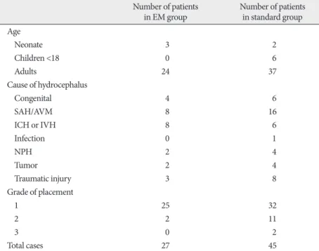
A total of 72 patients were participated in this study, 27 with the EM navigated shunts and 45 with the standard shunts. Eleven patients were children (11% of in the EM group. as 17% of the standard group) and 61 patients were adults. Patients had numer- ous causes from congenital anomaly to spontaneous or traumatic intracranial hemorrhage. Time interval of follow-up period was various from a day to 20 months. The baseline characteristics of the study patients are shown in Table 1.
Grade of shunt placement
Grade I was found in 25 patients (93% of 27 patients) from the EM guided group and 32 patients (71% of 45 patients) from the standard group. The EM navigated shunts had catheter tip more free floating in CSF (Grade I) compared with one of the standard shunts (p=0.03, chi-square test). Eleven patients (24%) in the standard shunts were classified as grade II compared with 2 pa- tients (7%) in the EM navigated group. Only 2 shunts (4%) were of electromagnetic (EM) navigation on surgery. The first group
received surgery using EM navigation (Stealth Station Axiem navigation system, Medtronic Incorporation, Louisville, USA) for routine shunt placement. The second group had catheters in- serted using manual method with anatomical landmark. Each of shunt revision cases was reconsidered focusing on the causes of shunt failure and catheter position. Patients with shunts were fol- lowed up for more than 3 months or until the shunt failure in both groups.
Statistics were generated such as percentages and mean/me- dian values by the data collected from the patients’ medical re- cords. Data were analyzed by using statistical methods on the Statistical package for the social sciences version 19. Statistical significance was set at p<0.05.
Surgical techniques
Shunt procedures were performed by neurosurgeons with various training levels. The valve type and catheter were deter- mined depending on the surgeon’s preference. In the standard group, skin incision and trepanation of the cranium were done by using anatomical landmarks like Kocher’s point or Frazier’s point. Ventricular catheter length was determined based on surgeon’s measurement using brain
computed tomography (CT) imaging.
In the EM group, preoperative data ac- quisition in 3 planes was obtained by CT or magnetic resonance imaging.
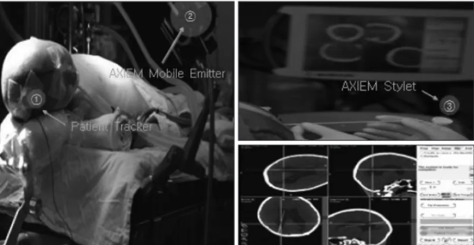
The dynamic reference frame (DRF), a magnetic field detector within the navi- gational field, was implanted in an area away from the proposed catheter entry site like Kocher’s point or Frazier’s point (Fig. 1). After system registration, the surgical plan could be created from the entry site to the final tip position of catheter. The ventricular catheter was placed over the guiding stylet which the detector coils wrapped around the tip of it. The complex was intended to pen- etrate the ventricular wall along the planned pathway.
Grading system of shunt placement
Proximal catheter position was graded on postoperative brain CT imaging on each case. All patients were graded ac- cording the following 3-point scale de- veloped for this study (Fig. 2) : 1) Grade I; catheter tip position free-floating in CSF, away from ventricular wall or cho- roid plexus, 2) Grade II; catheter tip touching choroid plexus or ventricular
Fig. 1. The operation with electromagnetic navigation system. A noninvasive dynamic reference frame applied to the scalp is shown to identify the location of anatomy within the frame of refer- ence. The bur hole and catheter trajectory were planned preoperatively in three planes to achieve optimal catheter tip position. The navigation stylet is used as the catheter trocar.
Fig. 2. Grading of ventricular catheter position (three-point scales). 1) Grade I; optimal catheter tip position free-floating in CSF. 2) Grade II; catheter tip touching choroid plexus or ventricular lining wall. 3) Grade III; tip within parenchyma or failure to reach the intraventricular space. CSF : cerebro- spinal fluid.
Grade 1 Grade 2 Grade 3
statistically significance (distal obstruc- tion p=0.879, infection p=0.905).
Shunt failure rate according to catheter position
Fig. 4 shows comparison of failure rate among the grades. In grade I, about 16%
of cases received revision surgery, as 46%
of grade II. Two shunts were included in grade III and those cases all required shunt revision (p=0.002, chi-square test).
When compared in terms of proximal catheter position on each group, grade I tended to have lower risk of revision. 2 patients (8%) of grade I and 1 patient (50%) of grade II needed shunt revision on group 1. In group 2, there was grow- ing trend of revision rate according to in- crease of grade. Revision surgery was performed on 7 patients (21%) of grade I, 5 patients (47%) of grade II and 2 pa- tients (100%) of grade III.
Proximal obstruction was seen in 8 patients in the standard group, especially 100% (two of two pa- tients) in grade III, 15% (2 of 13 patients) in grade II and 5% (3 of 57 patients) in grade I. In the other hand, there was no proxi- mal obstruction in the EM group.
Statistical considerations
This study has several statistical limitations. The number of the samples was too small to represent the population of shunt patients. And it was necessary to consider other factors that might have influenced the results. However, we tried to include patients who had various factors as possible. Other factors were already proven to have no statistical significance except catheter tip position by using multiple regression analysis (age : p=0.138, sex : p=0.430, cause : p=0.226).
The type of shunt could be an important factor to affect the catheter function, but there was insufficient consideration about that point in this study. There has been no specific study to con- firm the relationship between shunt types and shunt obstruc- tion up to the present except Tuli et al.21) assumed many factors to have effect on shunt failure including valve type and shunt type. They verified that shunt valve itself could not be a predic- tor of shunt revision using the multivariable model. However, it would be challenging to prove that shunt type is related with catheter obstruction on the basis of valve’s composition. And even if same type of valve was used, shunt valve pressure could make another bias.
DISCUSSION
According to the results of the previous studies, various meth- determined to be grade III in the standard group. This result
showed that the EM navigated group can get the high potential to have optimal catheter tip position within ventricle (Fig. 3).
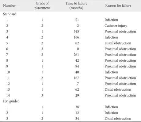
Shunt failure rate and characteristics of failure cases The Table 2 showed characteristics of shunt failure cases. There were 14 shunt failure cases (31%) in the standard group com- pared with 3 cases (11%) in the EM group. The most common cause of shunt failure in the standard group was proximal ob- struction comparing with no revision case from proximal ob- struction in the EM navigated group (p=0.020, chi-square test).
For distal obstruction, there was higher rate of revision in the standard group (6.7%) than the EM group (3.7%). Similar result of shunt failure rate between 2 groups was revealed with infec- tion. Comparison of distal obstruction and infection showed no Table 1. The baseline characteristics of the patients in this study
Number of patients
in EM group Number of patients
in standard group Age
Neonate 3 2
Children <18 0 6
Adults 24 37
Cause of hydrocephalus
Congenital 4 6
SAH/AVM 8 16
ICH or IVH 8 6
Infection 0 1
NPH 2 4
Tumor 2 4
Traumatic injury 3 8
Grade of placement
1 25 32
2 2 11
3 0 2
Total cases 27 45
SAH : subarachnoid hemorrhage, AVM : arteriovenous malformation, ICH : intracerebral hemorrhage, IVH : intra- ventricular hemorrhage, EM : electromagnetic
Fig. 3. Grade of shunt catheter placement. This paragraph demonstrates that EM-navigated group can get the optimal catheter tip position within ventricle. EM : electromagnetic.
0%
20%
40%
60%
80%
100%
EM navigated group Standard group 93%
71%
7%
24%
0% 4%
Grade 1 Grade 2 Grade 3
ship of tip position and shunt failure rate has already reported in our study. There might be several other factors to affect shunt catheter obstruction like type of catheter.
Although it is already demonstrated that endoscopically guided catheter insertion could get intended position of the tip with con- troversy, image guidance like neuronavigation system may be helpful in determining the entry point and approach trajectory before and during the procedure17). The EM group of this study has been found more in grade I, representing the close relation- ship of navigation and catheter position. Therefore, the formula can be established that use of EM navigation lead an ultimate catheter placement, resulting decrease of shunt failure rate.
There are several disadvantages to frameless neuronavigation which include the prolongation of operation time and depen- dence on expensive technology8,23,25). More operation time can ods have been suggested to improve the
accuracy of catheter tip position. Pang and Grabb15) described a method of calculating catheter length based on bony landmarks on skull radiographs and a shunt placement by free-hand passage using simple stereotactic guid- ance and palpable surface anatomy.
Serlo et al.18) ascertained the catheter position using X-ray imaging by filling the catheter with contrast medium.
Neuroendoscopic procedures have also been used effectively16,24). Kestle et al.13) compared the revision rates after endo- scopically versus non-endoscopically placed ventricular catheter. Despite placement of catheter tip under direct vision, that study concluded no differ- ences in failure rates between 2 cohorts because burr hole and catheter trajecto- ry were still based on anatomical land- marks1,11).
Frameless stereotaxy has been shown to be helpful for precise location of
catheter tip and avoiding unnecessary injury to brain tissue, leading to prevent repeated shunt revisions in children with slit ventricles8,9,18). This study has demonstrated that neuronaviga- tion in the placement of ventricular catheters assures optimal catheter position within ventricles, reducing revision rate in not only special cases, but also general shunt surgeries.
VP shunts are still often complicated by malfunction, predom- inantly with proximal catheter obstruction despite improved surgical equipment and operative skill10,14,19,25). About 30% to 50% of shunts fail within the first year and only 30% to 37% of shunts survive for 10 years without revision3,21,22,25). Choroid plexus has generally been considered to be the most frequent cause of ventricular catheter obstruction4,12,14,17). It is well known that the ideal placement of the proximal catheter tip is the fron- tal horn away from the choroid plexus5,7). The obstruction of catheter tip is caused by connective tissue, inflammatory chang- es, and foreign bodies which are found frequently at the ven- tricular end. Astrocytes and fibroconnective tissues are espe- cially capable of proliferation. They may fill the side holes in the 15 mm from their tip and the inside of the catheter, leading ob- struction of catheters2). Choroid plexus and ependymal cells also have proliferative capability under some conditions. Col- lins et al.6) frequently found choroid plexus in catheter tips placed in behind the foramen of Monro, but ependyma pre- dominated in catheters placed in front of it.
A ventricular catheter tip surrounded by CSF decreased the risk of shunt failure to one-fifth, whereas a catheter tip touching the brain decreased the risk to one-third, compared with a cath- eter tip surrounded by brain tissue21,25). The significant relation-
Table 2. The characteristics of the shunt failure cases
Number Grade of
placement Time to failure
(months) Reason for failure
Standard
1 1 51 Infection
2 2 2 Catheter injury
3 1 545 Proximal obstruction
4 2 166 Infection
5 2 62 Distal obstruction
6 3 0 Proximal obstruction
7 2 261 Proximal obstruction
8 1 42 Proximal obstruction
9 1 94 Proximal obstruction
10 1 40 Infection
11 2 167 Proximal obstruction
12 1 7 Proximal obstruction
13 1 62 Distal obstruction
14 3 29 Proximal obstruction
EM guided
1 1 38 Infection
2 1 12 Infection
3 2 34 Distal obstruction
EM : electromagnetic
Fig. 4. Comparison of revision rate between grades. All cases in grade 3 required shunt revision surgery.
0%
20%
40%
60%
80%
100%
120%
16%
46%
100%
Grade 1 Grade 2 Grade 3
7. Dickerman RD, McConathy WJ, Morgan J, Stevens QE, Jolley JT, Sch- neider S, et al. : Failure rate of frontal versus parietal approaches for proximal catheter placement in ventriculoperitoneal shunts : revisited. J Clin Neurosci 12 : 781-783, 2005
8. Gil Z, Siomin V, Beni-Adani L, Sira B, Constantini S : Ventricular cathe- ter placement in children with hydrocephalus and small ventricles : the use of a frameless neuronavigation system. Childs Nerv Syst 18 : 26-29, 2002
9. Haase J : Neuronavigation. Childs Nerv Syst 15 : 755-757, 1999 10. Hakim S : Observations on the physiopathology of the CSF pulse and
prevention of ventricular catheter obstruction in valve shunts. Dev Med Child Neurol Suppl 20 : 42-48, 1969
11. Hayhurst C, Beems T, Jenkinson MD, Byrne P, Clark S, Kandasamy J, et al. : Effect of electromagnetic-navigated shunt placement on failure rates : a prospective multicenter study. J Neurosurg 113 : 1273-1278, 2010 12. Kang JK, Lee IW : Long-term follow-up of shunting therapy. Childs
Nerv Syst 15 : 711-717, 1999
13. Kestle JR, Drake JM, Cochrane DD, Milner R, Walker ML, Abbott R 3rd, et al. : Lack of benefit of endoscopic ventriculoperitoneal shunt in- sertion : a multicenter randomized trial. J Neurosurg 98 : 284-290, 2003 14. Lazareff JA, Peacock W, Holly L, Ver Halen J, Wong A, Olmstead C :
Multiple shunt failures : an analysis of relevant factors. Childs Nerv Syst 14 : 271-275, 1998
15. Pang D, Grabb PA : Accurate placement of coronal ventricular catheter using stereotactic coordinate-guided free-hand passage. Technical note.
J Neurosurg 80 : 750-755, 1994
16. Rhoten RL, Luciano MG, Barnett GH : Computer-assisted endoscopy for neurosurgical procedures : technical note. Neurosurgery 40 : 632- 637; discussion 638, 1997
17. Sekhar LN, Moossy J, Guthkelch AN : Malfunctioning ventriculoperito- neal shunts. Clinical and pathological features. J Neurosurg 56 : 411- 416, 1982
18. Serlo W, Heikkinen E, Saukkonen AL, von Wendt L : Classification and management of the slit ventricle syndrome. Childs Nerv Syst 1 : 194- 199, 1985
19. Stein SC, Guo W : Have we made progress in preventing shunt failure?
A critical analysis. J Neurosurg Pediatr 1 : 40-47, 2008
20. Stieglitz LH, Giordano M, Samii M, Luedemann WO : A new tool for frameless stereotactic placement of ventricular catheters. Neurosurgery 67 (3 Suppl Operative) : ons131-ons135; discussion ons135, 2010 21. Tuli S, Drake J, Lawless J, Wigg M, Lamberti-Pasculli M : Risk factors
for repeated cerebrospinal shunt failures in pediatric patients with hy- drocephalus. J Neurosurg 92 : 31-38, 2000
22. Tuli S, O’Hayon B, Drake J, Clarke M, Kestle J : Change in ventricular size and effect of ventricular catheter placement in pediatric patients with shunted hydrocephalus. Neurosurgery 45 : 1329-1333; discussion 1333-1335, 1999
23. Tulipan N, Lavin PJ, Copeland M : Stereotactic ventriculoperitoneal shunt for idiopathic intracranial hypertension : technical note. Neuro- surgery 43 : 175-176; discussion 176-177, 1998
24. Wagner W, Gaab MR, Schroeder HW, Sehl U, Tschiltschke W : Experi- ences with cranial neuronavigation in pediatric neurosurgery. Pediatr Neurosurg 31 : 231-236, 1999
25. Wan KR, Toy JA, Wolfe R, Danks A : Factors affecting the accuracy of ventricular catheter placement. J Clin Neurosci 18 : 485-488, 2011 have risk to increase intraoperative infection rate. In fact, 5 to
10 minutes can be required to install and to register the naviga- tion by skilled person11). Stieglitz et al.20) described that the addi- tional time needed for preparations did not exceed 15 minutes with their experiences. The DRF can be placed while anesthesia personnel are obtaining intravenous access1). The other problem is that frameless stereotaxy is expensive program. It requires ad- ditional imaging to be used for navigation with additional radia- tion dosage11). It also adds cost to the procedure of neuronavi- gation and to the use of a navigation stylet. As a practice matter, average 40% of total sum of money was charged to operation fee at our institution with EM navigation shunts. But, revision surgery needs more cost added on the first surgery11). Hayhurst et al.11) measured that cost of a shunt revision was ten times greater than the cost of a using navigation in their own country.
Furthermore, the time span between revisions shortens pro- gressively after second revision, indicating that economical dis- advantages can be replicated14). Therefore, the cost-benefit ratio must be carefully established to obviate its need.
CONCLUSION
Mechanical malfunction and infection are the most signifi- cant problems associated with shunts for the treatment of hy- drocephalus. Above all, a significant proportion of shunt fail- ures were due to obstruction of the ventricular catheter, and accurate placement of the shunt catheter is highly important to reduce the incidence of shunt malfunction.
In conclusion, the use of EM navigated system in routine shunt surgery can eliminate poor shunt placement resulting in a dramatic reduction in shunt revision rates.
References
1. Azeem SS, Origitano TC : Ventricular catheter placement with a frame- less neuronavigational system : a 1-year experience. Neurosurgery 60 (4 Suppl 2) : 243-247; discussion 247-248, 2007
2. Bierbrauer KS, Storrs BB, McLone DG, Tomita T, Dauser R : A prospec- tive, randomized study of shunt function and infections as a function of shunt placement. Pediatr Neurosurg 16 : 287-291, 1990-1991 3. Browd SR, Ragel BT, Gottfried ON, Kestle JR : Failure of cerebrospinal
fluid shunts : part I : Obstruction and mechanical failure. Pediatr Neu- rol 34 : 83-92, 2006
4. Cho TH, Park JY, Lee JK, Park YK, Chung HS, Lee KC, et al. : Radiolog- ic location of ventricular tip and the patency of ventriculoperitoneal shunt. J Korean Neurosurg Soc 26 : 513-517, 1997
5. Choudhury AR : Avoidable factors that contribute to the complications of ventriculoperitoneal shunt in childhood hydrocephalus. Childs Nerv Syst 6 : 346-349, 1990
6. Collins P, Hockley AD, Woollam DH : Surface ultrastructure of tissues occluding ventricular catheters. J Neurosurg 48 : 609-613, 1978