늑간 동맥의 급성 출혈은 매우 드물지만 외상이나 늑간 천 자 등의 의인성 요인으로 주로 발생하며(1-6), 대량출혈에 의 한 생체징후 저하, 쇼크, 사망의 합병증이 생길 가능성이 높으 므로(2) 이에 대한 진단 및 치료방법을 알고 빨리 대처하는 것이 중요하다. 저자들은 늑간동맥 출혈을 혈관 조영술로 진 단하고 경동맥 색전술로 치료한 그 동안의 사례를 분석하여, 늑간 천자로 인한 동맥손상 가능성을 최소화 할 수 있는 방법 과, 늑간 동맥 출혈에 대한 적절한 색전치료방법을 제시하고 자 한다.
대상과 방법
2000년 1월부터 2005년 3월까지 혈관조영 시술기록을 모 두 조사하여 혈관조영술상 늑간동맥으로부터 출혈이 확인 된 8예를(남:여= 6:2, 29-77세, 평균 58세) 대상으로 하여, 원 인, 임상 소견, 혈관조영 소견, 출혈부위, 색전 방법, 색전술의 결과 및 환자의 임상경과 등을 후향적으로 분석하였다. 원인 은 의무기록과 시술기록을 기준으로 조사하였는데, 자발성, 의
인성, 외상성으로 구분하였다. 임상소견과 임상경과는 환자에 대한 의무기록을 기준으로 조사하였다. 급성늑간동맥출혈의 정 의는 혈관조영상 늑간동맥과 그 분지로부터 직접 혈액누출이 보이거나 가성동맥류를 형성하고 있는 경우로 하였다. 출혈부 위는 어떤 늑간동맥의 어떤 분지에서 생겼는지, 그리고 늑간 의 상연, 중간, 하연 중 어느 부위에서 출혈하였는지를 조사하 였다. 색전술의 방법으로는 미세도관의 사용 여부, 출혈부위까 지의 접근 여부, 사용한 색전물질과 색전부위를 조사하였다.
지혈의 성공은 색전술 후 시행한 혈관조영상 출혈의 소견이 소실되고, 임상적 출혈소견도 소실되는 경우로 하였다.
결 과
늑골 동맥 손상의 원인으로는, 흉수에 대한 천자나 삽관에 의한 경우가 3예, 경피경간담도배액관 삽입에 의한 경우가 1 예, 경피적 간 농양 배액관 삽입에 의한 경우가 1예, 간암 고 주파 치료와 관련한 경우가 1예, 간암의 경피적 에탄올 주입 술에 의한 경우가 1예, 외상에 의한 늑골골절이 1예로서 모두 의인성(n=7)이나 외상성(n=1)이었다. 의인성 7예 중 6예는 바늘이나 관 삽입에 의한 직접 혈관 손상이 원인이었고, 간 암 고주파 치료와 관계된 1예는 시술과 연관되어 천자부 흉
급성 늑간 동맥 출혈의 경동맥 색전 치료 1
배재익・박오환・이선주・고기영2・윤현기2・윤창진3・신태범4・김영환5
목적 : 급성 늑간동맥 출혈을 경동맥 색전 치료한 사례를 분석하였다.
대상과 방법: 혈관조영술상 늑간동맥출혈이 확인된 8예를 대상으로(남:여= 6:2, 29-77세, 평
균 58세), 원인, 출혈부위, 혈관조영 소견, 치료 방법, 치료 결과 및 환자의 임상경과 등을 후 향적으로 분석하였다.
결과: 늑간동맥 출혈의 원인은 모두 의인성(88%) 이나 외상성(12%) 이었다. 출혈부위는 늑 골의 상연을 따라 주행하거나 늑간을 가로지르는 부행늑간동맥(collateral intercostal artery) 인 경우가 75%였고, 후늑간동맥(posterior intercostal artery)인 경우가 25%였다. 전 예에서 경동맥 색전술로 성공적으로 지혈 하였지만, 많은 양의 혈흉이 있었던 2예에서는 색전치료에 도 불구하고 사망하였다.
결론: 늑간동맥 손상을 줄이기 위해서는 부행늑간동맥이 있는 늑간의 상연 보다는 중간부위를 천자하는 것이 바람직하다. 경동맥색전술은 늑간동맥출혈의 치료에 매우 효과적인 방법이다.
1인제대학교 의과대학 부산백병원 영상의학과
2울산대학교 서울아산병원 영상의학과
3서울대학교 분당병원 영상의학과
임상 소견으로, 전 예에서 혈색소 감소와 저혈압이 나타났 고, 6예에서는 늑간 동맥 출혈로 많은 양의 혈흉이 발생하였 는데, 그 중 2예의 경우는 저혈압성 쇼크 상태였다. 나머지 2 명에서는 상처부위에서 직접 출혈이 보이거나 삽입된 관을 통
하여 급성 출혈이 보였다.
전 예에서 천자부나 외상부위 근처의 늑간 동맥에 대한 선 택적 혈관조영술을 시행하여 출혈을 확인하였는데, 3예에서는 혈관에서 직접 조영제가 누출되는 소견을 보였고 5예에서는
A B
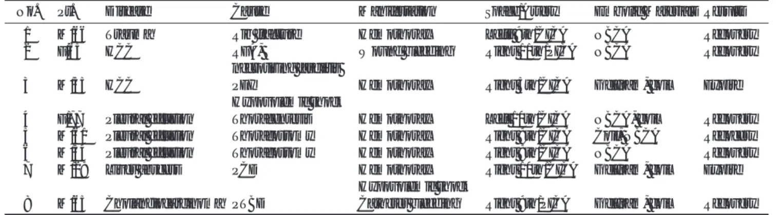
Fig. 1. A 29-year-old man (case7) pre-
sented with Hemothorax occurred af- ter a percutaneous drainage of a hepat- ic abscess.
A. Angiography of the right 9th poste-
rior intercostal artery demonstrated multiple collateral branches and an ex- travasation of contrast (arrow) from a collateral artery ran along the superior border of the right 10th rib.
B. Although the collateral artery was
selected with a microcatheter, ad- vancement of the microcatheter to the bleeding point was not possible, thus the artery was embolized using ab- sorbable gelatin sponge and the poste- rior intercostal artery (arrows) was embolized using microcoils.
Fig. 2. Hemothorax occurred after tho-
racentesis in a 54-year- old man (case 6).
Table 1. Patient Data
No. Pt. Disease Cause Manifestation Space/Artery Embolic Materials Results
1 M/66 Trauma Rib fracture Hemothorax Leftt 9th/CICA NBCA Recovery
2 F/65 HCC RFA, Wound bleeding Right 11th/PICA NBCA Recovery
necrotizing fasciitis
3 M/55 HCC PEI Hemothorax Right 5th/CICA Gelfoam, coil Expire
Hypovolemic shock
4 F/77 Pleural effusion Thoracentesis Hemothorax Left 10th/CICA NBCA, coil Recovery 5 M/61 Pleural effusion Thoracostomy Hemothorax Right 9th/CICA Coil, NBCA Recocery
6 M/54 Pleural effusion Thoracostomy Hemothorax Right 9th/CICA NBCA Recovery
7 M/29 Liver abscess PCD Hemothorax Right 10th/CICA Gelfoam, coil Expire
Hypovolemic shock
8 M/65 Cholangiocarcinoma PTBD Catheter bleeding Right 9th/PICA Gelfoam, coil Recovery
HCC = hepatocellular carcinoma, RFA = radiofrequency ablation, PEI = percutaneous ethanol injection, PCD = percutaneous catheter
drainage, PTBD = percutaneous transhepatic biliary drainage
가성동맥류 형성과 주변으로의 조영제 누출이 보였다.
8예 중 6예(75%)에서 부행늑간동맥(collateral intercostal arteries)에서 출혈하였는데, 그 중 3예는 늑골의 상연을 따라 주행하는 분지에서 출혈이 생긴 경우였고(Figs. 1, 2), 나머지 3예는 늑간의 중앙에서 늑간을 가로지르는 분지로부터 출혈 이 생긴 경우였다. 8예 중 2예(25%)에서는 후늑간동맥 (posterior intercostal artery)에서 출혈하였는데, 그 중 1예는 괴사성 근막염이 원인이었고(Fig. 3), 삽관에 의한 직접 혈관 손상은 1예 밖에 없었다.
색전 방법으로는 전 예에서 혈관조영도관 및 미세도관으로 출혈혈관을 선택한 후 경동맥 색전치료를 시행하였다. 부행늑 간동맥에서 출혈한 6예 중 4예에서는 혈관이 너무 가늘어 미 세도관으로 초 선택(superselection) 할 수 없어 해당 부행늑 간동맥에 연결되는 전, 후늑간동맥들을 젤폼(Spongostan, Johnson & Johnson, Soeborg, Denmark), N-butyl- cyanoacrylate(NBCA) (Histoacryl, B Braun, Melsungen, Germany), 코일(Tornado, Cook, Bloomington, IN, U.S.A.) 등을 단독으로 또는 같이 사용하여 색전하였고(Fig. 2.), 나머 지 2예에서도 해당 부행늑간동맥을 초 선택 할 수는 있었지 만 혈관이 너무 가늘어 출혈지점까지 미세도관의 진입이 불가
능하여 원거리에서 젤폼과 코일로 색전하였다(Fig. 1). 후늑 간동맥에서 출혈한 2예는 출혈부위 까지 미세 도관 진입이 가 능하였고 출혈부위의 근, 원위부를 코일이나 NBCA로 색전하 였다 그 중 한 예에서는 색전 후 혈관조영 검사에서 부행늑간 동맥을 통한 혈류로 가성동맥류에서 출혈이 지속되었으며 이 부행혈류 분지들을 NBCA로 추가로 색전하였다(Fig. 3). 전 예에서 경동맥색전술로 성공적으로 지혈되었고 재 출혈은 없 었다.
색전술 후 혈흉과 함께 저혈압성 쇼크 상태였던 2명의 환 자는 더 이상의 출혈은 없었으나 쇼크의 진행으로 사망하였고 나머지 4명의 혈흉 환자에서는 혈흉의 배액 치료가 이미 시 행된 상태이거나 혈관 색전과 동시에 시행하여, 모두 출혈이 멈추고 혈흉이 배액 되면서 임상적으로 회복되었다. 천자부나 배액관을 통하여 직접 출혈이 있었던 경우에도 출혈에 의한 문제는 모두 회복되었다.
고 찰
늑간동맥의 급성 출혈은 매우 드물어 외상이나 늑간 천자 등 의인성 요인에 의하여 발생한 예의 단편적 보고가 대부분
A B
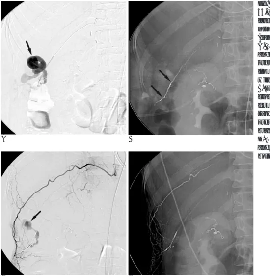
Fig. 3. Wound bleeding occurred in a
55-year-old woman with necrotizing fasciitis caused by radiofrequency ab- lation of a hepatocellular carcinoma (case 2).
A) The right 11th posterior intercostal
angiography demonstrated a large pseudoaneurysm (arrow) originated from the posterior intercostal artery with extravasation of contrast.
B. Embolization of the artery using mi-
crocoils and NBCA (arrows) was per- formed, (C) Post-embolization angiog- raphy revealed contrast filling into the pseudoaneurysm (arrow) via collateral branches.
D. The 10th posterior intercostal artery
and collateral branches were em-
bolized using NBCA (arrows).
이며(1-6), 원인적 요소에 대한 분석은 거의 없다(7). 영상 의학과 의사는 폐 질환에 대한 조직 생검 이나, 흉막 질환의 진단과 치료, 간 종양의 초음파 소작술, 상복부의 저류액체배 액, 경피적 담도 배액 등으로 늑간 천자를 많이 시행하고 있 어 늑간 동맥 출혈을 경험할 가능성이 높으므로 이의 발생을 최소화하는 방법과 치료법을 아는 것은 매우 중요하다.
늑간 동맥은 대동맥으로부터 직접 기시하는 후늑간동맥과 내흉동맥(internal thoracic artery)에서 기시하는 전늑간동맥, 그리고 이들을 연결하는 부행늑간동맥으로 구성된다. 후늑간 동맥과 전늑간동맥은 서로 만나 하나의 혈관 고리를 이루는데 이는 굵고 주된 혈행 경로이며 늑골의 하연을 따라서 늑간 정 맥 및 늑간 신경과 같이 주행한다. 부행늑간동맥은 후늑간동 맥에서 분지하는데 분지 지점은 다양하지만 늑골의 각(angle) 부위에서 주로 분지 하여 늑간을 가로지른 후 아래쪽 늑골의 상연을 따라서 주행하다가 전늑간동맥과 합쳐진다(7, 8). 따 라서 중액와선(mid-axillary line) 상이나 후액와선(posterior axillary line) 상에서 늑골의 상연으로는 부행늑간동맥이 존재 하고 있는 경우가 대부분이라고 할 수 있다. da Rocha 등(7) 이 90개의 늑간에 대하여 해부학적 분석을 시행하였는데, 제 5번과 제 8번 늑간의 중액와선상에서 후늑간동맥의 평균굵기 는 각각 1.5 mm 와 3.8 mm였고, 부행늑간동맥의 굵기는 각 각 0.6 mm 와 0.5 mm로서, 하부 늑간일수록 부행늑간동맥 이 가늘어 지기는 하지만 그 굵기가 상당한 것을 알 수 있다.
저자들의 예에서 주된 출혈 부위(75%)는 전, 후늑간동맥이 아니라 부행늑간동맥이였다. 일반적으로 늑간천자 시 늑골의 상연에 매우 근접하여 바늘을 진행시키는 것이 후 늑간 동맥 이 주행하는 경로를 피하는 방법으로 알려져 있으나(9), 해부 학적으로 늑골의 상연을 따라 주행하는 부행늑간동맥이 이미 널리 알려져 있고 이의 굵기도 상당할 수 있으므로 늑골 상연 에 근접하여 천자하는 방법은 재고되어야 할 것으로 생각된 다. 늑간을 가로지르는 부행늑간동맥의 경우는 예측을 할 수 없으므로 이를 피하는 뚜렷한 방법은 없겠지만, 늑골의 상연 으로는 거의 항상 부행늑간동맥이 지나고 있음을 고려한다면 늑간의 중간을 천자하는 것이 가장 바람직할 것으로 생각된 다. 후늑간동맥에서 출혈이 생긴 경우는 2예 였지만 천자나 삽관에 의한 직접 혈관 손상은 1예 밖에 없었다. 이는 늑골 하 연으로 천자하는 경우는 거의 없음에 기인하는 것으로 여겨진 다.
각된다. 또한, 반드시 인접 늑간동맥을 같이 검사하여 부행 혈 류 여부를 확인하고 필요하면 이를 같이 치료하여야 한다. 부 행늑간동맥 출혈의 경우, 출혈 지점까지 미세 도관 진입이 가 능하다면 해당 부위만을 색전하면 되나, 혈관이 너무 가늘어 출혈 지점까지 미세도관의 진입이 불가능한 경우가 더 많았고 (7예 중 6예: 86%), 이때, 혈관의 초 선택이 가능한 상태라면 원거리에서 젤폼이나 polyvinyl alcohol particle등의 입자성 색 전 물질로 색전하면 되고, 혈관의 초 선택 자체가 불가능 하 다면 입자성 색전 물질, 또는 NBCA등의 액체성 색전물질로 후늑간동맥 및 부행늑간동맥들을 같이 색전하는 방법을 사용 하여야 한다.
연구의 제한점으로는 증례의 수가 적고, 후향적 연구이며 여 러 병원에서 다양한 방법으로 색전치료가 시행되어 색전술의 방법이 통일되어 있지 않다는 점이다. 본 연구만으로 늑간의 중간을 천자하는 것이 동맥출혈의 가능성을 줄이는 방법인지 를 확실하게 규명할 수는 없으며, 이를 위해서는 여러 병원의 늑간 천자 환자를 대상으로 전향적인 연구가 필요할 것으로 사료된다.
결론적으로,늑간 천자 시 늑골 상연에는 부행늑간동맥이 있 기 때문에 가능하면 늑간의 중간부위를 천자하는 것이 바람직 하다. 늑간동맥 출혈이 흉곽 내로 고일 때에는 많은 양의 실 혈이 있을 수 있으며 경동맥색전술은 늑간동맥출혈의 치료에 매우 효과적인 방법이다.
참 고 문 헌
1. Casas JD, Perendreu J, Gallart A, Muchart J. Intercostal artery pseudoaneurysm after a percutaneous biliary procedure: diagnosis with CT and treatment with transarterial embolization. J Comput Assist Tomogr 1997;21:729-730
2. Muthuswamy P, Samuel J, Mizock B, Dunne P. Recurrent massive bleeding from an intercostal artery aneurysm through an empye- ma chest tube. Chest 1993;104:637-639
3. Bluebond-Langner R, Pinto PA, Kim FJ, Hsu T, Jarrett TW.
Recurrent bleeding from intercostal arterial pseudoaneurysm after retroperitoneal laparoscopic radical nephrectomy. Urology 2002;
60:1111-1112
4. Barbaric ZL, Luka NL. Angiographic demonstration and tran- scatheter embolic control of post-traumatic intercostal arterial he- morrhage. Surgery 1977;81:409-412
5. Carney M, Ravin CE. Intercostal artery laceration during thoracen-
J Korean Radiol Soc 2005;53:169-173
Address reprint requests to : Jae-Ik, Bae, M.D., Department of Radiology, Busan Paik Hospital, Inje University College of Medicine, 633-165, Gaegeum2-dong, Busanjin-gu, Busan 614-735, Republic of Korea.
Tel. 82-51-890-6549 Fax. 82-51-896-1085 E-mail: [email protected]
Transarterial Embolization of Acute Intercostal Artery Bleeding
1Jae-Ik Bae, M.D., Auh Whan Park, M.D., Seon-Joo Lee, M.D., Gi-Young Ko, M.D.2, Hyun-Ki Yoon, M.D.2, Chang Jin Yoon, M.D.3, Tae Beom Shin, M.D.4, Young Hwan Kim, M.D.5
1
Department of Radiology, Busan Paik Hospital, Inje University College of Medicine, Busan, Korea
2
Department of Radiology, Asan Medical Center, University of Ulsan College of Medicine, Seoul, Korea
3
Department of Radiology, Seoul National University Bundang Hospital, Seongnam, Korea
4
Department of Radiology, Dong-A University College of Medicine, Busan, Korea
5
Department of Radiology, Dongsan Medical Center, Kyimyung University School of Medicine, Daegu, Korea
Purpose:
To report our experiences of transarterial embolization for acute intercostal artery bleeding.Materials and Methods:
A retrospectively analysis of the causes, clinical manifestations, angiographic findings and transarterial embolization technique in 8 patients with acute intercostal artery bleeding, with a review of the anatomical basis.Results:
The causes of intercostal artery bleeding were iatrogenic and traumatic in 88 and 12% of cases, re- spectively. Active bleeding from the collateral intercostal or posterior intercostal arteries was angiographically demonstrated in 75 and 25% of cases, respectively. Transarterial embolization successfully achieved hemosta- sis in all cases. However, two patient with hypovolemic shock expired due to a massive hemothorax, despite successful transarterial embolization.Conclusion:
Intercostal access should be performed through the middle of the intercostal space to avoid injury to the collateral intercostal artery. Transarterial embolization is an effective method for the control of inter- costal artery bleeding.Index words :
Arteries, therapeutic embolization Interventional proceduresHemothorax