조직 배양한 에키네시아 추출물에 관한 효능 연구
박 창 민†⋅정 민 석⋅최 종 완⋅백 기 엽* 한국화장품(주) 기술개발연구소, *충북대학교 (2008년 3월 3일 접수, 2008년 4월 28일 채택)
The Study on Tissue-Cultured Echinacea purpurea Adventitious Roots Extract for Application as a Cosmetic Ingredient
Chang-Min Park†, Min-Seok Joung, Jong-Wan Choi, and Kee-Yoeup Paek* R&D Center, Hankook Cosmetics Co., Ltd., 76-1, Yongseong-ri, Samseong-myeon,
Eumseong-gun, Chungbuk 369-834, Korea
*Research Center for The Development of Advanced Horticultural Technology, Chungbuk National University (Received March 3, 2008; Accepted April 28, 2008)
요 약: 에키네시아는 미국 북미 대평원에 서식하는 야생 식물로 수세기 동안 감기나 또 다른 바이러스 감염에 의한 질병에 대하여 면역 기능을 증진 시키는 전통 식물 약재로 널리 사용되어 왔다. 최근에 에키네시아 추출물은 피부 보호 를 위한 화장품 원료로서 응용되어 오고 있다. 이에 본 연구에서는 에키네시아를 인공적으로 조직배양하여 얻어낸 캘러 스로부터 부정근을 유도하여, 대단위 배양을 한 부정근을 추출하여 화장품 성분으로서의 응용 가치를 평가하였다. 이미 몇몇 보고된 논문에서 에키네시아 추출물은 항산화 효과 및 면역증진효과는 보고되었지만 다른 효능에 대한 연구는 충분하지 않다. 따라서 본 연구에서는 피부 보호에 대한 화장품 원료로서의 응용을 위한 항산화, 미백 및 주름과 관련한 효능 효과를 평가하였다. 조직 배양한 에키네시아 추출물의 효능 효과 평가 결과, 추출물 농도 2 %까지 세포독성이 나타나지 않았으며, 외부환경에 의한 피부노화 및 피부색, 기미, 주근깨 등과 같은 피부현상에 중요한 영향을 미치는 인자인 활성산소종의 소거 효과가 우수함을 확인하였으며, 또한 B16 melanoma 세포 내에서 tyrosinase의 발현을 농도 의존적으로 감소시키고 멜라닌 합성을 억제하였다. 피부 주름과 관련하여 진피층의 extracellular matrix (ECM) degra- dation에 관여하는 콜라겐 분해효소인 MMP-1, MMP-2의 발현을 억제하였다.
Abstract: Echinacea purpurea, an indian traditional plant medicine has been widely used as herbal remedy for the treatment of disease such as colds or other infections. However, Echinacea purpurea extracts recently have been ap- plied as a cosmetic ingredient for skin care. We artificially cultured Echinacea purpurea by using the bioreactor cul- ture system for this study. We induced callus from Echinacea purpurea and separated adventitious roots, harvested and extracted after cultured in bioreactors. Previously, several studies have been reported on anti-oxidant and im- muno-enhancing effects of Echinacea purpurea extract but other efficacies were not well known. In this study, we investigated the whitening, anti-wrinkle and anti-oxidant effects to know applicable value of tissue-cultured Echinacea purpurea adventitious roots extract (TCEPARE) as a cosmetic ingredient. TCEPARE did not show cyto- toxicity until a concentration of 2 % and showed the anti-oxidative effect in DPPH and NBT tests. Also, the extract decreased tyrosinase expression in a dose-dependent manner and inhibited melanin synthesis in B16 melanoma cells.
TCEPARE reduced protein levels of MMP-1, 2 secreted in culture medium or in cell lysates. From these results we suggest that TCEPARE has potential benefits applicable as to cosmetic ingredient for skin care products.
Keywords: Echinacea purpurea, whitening, anti-wrinkle, matrix metalloproteinase, B16 melanoma cell 1)
† 주 저자 (e-mail: [email protected])
1. Introduction
Echinacea purpurea, commonly called as ‘purple coneflower’ is a member of the Asteraceae family and herbaceous perennial. It’s original to South America and American Indians have long used Echinacea purpurea as herbal remedy against colds, wound, sores, snakes and insects bites. In addition, the flower of Echinacea species are used as the herbal tea to help the strength of immune system[1,2]. Several studies previously have been reported anti-oxidant and immuno-enhanc- ing effects of Echinacea purpurea extract[3,4]. Nowadays, the abilities of Echinacea purpurea were well known by many studies. It has the capacity to activate human macrophages and stimulate the phagocytosis[5,6]. The roots of Echinacea purpurea have many active func- tions for the human body[7,8]. However, the study of Echinacea purpurea has a limit because the Echinacea purpurea is very expensive and rare. Therefore, we ar- tificially cultured Echinacea purpurea adventitious roots using the bioreactor culture system. We induced callus from Echinacea purpurea directly. Separated adventi- tious roots were precultured and then, transferred in bioreactors. In this study, we investigated the whiten- ing effect, anti-wrinkle effect and the safety of tis- sue-cultured adventitious roots extract of Echinacea purpurea in order to identify the merit as a cosmetic ingredient.
2. Materials and Methods
2.1. Reagents and Cell Culture
Antibodies against MMP-1, MMP-2, tyrosinase, and β-actin were purchased from Santa Cruz Biotechnology (Santa Cruz, CA, USA). All reagents were purchased from Sigma-Aldrich (St. Louis, MO, USA). Other commercially available reagents and solvents were used as received. Mouse fibroblast cell lines NIH3T3-L1 and B16 melanoma cells (obtained from American Type Culture Collection) were maintained in Dulbecco's modified Eagle's medium (DMEM) supplemented with 10 % heat-inactivated FBS, penicillin (100 U/mL) and streptomycin (100 µg/mL) at 37 ℃ in a humidified
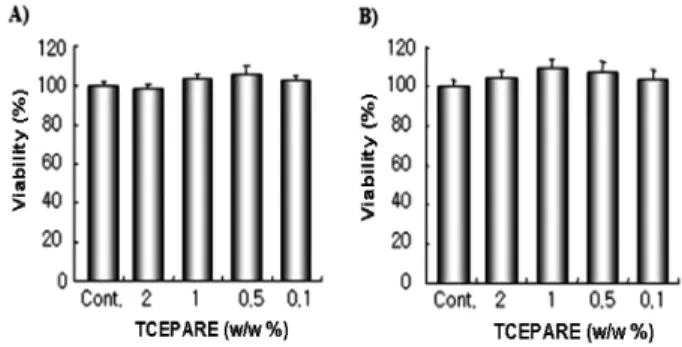
Figure 1. Cell viability of tissue-cultured Echinacea purpurea adventitious roots extract. Cells were treated with various concentrations of the extract for 48 h and cell viability meas- ured by A, NR assay and B, MTT assay as described in ma- terials and methods. The results were represented as mean of standard deviation (S.D.) of three independent experiments.
atmosphere containing 5 % CO2.
2.2. Preparation of Tissue-cultured Echinacea purpurea Adventitious Roots Extract (TCEPARE)
2.2.1. Cultivation
The adventitious roots were induced from the callus on Murashige & Skoog medium (Duchefa, Netherland) supplemented with 1.0 mg/L IBA (Duchefa, Netherland) and 5 % sucrose, and were subcultured in MS liquid medium with the same plant growth regulators.
2.2.2. Extraction
After dried, 1 kg of tissue-cultured Echinacea pur- purea adventitious roots were immersed in 27 kg of 70
% ethanol solution and 3 kg of 1,3-butylene glycol (1,3-BG) for sufficient time and mixed enough for 48 h in 45 ± 5 ℃ using agitator. After filtering with a fil- ter paper (5C, TOYO, Japan), tissue-cultured Echinacea purpurea adventitious roots extract was obtained.
2.3. Cell Viability Assay
2.3.1. NR Assay
3 × 10³ cells/well were seeded in 10 % FBS/DMEM medium. Cells were treated with sample for indicated times and incubated at 37 ℃ for 48 h in 5 % CO2
incubator. 0.1 mL of neutral red diluted at concen- tration of 50 µg/mL in DMEM was added into each
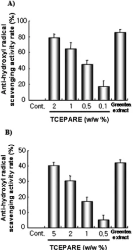
Figure 2. Anti-oxidant effect of tissue-cultured Echinacea purpurea adventitious roots extract by A, DPPH test and B, NBT test. We examined the various concentrations and com- pared with green tea extract (1 % in DPPH assay and 5 % in NBT assay) as positive control as described in materials and methods. The results were represented as mean of stand- ard deviation (S.D.) of three independent experiments.
well and incubated for 3 h, the media were removed from each well. After fixed the cells by 1 % of formal- in, 1 % of CaCl2, the accumulated neutral red in the cells was extracted with 1 % of acetic acid in 50 % ethanol and it was measured by microplate reader (Molecular Devices, USA) in dual ranges of 540 nm and 630 nm.
2.3.2. MTT Assay
The test materials were added and incubated in 5 % CO2 incubator at 37 ℃ for 48 h by a same procedure of NR assay and 0.01 mL of MTT at the concentration
of 5 mg/mL in PBS was added into each well and in- cubated for 4 h. Measurement of mitochondrial activity from purple formazan by MTT was used to assess the viability of cells following sample treatment: MTT (0.5 mg/mL), one tenth of the original culture volume, was added to each culture and incubated for 3 h at 37 ℃, in 5 % CO2. The purple formazan formed by viable cells was dissolved by the addition of DMSO, and absorb- ance at 540 nm was measured by using microplate reader (Molecular Devices, USA).
2.4. UV Irradiation
Cells were plated in 6-well cell culture dishes and in- cubated at 37 ℃ under humidified 5 % CO2 and 95 % air in culture medium until 70 % to 80 % confluent.
Then cells were exposed 5.8 J/cm2 (UVA) from high intensity UV lamp (UVGL-58, San Gabriel, USA).
2.5. Inhibition Assay of Melanization
B16 melanoma cells were cultured in DMEM supple- mented with 10 % FBS in humidified incubator at 37
℃ under 5 % CO2 in 6 well plate at density of 2.0 × 10⁴ cells/well. After cells were attached, medium was replaced with DMEM containing 10 % FBS, 0.2 µM α -MSH, 2 mM theophylline and samples addition. After 4 days, trypsin was added and suspended cells were collected by centrifugation. Then cell pellets were dried and dissolved in 1 N NaOH. Melanin synthesis in- hibition rates were measured at 490 nm using enzyme linked immunosolvent assay (ELISA) reader.
2.6. Free Radical Scavenging Activity Assay (DPPH Test)
1,1-diphenyl-2-picrylhydrazyl (DPPH) is a stable free radical using determination of radical scavenging assay. The radical scavenging activity was determined by a previous report of Haraguchi et al.[9]. Then, the anti-oxidative activity (%) was calculated as compared with blank control.
2.7. Superoxide Radical Scavenging Activity Assay (NBT assay)
Superoxide dismutase (SOD) activity was measured
using xanthine-xanthine oxidase system for a source of superoxide and nitroblue tetrazolium (NBT) as a scav- enger for this radical. The superoxide anion scavenging activity was determined by a previous report of K.
Furuno[10].
2.8. Gelatin Zymography
Fibroblast cells in subconfluent culture were washed and refreshed with serum-free DMEM, and treated for 18 h. The enzymatic activity and molecular weight of eletrophoretically separated gelatinolytic enzymes in the conditioned medium of cells were determined by SDS-PAGE as follows. 20 µL of serum free culture medium per sample was prepared in non-denaturing loading buffer and size fractionated in 10 % SDS-poly- acrylamide gel impregnated with 0.1 % gelatin. The gels were washed with 2.5 % Triton X-100 for 1 h at room temperature to remove SDS, rinsed twice with water, and then incubated in developing buffer (50 mM Tris-HCl buffer, pH 7.4, 20 mM NaCl, 10 mM CaCl2 and 0.1 % NaN3) for 18 h at 37 ℃. Subsequently, gels were fixed and stained with 10 % acetic acid con- taining 0.5 % Coomassie Blue R250. Zones of gelati- nolytic activity were detected as clear bands against a blue background. Densitometric analysis was done us- ing Scion Image NIH Image program.
2.9. Western Blot Analysis
Cells were irradiated at various dose and lysed in ly- sis buffer (20 mM Tris, pH 7.4, 150 mM NaCl, 1 mM EDTA, 1 mM β-glycerolphosphate, 1 mM Na3VO4, 1 mg/mL leupeptin and 1 mM PMSF). The lysates were clarified by centrifugation at 12,000 × g for 15 min at 4 ℃, and protein content was measured by SDS-PAGE and blotted to nitrocellulose membrane (0.2 mm, Amersham, Arlington Heights, IL, USA). The mem- brane was blocked with 5 % nonfat skim milk in TBS-T and incubated with the primary and secondary antibodies. Immunoblots were visualized by enhanced chemiluminescence (Amersham, UK) according to the manufacturer’s protocol. Conditioned medium from each sample was also subjected to protein analysis of MMP-1, MMP-2 and collagen. For this purpose, cul-
ture medium in each tissue culture dish was collected and concentrated using a Centricon 10 microconcentrator (Amicon, Beverly, MA, USA) and 5-fold concentrated conditioned medium (20 µL) was used for SDS-PAGE analysis. Densitometric analysis for protein bands was done using Scion image NIH image program.
3. Results
3.1. Cell Viability Assay
To investigate the cytotoxicity of tissue-cultured Echinacea purpurea adventitious roots extract, we first treated the cells with TCEPARE extract in indicated concentrations for 48 h and cell cytotoxicity was meas- ured by NR assay and MTT assay. As shown in Figure 1A and B, TCEPARE extract did not show a cytotox- icity in a dose-dependent manner until 2 % concentration.
3.2. Free Radical Scavenging Activity of TCEPARE To investigate the scavenging effect of hydroxyl and superoxide radical known as the strongest free radical in cellular oxidation reactions, we studied the effect of TCEPARE extract on the free radical scavenging activity. As shown in Figure 2A and B, the TCEPARE extract enhanced the anti-oxidative effect in DPPH and NBT test.
3.3. Whitening Effect of TCEPARE
To investigate whether TCEPARE extract inhibits melanin synthesis and tyrosinase expression in B16 melanoma cells, we analyzed inhibition rate of melanin synthesis and tyrosinase expression for TCEPARE. As shown in Figure 3A and B, The extract decreased ty- rosinase expression in a dose-dependent manner and inhibited a melanin synthesis in B16 melanoma cells.
3.4. Anti-wrinkle Effect of TCEPARE
To further indentifiy the effect of TCEPARE ex- tract, we next examined the expression of MMP-1 and MMP-2 enhancing the degradation extracellular matrix (ECM) components relating with skin wrinkle. As shown in Figure 4A and B, the tissue-cultured Echinacea purpurea adventitious roots extract (TCEPARE)
Figure 3. Inhibition of melanin synthesis and tyrosinase by tissue-cultured Echinacea purpurea adventitious roots extract.
A, western blot analysis of tyrosinase and B, melanin syn- thesis assay in B16 melanoma cell treated with melanization inducer (0.2 µM α-MSH, 2 mM theophylline). Cells were lysed in lysis buffer and subjected to Western blot for the de- tection of tyrosinase expression as described in materials and methods. B16 melanoma cells were treated with indicated concentration and compared with arbutin (0.05 %). The re- sults were represented as mean of standard deviation (S.D.) of three independent experiments.
reduced the expression of MMP-1 and MMP-2 se- creted in culture medium or cell lysates.
4. Discussion
Tissue-cultured Echinacea purpurea adventitious roots extract (TCEPARE) showed the whitening effect, anti-oxidant effect and anti-wrinkle effect. In the inhibition assay of melanization in B16 melanoma cells, TCEPARE extract showed inhibitory effects of melanin synthesis in B16 melanoma cells. The extract showed anti-oxidant effect similar to green tea extract on investigation by DPPH and NBT assay. Also
Figure 4. Effect of tissue-cultured Echinacea purpurea ad- ventitious roots extract on the expression of MMP-1 and MMP-2 in NIH3T3-L1 fibroblast cell lines. A, gelatin zymog- raphy analysis of serum-free conditioned medium and B, Western blot analysis of MMP-1 and MMP-2 in cell lysates from NIH3T3-L1 cells. Cells were incubated after exposure to UVA and treatment with indicated concentration in se- rum-free medium for 18 h. Conditioned medium was collected and used for gelatin zymography. Cells were lysed in lysis buffer and subjected to Western blot for the detection of ty- rosinase expression as described in materials and methods.
Zymography bands were subjected to densitometric scanning using the Scion image NIH image software. Cells were treated with indicated concentrations for 24 h.
TCEPARE extract inhibited the expression of MMP-1 and MMP-2. Thus, we expect that TCEPARE extract has potential merits applicable to cosmetic ingredient for skin care. Although further work will be required to elucidate whether this TCEPARE extract has also desirable effects on clinical test, these findings provide
the possibility of cosmetic ingredient for skin care products.
References
1. R. A. Burger, A. R. Torres, R. P. Warren, and V.
D. Caldwell, Echinacea-induced cytokine production by human macrophages, Int. J. Immunopharmacol., 19(7), 371 (1997).
2. M. Stimpel, A. Proksch, H. Wagner, and M. L.
Lohmann-Matthes, Macrophage activation and in- duction of macrophage cytotoxicity by purified pol- ysaccharide fractions from the plant Echinacea purpurea, Infect Immun., 46(3), 845 (1984).
3. H. Haraguchi, H. Ishikawa, Y. Sanchez, T. Ogura, Y. Kubo, and I. Kubo, Antioxidative constituents in Heterotheca inuloides, Bioorg. Med. chem., 5, 865 (1997).
4. K. Furuno, T. Akasako, and N. Sugihara, The con- tribution of the pyrogallol moiety to superoxide rad- ical scavenging activity of flavonoids, Biol. Pharm.
Bull., 25, 19 (2002).
5. S. Mishima, K. Saito, H. Maruyama, M. Inoue, T.
Yamashita, T. Ishida, and Y. Gu, Antioxidant and immuno-enhancing effects of Echinacea purpurea, Biol. Pharm. Bull., 27(7), 1004 (2004).
6. F. Pellati, S. Benvenuti, L. Magro, M. Melegari, and F. Soragni, Analysis of phenolic compounds and radical scavenging activity of Echinacea spp., J.
Pharm. Biomed. Ana., 35(2), 289 (2004).
7. B. D. Sloley, L. J. Urichuk, C. Tywin, R. T. Coutts, P. K. Pang, and J. J. Shan, Comparison of chemical components and antioxidants capacity of different Echinacea species, J. Pharm. Pharmacol., 53(6), 849 (2001).
8. C. Hu and D. D. Kitts, Studies on the antioxidant activity of Echinacea purpurea root extract, J.
Agric. Food. Chem., 48(5), 1466 (2000).
9. H. Haraguchi, H. Ishikawa, Y. Sanchez, T. Ogura, Y. Kubo, and I. Kubo, Antioxidative constituents in Heterotheca inuloides, Bioorg. Med. Chem., 5, 865 (1997).
10. K. Furuno, T. Akasako, and N. Sugihara, The con- tribution of the pyrogallol moiety to superoxide rad- ical scavenging activity of flavonoids, Biol. Pharm.
Bull., 25, 19 (2002).