Inhibition and Chemical Mechanism of Protocatechuate 3,4-dioxygenase from Pseudomonas pseudoalcaligenes KF707
Taekyeong Kang
†, Sang Ho Kim
†, Mi Ja Jung and Yong Kweon Cho*
Department of Biochemistry and Health Science, College of Natural Science, Changwon National University, Changwon, Gyeongnam 641-773, Korea
Received January 27, 2015 /Revised May 12, 2015 /Accepted May 21, 2015
We carried out pH stability, chemical inhibition, chemical modification, and pH-dependent kinetic pa- rameter assessments to further characterize protocatechuate 3,4-dioxygenase from Pseudomonas pseu- doalcaligenes KF707. Protocatechuate 3,4-dioxygenase was stable in the pH range of 4.5~10.5. L-ascor- bate and glutathione were competitive inhibitors with K
isvalues of 0.17 mM and 0.86 mM, respectively. DL-dithiothreitol was a noncompetitive inhibitor with a K
isvalue of 1.57 mM and a K
iivalue of 8.08 mM. Potassium cyanide, p-hydroxybenzoate, and sodium azide showed a noncompetitive inhibition pattern with K
isvalues of 55.7 mM, 0.22 mM, and 15.64 mM, and K
iivalues of 94.1 mM, 8.08 mM, and 662.64 mM, respectively. FeCl
2was the best competitive inhibitor with a K
isvalue of 29 μM. FeCl
3, MnCl
2, CoCl
2, and AlCl
3were also competitive inhibitors with K
isvalues of 1.21 mM, 0.85 mM, 3.98 mM, and 0.21 mM, respectively. Other metal ions showed noncompetitive inhibition patterns. The pH-dependent kinetic parameter data showed that there may be at least two catalytic groups with pK values of 6.2 and 9.4 and two binding groups with pK values of 5.5 and 9.0. Lysine, cysteine, tyrosine, carboxyl, and histidine were modified by their own specific chemical modifiers, in- dicating that they are involved in substrate binding and catalysis.
Key words : Chemical modification, pH-dependent kinetic parameters, protocatechuate 3,4-dioxygenase,
Pseudomonas pseudoalcaligenes KF707
†Authors contributed equally.
*Corresponding author
*Tel : +82-55-213-3551, Fax : +82-55-213-3551
*E-mail : [email protected]
This is an Open-Access article distributed under the terms of the Creative Commons Attribution Non-Commercial License (http://creativecommons.org/licenses/by-nc/3.0) which permits unrestricted non-commercial use, distribution, and reproduction in any medium, provided the original work is properly cited.
Journal of Life Science 2015 Vol. 25. No. 5. 487~495 DOI : http://dx.doi.org/10.5352/JLS.2015.25.5.487
Introduction
Polyphenol oxidase (monophenol, dihydroxy-L-phenyl- alanine: oxygen oxidoreductase; EC 1.14.8.1) is a copper-con- taining enzyme widely distributed in nature and responsible for browning in fruits and vegetables. Polyphenol oxidase is a mixed-function oxidase that catalyzes both the ortho hy- droxylation of monophenols to o-diphenols (cresolase activ- ity) and the further oxidation of o-diphenols to o-quinones (catecholase activity). The o-quinones are highly reactive compounds that polymerize spontaneously to form brown pigment (melanin) or react with amino acids and proteins [34]. Enzymatic browning is a significant problem during processing of raw agro-materials such as banana, pear, grape, peach, apple, mushroom, potato, lettuce, shrimps,
lobster, and crab [12, 23]. Therefore, development of high
performance tyrosinase inhibitors is important to prevent
browning of vegetables and fruits. Many methods to prevent
the browning reaction have been proposed [1], including
high temperature [22], sulfur compounds such as sodium
sulfite, sodium bisulfite, and sodium metabisulfite [19], acids
such as citric acid, malic acid, phosphoric acid, and ascorbic
acid [14, 28], reducing agents such as L-cysteine or DL-me-
thionine [24], and boric acid and borates [2]. Although nu-
merous studies have reported attempts to control various
aspects of browning reactions by phenolase, little attention
has been paid to reactions that can modify its phenolic sub-
strates so that they become unavailable for the darkening
reactions. Bacterial oxygenase, which carries out oxidative
ring-opening reactions, cleaves the catechol ring of plant-
darkening constituents [16]. Use of such ring-opening re-
actions is a simple method for controlling phenolase darken-
ing of compounds. In a previous study [30], we reported
the purification and characterization of protocatechuate
3,4-dioxygenase from Pseudomonas pseudoalcaligenes KF707 as
a possible anti-browning agent. Here, we describe the in-
hibition mode of chemicals and the chemical mechanism by
evaluating chemical modification and pH-dependent kinetic
parameters.
Materials and Methods
Materials
MES (2-(N-morpholino)-ethanesulfonic acid), Tris (Tris (hydroxymethyl)-aminomethane), CHES ((2-N-cyclohexyl- amino)-ethanesulfonic acid), and CAPS (3-(cyclohexylamino)- 1-propanesulfonicacid) were obtained from Research Organics Inc. (Cleveland, OH, USA). Pyridoxal-5'-phosphate (PLP), 2,4-dinitrofluorobenzoic acid (FDNB), 5,5'-dithio- bis(2-nitrobenzoic acid) (DTNB), ninhydrin, 2,3-butanedione, N-acetylimidazole, iodoacetamide, (1-ethyl-3-(3-dimithyla- minopropyl)-carbodiimide) (EDAC), diethylpyrocarbonate (DEPC), and o-methylisourea were purchased from Sigma- Aldrich (St. Louis, MO, USA). All other chemicals and re- agents were of analytical grade and used without further purification. All solutions were made using distilled water deionized with a 0.22 μm filter and a Mili-Q plus reagent water system (Millipore Co., Molsheim, France).
Protocatechuate 3,4-dioxygenase assay
All assays were carried out using a CARY 3 Bio Spectrophotometer (Varian Australia Pty. Ltd., Melbourne, Australia). Kinetic parameters of protocatechuate 3,4-dioxy- genase was determined as described previously [30]. The as- say mixture contained 50 mM air-saturated buffer (Tris, pH 7.5) and 48 μM protocatechuate in the total volume of a 1 mL quartz cuvette (1 cm width). The reaction was initiated by adding 10 μl enzyme solution (1 unit) with an add- er-mixer. One unit of enzyme activity was defined as the amount of enzyme that converts 1 mmol of substrate per minute. Enzymatic activity was determined with 3,4-dihy- droxyphenylacetate at 258 nm (ε = 9.4 mM
-1cm
-1) for the con- version of protocatechuic acid to 3-carboxy-cis,cis-muconic acid [13].
Protein determination
Protein concentration was determined with the Bio-Rad Protein Assay Kit (Hercules, CA, USA), according to the Bradford method. Crystalline bovine serum albumin (BSA) was used as the standard, as described previously [3].
pH stability
Buffers were chosen for adequate buffer capacity, and more than one buffer was used to examine the effect of buf-
fer on activity. Buffers at a 50 mM final concentration were used over the following pH ranges: Acetate, pH 4.5~5.5;
MES, pH 5.5~6.5; phosphate, pH 6.5~7.5; Tris, 7.5~8.8; CHES, pH 8.8~10; and CAPS, pH 10~10.5. The pH of the reaction mixtures was measured before and after sufficient data were collected to determine initial velocities. Negligible pH changes were observed before and after the reaction. A con- centrated enzyme solution (10 units) was mixed with 980 μl of each 50 mM buffer. An aliquot was taken out at differ- ent times (every 5 to 10 minutes) and assayed for remaining activity at 25°C. Protocatechuate 3,4-dioxygenase activity was measured with 48 μM protocatechuate in a 50 mM air-saturated buffer (Tris, pH 7.5). The pH of the reaction mixtures were checked before and after the reaction. All as- says were performed in duplicate.
pH studies
All buffers were titrated to the desired pH with KOH.
In all cases, overlaps were obtained when buffers were changed so that a correction could be made for spurious buffer effects. Negligible pH changes were observed before and after the reaction. All assays were performed in dupli- cate.
Effect of inhibitors
Organic (L-cysteine, glutathione, L-ascorbate, β-mercap- toethanol, DL-dithiothreitol, and p-hydroxybenzoic acid) and inorganic (sodium azide, potassium cyanide, thiourea, FeCl
2, AgNO
3, CuCl
2, FeCl
2, ZnCl
2, SnCl
2, AlCl
2, MnCl
2, CoCl
2, NiCl
2, MgSO
4, KCl, NaCl, and CaCl
2) compounds were used for the inhibition study. The materials selected for the inhibition study were added to a reaction mixture (1 mL) containing 50 μM protocatechuate and 50 mM Tris buffer (pH 7.5). The reaction was initiated by adding 10 μl enzyme solution (1 unit) and monitored at 258 nm. All as- says were performed in duplicate.
Chemical modification
Enzyme solution (10 unit) was incubated with different
concentrations of chemical modifiers in 100 μl of 50 mM Tris
buffer (pH 7.5), as described previously [4]. The chemical
modifiers used in this study were PLP, FDNB, DTNB, ninhy-
drin, 2,3-butanedione, N-acetylimidazole, iodoacetamide,
EDAC (1-ethyl-3-(3-dimithylaminopropyl)-carbodiimide), di-
ethylpyrocarbonate, and o-methylisourea. A 20 μl aliquot
was withdrawn at specified time intervals and assayed for
Table 1. Inhibition parameters of antioxidants and chemicals for the protocatechuate 3,4-dioxygenase
Inhibitor Inhibition
Pattern
Inhibition Constants Kis (mM) Kii (mM) L-Ascorbic acid
DL-Dithiothreitol Glutachione P-Hydroxybenzoate Sodium azide Potassium cyanide
C NC
C NC NC NC
0.17 1.57 0.86 0.22 15.64 55.7
- 9.28
- 9.28 662.4 94.1 C: competitive inhibition, NC: noncompetitive inhibition, Kis:
slope inhibition constant, Kii: intercept inhibition constant.
Table 2. Inhibition parameters of metal ions for the proto- catechuate 3,4-dioxygenase
Inhibitor Inhibition Pattern
Inhibition Constants Kis (mM) Kii (mM) FeCl2
FeCl3
MnCl2
CoCl2
AlCl3
AgNO3
CaCl2
ZnCl2
SnCl2
NiCl2
KCl NaCl MgSO4
C C C C C NC NC NC NC NC NC NC NC
2.9×10⁻² 1.21 0.82 3.98 0.21 3×10⁻⁸ 8.9×10⁻²
0.65 1.52 4.5×10⁻²
109 21 0.17
- - - - - 1.6×10⁻⁷ 7.6×10⁻² 0.14 0.34 0.34 566 193 5.36 C: competitive inhibition, NC: noncompetitive inhibition, Kis:
slope inhibition constant, Kii: intercept inhibition constant.
enzyme activity. All assays were performed in duplicate.
Data processing
All kinetic data were fitted using the appropriate rate equations [5]. Data for initial velocity studies were fit to Eq.
1. Data for competitive, noncompetitive, uncompetitive in- hibition were fit to Eq. 2-3, respectively. In Eq. 1-4,
v = V
maxA/[K
m+ A] (1) v = V
maxA/[K
m(1+I/K
is)+A] (2) v = V
maxA//[K
m(1+I/K
is)+A(1+I/K
ii)] (3) where v is observed velocity; V
maxis maximum velocity;
A is substrate concentration; I is inhibitor concentration; K
mis the Michaelis constant; K
isis the slope of the inhibition constant; and K
iiis the intercept of the inhibition constant.
In the pH studies, the individual saturation curves used to obtain pH profiles were fit to Eq. 1. Data for the pH profiles that decreased with slopes of +1 and -1 were fit using Eq.
4.
log Y = log C/[1+(H/K
1)+(K
3/H)] (4) where A and H are the reactant and hydrogen ion concen- trations, respectively. The constant K
1represents the acid dissociation constant for the enzyme or reactant functional groups reflected on the acid side of the pH profiles; K
3repre- sents the acid dissociation constant for the enzyme or re- actant functional groups reflected on the basic side of the pH profiles. Y is the value of the parameter observed as a function of pH, and C is the pH-independent value of Y.
Results and Discussion
Antioxidant and chemical inhibitory modes
Kinetic data obtained using different concentrations of L-ascorbate, DL-dithiothreitol, glutathione, potassium cya- nide, p-hydroxybenzoate, and sodium azide were subjected to a double-reciprocal plots analysis. To establish the type of inhibition and to determine the kinetic parameters K
isand K
ii, the kinetic program of Cleland [5] was used, and the results are given in Table 1. L-ascorbate and glutathione were competitive inhibitors with K
isvalues of 0.17 mM and 0.86 mM, respectively. DL-dithiothreitol was a non- competitive inhibitor with a K
isvalue of 1.57 mM and a K
iiof 8.08 mM. Durham et al. [8] reported that glutathione and dithiothreitol do not inhibit protocatechuate 3,4-dioxygenase from Azotobacter vinelandii. L-Cysteine does not act as an in- hibitor of protocatechuate 3,4-dioxygenase, whereas it is an
inhibitor of phenolase [15]. Potassium cyanide, p-hydrox- ybenzoate, and sodium azide were noncompetitive in- hibitors with K
isvalues of 55.7 mM, 0.22 mM, and 15.64 mM, and K
iivalues of 94.1 mM, 8.08 mM, and 662.64 mM, respectively.
Metal ion inhibitory modes
FeCl
2, FeCl
3, MnCl
2, CoCl
2, AgNO
3, CaCl
2, AlCl
3, MgSO
4, CuCl
2, ZnCl
2, SnCl
2, NiCl
2, KCl, and NaCl were used to in- vestigate their effects on protocatechuate 3,4-dioxygenase ac- tivity from Pseudomonas pseudoalcaligenes KF707, and the re- sults are shown in Table 2. Most metal ions showed in- hibitory activity. FeCl
2was the best competitive inhibitor with a K
isvalue of 29 μM. FeCl
3, MnCl
2, CoCl
2, and AlCl
3were also competitive inhibitors with K
isvalues of 1.21 mM,
0.85 mM, 3.98 mM, and 0.21 mM, respectively. Ferrous ion
Fig. 1. pH Stability of the protocatechuate 3,4-dioxygenase. A concentrated enzyme solution (10 units) was mixed with 980 μl of each 50 mM buffer. An aliquot was taken out at different times and assayed for remaining activity at 25°C. Protocatechuate 3,4-dioxygenase activity was measured with 48 μM protocatechuate in a 50 mM air-saturated buffer (Tris, pH 7.5). (A) pH 7.0, (B) pH 8.5, (C) pH 6.4, (D) pH 5.7, (E) pH 4.6, (F) pH 10.5
A
B
Fig. 2. pH-Dependent kinetic parameters of the protocatechuate 3,4-dioxygenase. Kinetic parameters were measured in a 50 mM air-saturated buffer at different pHs. Points are experimental values obtained from the double reciprocal plots of each pH. (A)log (Vmax) versus pH, (B)log (Vmax/Km) versus pH.
had a low K
isvalue of 29 μM, indicating that the active site ferric ion is not tightly bound in this enzyme. Kurahashi et al. [17] reported that the X-ray crystal structure of proto- catechuate 3,4-dioxygenase from Pseudomonas putida has a trigonal-bipyramidal ferric ion center with four endogenous protein ligands of Tyr408, Tyr447, His460, and His462 and a solvent-derived ligand. However, no reports are available on the ferrous ion inhibition of protocatechuate 3,4-dioxyge- nase from Pseudomonas putida. Dagley [6] reported that anions such as phosphate, acetate, and chloride are com- petitive inhibitors of the protocatechuate 3,4-dioxygenase from Pseudomonas aeruginosa. AgNO
3, CaCl
2, ZnCl
2, NiCl
2, KCl, NaCl, and MgSO
4are noncompetitive inhibitors.
AgNO
3was the best noncompetitive inhibitor with a K
isval- ue of 0.03 pM and a K
iiof 0.16 pM. The K
isand K
iivalues for other metal ions were as follows: 89 μM and 76 μM for CaCl
2, 0.65 mM and 0.14 mM for ZnCl
2, 1.52 mM and 0.34 mM for SnCl
2, 45 μM and 0.34 mM for NiCl
2, 109 mM and 566 mM for KCl, 21 mM, and 193 mM for NaCl, 0.17 mM and 5.36 mM for MgSO
4, respectively.
pH stability
The pH stability profile is shown in Fig. 1, indicating that the enzyme was stable in the pH range of 4.5~10.5. This means that there was only a change in ionization state of functional amino acids for substrate binding and catalysis, not in the three-dimensional structure of the enzyme.
pH profiles of the kinetic parameters
Changes in V
max, and V
max/Kmvalues depend on ionization of both the free enzyme and the enzyme-substrate complex [10, 29]. Protocatechuate 3,4-dioxygenase from Pseudomonas pseudoalcaligenes KF707 was stable over the pH range 4.5~
10.5 for at least 50 min, as mentioned above. The pH profiles for the V
maxand V
max/Kmvalues are shown in Fig. 2, respectively. The V
maxvalues decreased at low and high pHs with slopes of + 1 and - 1, respectively. Fitting Eq. 4 to the data provided pK values of 6.2 and 9.4 on the acidic and basic sides, respectively. The V
max/K
mvalues also decreased at low and high pHs with slope of + 1 and - 1, respectively.
Fitting Eq. 4 to the data provided pK values of 5.5 and 9.0 on the acidic and basic sides, respectively. It was assumed that there are at least two catalytic groups with pK values of 6.2 and 9.4 and two binding groups with pK values of 5.5 and 9.0 based on the result that the V
maxprofile exhibited pK values for the catalytic group and V
max/K
mprofile for binding and catalytic groups of enzyme and reactants.
However, no reports on pH profiles of the kinetic parame- ters for protocatechuate 3,4-dioxygenases are available.
Chemical modification of protocatechuate 3,4-diox- ygenase
To confirm the amino acid residues that were proposed
to be involved in binding and catalysis at the active site of
A B
C D
E F
G
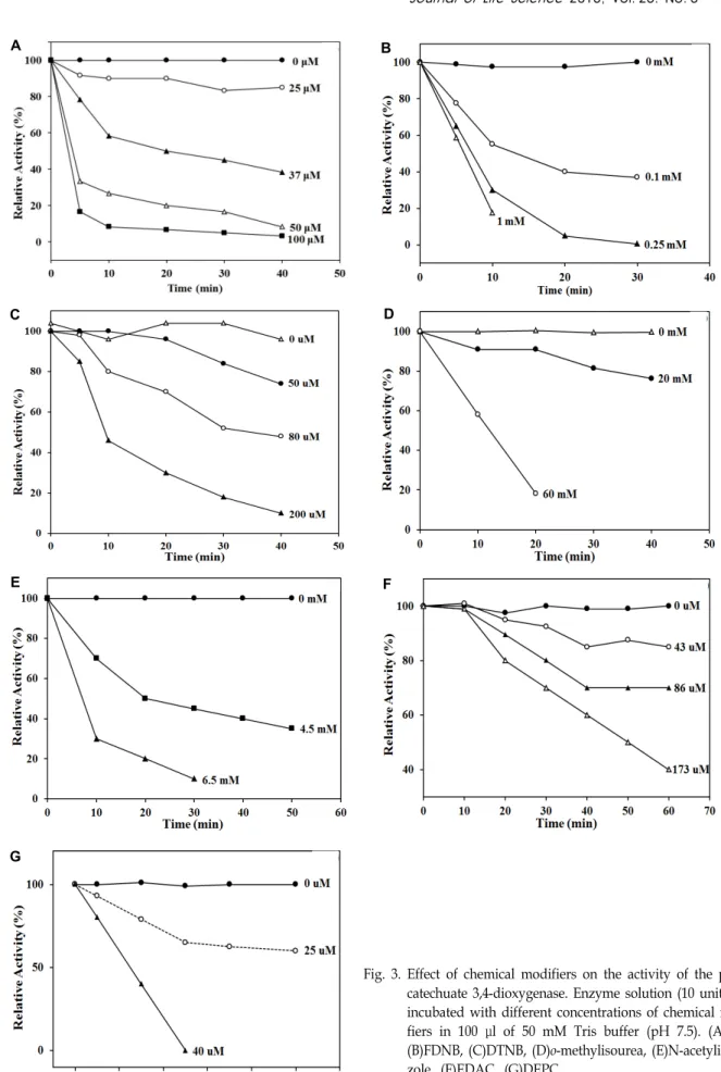
Fig. 3. Effect of chemical modifiers on the activity of the proto- catechuate 3,4-dioxygenase. Enzyme solution (10 unit) was incubated with different concentrations of chemical modi- fiers in 100 μl of 50 mM Tris buffer (pH 7.5). (A)PLP, (B)FDNB, (C)DTNB, (D)o-methylisourea, (E)N-acetylimida- zole, (F)EDAC, (G)DEPC
Fig. 4. Proposed active site map of the protocatechuate 3,4-dioxygenase. The data show that cysteine, ly- sine, histidine, carboxylate, and tyrosine are in- volved in binding or catalysis of protocatechuate 3,4-dioxygenase from Pseudomonas pseudoalcaligenes KF707.
the enzyme, the chemical modifiers: PLP and FDNB, DTNB and o-methylisourea, ninhydrin and 2,3-butanedione, N-ace- tylimidazole, 1-ethyl-3-(3-dimethylaminoptopyl)-carbodiimide (EDAC), diethylpyrocarbonate, and iodoacetamide were used to modify the lysine, cysteine, arginine, tyrosine, car- boxylate, histidine, and methionine side chains, respectively [20, 21]. PLP inactivated the enzyme in a biphasic pattern with 100% inactivation at > 50 μM. FDNB also inactivated the enzyme in a biphasic pattern with 100% inactivation at
> 250 μM. These data strongly suggest that a lysine residue is involved in binding and/or catalysis. DTNB and o-methyl- isourea inactivated the enzyme in a linear pattern with 100%
inactivation at > 0.2 mM and 60 mM, respectively. This result suggests that a cysteine residue plays a role as a binding or catalytic amino acid. However, ninhydrin and 2,3-butane- dione did not inhibit enzyme activity, indicating that the ar- ginine residue is absent at the active site. N-acetylimidazole inactivated the enzyme in a biphasic pattern with 100% in- activation at > 6.5 mM, suggesting that a tyrosine residue is present at the active site. Chemical modification by EDAC resulted in a linear time-dependent inactivation pattern, in- dicating that carboxylate may be involved in the binding or catalytic step. Diethylpyrocarbonate inactivated the en- zyme in a linear pattern with 100% inactivation at > 40 μM, suggesting that a histidine residue is present at the active site. However, the enzyme was not inactivated by iodoaceta- mide, indicating that methionine is not a part of the active site. All data are shown in Fig. 3. Ohlendorf et al. [25] re- ported that tyrosine and histidine side chains exist at the active site by X-ray crystallography. Thus, tyrosine and histi- dine residues are present in both enzymes. Taken together, the chemical modification data show that the side chains of cysteine, tyrosine, histidine, carboxylate, and lysine exist at the active site of protocatechuate 3,4-dioxygenase from Pseudomonas pseudoalcaligenes KF707.
Chemical mechanism
The pH profiles of the kinetic parameters show that at least two amino acid groups with pK
rvalues of 6.2 and 9.4 for catalysis and at least two amino acid groups with pK
rvalues of 5.5 and 9.0 for binding exist at the active site. The chemical modification data show that the side chains of ty- rosine, histidine, cysteine, and lysine are at the proto- catechuate 3,4-diooygenase active site. The combination of data from the pH study and chemical modification suggest that cysteine (pK
r6.2) and tyrosine (pK
r9.4) serve as catalytic
groups, and that histidine (pK
r5.5) and lysine (pK
r9.0) serve as binding groups. The possible reason for the lower pK val- ues for each side chain is that deprotonation may occur much faster in the hydrophobic condition. For example, Lim et al. [18] reported that the pK
rof the active site cysteine of mouse methionine sulfoxide reductase is 7.2 even in the absence of substrate. However, carboxylate was not shown up in the pH titration. It is carefully assumed that too low a carboxylate pK
rvalue could not be reflected in the tested pH range. Protocatechuate 3,4-dioxygenases isolated from Acinetobacter calcoaceticus ADP1, Brevibacterium fuscum [31], and Pseudomonas putida [32] are the best characterized structurally. Many X-ray crystallographic structures are available for protocatechuate 3,4-dioxygenases from Pseudo- monas putida and Acinetobacter calcoaceticus ADP1 [9, 11, 25- 27, 33]. The data show that the geometry of the high-spin ferric center is in a trigonal bipyramidal arrangement around the metal with histidine and tyrosinate as axial ligands and histidine, tyrosinate, and solvent-derived OH groups as equatorial ligands [25]. X-ray absorption spectroscopy of 3,4-PCD from Brevibacterium fuscum and Pseudomonas putida reveal that the water-based ligand is a hydroxide and thus charge neutrality of the active site was obtained [31, 7].
However, these studies show that histidine and tyrosine res-
idues are involved in binding and catalysis and not cysteine
and lysine. Based on the finding that cysteine, lysine, histi-
dine, carboxylate, and tyrosine are involved in binding or
catalysis of protocatechuate 3,4-dioxygenase from Pseudomonas
pseudoalcaligenes KF707, we propose the possible active site
map and chemical mechanism as shown in Fig. 4 and 5.
Fig. 5. Proposed chemical mechanism of the protocatechuate 3,4-dioxygenase. Catalysis occurs by binding of Fe3+ to the imidazole ring of histidine and carboxylate oxygen (Oδ–), and subsequent binding of O2 to Fe3+, and the carboxylate of proto- catechuate to the lysine residue.
Catalysis occurs by binding of Fe
3+to the imidazole ring of histidine and carboxylate oxygen (Oδ
–), and subsequent binding of O
2to Fe
3+, and the carboxylate of protocatechuate to the lysine residue. Then, the deprotonated sulfhydryl at- tacks C
3of protocatechuate with subsequent cleavage of the C
3–C
4bond. C
4obtains an electron from C
3and becomes C
4–by subsequent cleavage of the C
3–C
4bond. A sulfur electron attacks the hydroxyl proton of tyrosine and the C
3– S bond is broken with C
3+remaining. Oxygen dissociates into negative and positive oxygen elements and binds to C
3+and C
4–, respectively, with two resulting carboxylates. In contrast, the protonated sulfhydryl is attacked by a de- protonated hydroxyl of tyrosine and returns to its original state for another catalysis reaction.
Acknowledgment
This research was financially supported by Changwon National University in 2013-2015.
References
1. Ashie, I. N. A., Simpson, B. K. and Smith, J. P. 1996.
Mechanisms for controlling enzymatic reactions in foods.
Crit. Rev. Food Sci. Nutr. 36, 1-30.
2. Bedrosian, K., Nelson, A. I. and Seinberg, M. P. 1959. Effect of borates and other inhibitors on enzymatic browning in apple tissues. Food Technol. 13, 722-726.
3. Bradford, M. 1976. A rapid and sensitive method for the
quantitation of microgram quantities of protein utilizing the principle of protein-dye binding. Anal. Biochem. 72, 248-254.
4. Cho, Y. K. and Cook, P. F. 1988. Inactivation of py- rophosphate-dependent phosphofructokinase from Propioni- bacterium freudenreichii by pyridoxal 5’-phosphate. J. Biol.
Chem. 263, 5135-5140.
5. Cleland, W. W. 1979. Statistical analysis of enzyme kinetic data. Meth. Enzymol. 63, 103-138.
6. Dagleys, S. 1984. Microbial degradation of aromatic compounds. Devel. Ind. Microbiol. 25, 53-65.
7. Davis, M. L., Wasinger, E. C., Westre, T. E., Zaleski, J. M., Orville, A. M., Lipscomb, J. D., Hedman, B., Hodgson, K.
O. and Solomon, E. I. 1999. Spectroscopic investigation of reduced protocatechuate 3,4-dioxygenase: charge-induced alterations in the active site iron coordination environment.
Inorg. Chem. 38, 3676-3683.
8. Durham, D. R., Stirling, L. A., Ornston, L. N. and Perry, J. J. 1980. Intergeneric evolutionary homology revealed by the study of protocatechuate 3,4-dioxygenase from Azoto- bacter vinelandii. Biochemistry 19, 149-155.
9. Elgren, T. E., Orville, A. M., Kelly, K. A., Lipscomb, J. D., Ohlendorf, D. H. and Que, L. Jr. 1997. Crystal structure and resonance raman studies of protocatechuate 3,4-dioxygenase complexed with 3,4-dihydroxyphenylacetate. Biochemistry 36, 11504-11513.
10. Engel, P. C. 1996. Enzymology LabFax. pp. 175-190, Academic Press: San Diego, CA, USA.
11. Frazee, R. W., Orville, A. M., Dolbeare, K. B., Yu, H., Ohlendorf, D. H. and Lipscomb, J. D. 1998. The axial ty- rosinate Fe3+ ligand in protocatechuate 3,4-dioxygenase in- fluences substrate binding and product release: evidence for new reaction cycle intermediates. Biochemistry 37, 2131-2144.
12. Friedman, M. 1996. Food browning and its prevention: an
overview. J. Agric. Food Chem. 44, 631-653.
13. Fujisawa, H., Hiromi, K., Uyeda, M., Okuno, S., Nozaki, M.
and Hayaishi, O. 1972. Protocatechuate 3,4 dioxygenase III.
An oxygenated form of the enzyme as reaction intermediate.
J. Biol. Chem. 247, 4422-4428.
14. Joslyn, M. A. and Ponting, J. D. 1951. Enzyme-catalyzed oxi- dative browning of fruit products. Adv. Food Res. 3, 1-44.
15. Kahn, V. 1985. Effect of proteins, protein hydrolyzates and amino acids on o-dihydroxyphenolase activity of poly- phenol oxidase of mushroom, avocado, and banana. J. Food Sci. 50, 111-115.
16. Kelly, S. H. and Finkle, B. J. 1969. Action of a ring-cleaving oxygenase in preventing oxidase darkening of apple juice.
J. Sci. Food Agr. 20, 629-632.
17. Kurahashi, T., Oda, K., Sugimoto, M., Ogura, T. and Fujii, H. 2006. Trigonal-bipyramidal geometry induced by an ex- ternal water ligand in a sterically hindered iron salen com- plex, related to the active site of protocatechuate 3,4- dioxygenase. Inorg. Chem. 45, 7709-7721.
18. Lim, J. C., Gruschus, J. M., Kim, G., Berlett, B. S., Tjandra, N. and Levine, R. L. 2012. A low pKa cysteine at the active site of mouse methionine sulfoxide reductase A. J. Biol.
Chem. 287, 25596-601.
19. Lukes, B., O’brien, T. J. and Scanlan, R. A. 1980. Residual sulfur dioxide in finished malt: Colorimetric determination and relation to N-nitrosodimethylamine. Am. Soc. Brew.
Chem. J. 38, 146-148.
20. Lundblad, R. L. and Noyes, C. M. 1984a. Chemical reagents for protein modification. Vol I, pp. 1-166, CRC Press: Boca Raton, FL, USA.
21. Lundblad, R. L. and Noyes, C. M. 1984b. Chemical reagents for protein modification. Vol II, pp. 1-169, CRC Press: Boca Raton, FL, USA.
22. Mapson, L. W. and Tomalin, A. W. 1961. Preservation of peeled potato. Ⅲ. The inactivation of phenolase by heat. J.
Sci. Food Agr. 12, 54-58.
23. Martinez, M. V. and Whitaker, J. R. 1995. The biochemistry and control of enzymatic browning. Trends Food Sci. Technol.
6, 195-200.
24. Montgomery, M. W. 1983. Cysteine as an inhibitor of
browning in pear juice concentrate. J. Food Sci. 48, 951-952.
25. Ohlendorf, D. H., Orville, A. M. and Lipscomb, J. D. 1994.
Structure of protocatechuate 3,4-dioxygenase from Pseudo- monas aeruginosa at 2.15 Å resolution. J. Mol. Biol. 244, 586-608.
26. Orville, A. M., Lipscomb, J. D. and Ohlendorf, D. H. 1997a.
Crystal structures of substrate and substrate analog com- plexes of protocatechuate 3,4-dioxygenase: endogenous Fe3+
ligand displacement in response to substrate binding.
Biochemistry 36, 10052-10066.
27. Orville, A. M., Elango, N. E., Lipscomb, J. D. and Ohlendorf, D. H. 1997b. Structures of competitive inhibitor complexes of protocatechuate 3,4-dioxygenase: multiple exogenous li- gand binding orientations within the active site. Biochemistry 36, 10039-10051.
28. Ponting, J. D., Jackson, R. and Watters, G. 1971. Refrigerated apple slices. Effects of pH, sulfites and calcium on texture.
J. Food Sci. 36, 349-350.
29. Price, N. C. and Stevens, L. 1998. Fundamentals of Enzymology. Oxford University Press: New York, NY, USA.
30. Sim, H. W., Jung, M. J. and Cho, Y. K. 2013. Purification and Characterization of Protocatechuate 3,4-dioxygenase from Pseudomonas pseudoalcaligenes KF707. J. Kor. Soc. App.
Biol. Chem. 56, 401-408.
31. True, A. E., Orville, A. M., Pearce, L. L., Lipscomb, J. D.
and Que, L. Jr. 1990. An EXAFS study of the interaction of substrate with the ferric active site of protocatechuate 3,4-dioxygenase. Biochemistry 29, 10847-10854.
32. Valley, M. P., Brown, C. K., Burk, D. L., Vetting, M. W., Ohlendorf, D. H. and Lipscomb, J. D. 2005. Roles of the equatorial tyrosyl iron ligand of protocatechuate 3,4-dioxy- genase in catalysis. Biochemistry 44, 11024-11039.
33. Vetting, M. W., D’argenio, D. A., Ornston, L. N. and Ohlen- dorf, D. H. 2000. Structure of acinetobacter strain adp1 proto- catechuate 3,4-dioxygenase at 2.2 Å resolution: implications for the mechanism of an intradiol dioxygenase. Biochemistry 39, 7943-7955.
34. Whitaker, J. R. 1995. Polyphenol oxidase, pp. 271-307. In:
Wong, D. W. S. (ed), Food enzymes structure and mechanism.
Chapman & Hall: New York, NY, USA.
초록: Pseudomonas pseudoalcaligenes KF707에서 유래한 protocatechuate 3,4-dioxygenase 의 저해 및 화학적 메커니즘
강태경
†․김상호
†․정미자․조용권*
(창원대학교 자연과학대학 보건의과학과)