Evidence for Polyphyletic Origin of the Members of the Subsection IV Cyanobacteria as Determined by 16S rRNA Analysis
Yong Kook Shin
1and Pil-Soo Seo
2*
1School of Integrated Oriental Medical Bioscience, Semyung University, 65 Semyung-ro, Jecheon, Chungbuk 27136, Korea
2Environment-Friendly Agriculture Division, Sunchang Agricultural Development & Technology Center, 1548 Damsun-ro, Yudeung-myeon, Sunchang-gun, Jeonbuk 56025, Korea
Received September 13, 2016 /Revised October 7, 2016 /Accepted October 15, 2016
Unicellular cyanobacterial strains of Subsections I and II and filamentous cyanobacterial strains of Subsection III have been shown to be polyphyletic, heterocystous strains of Subsections IV and V, both of which were previously reported to be monophyletic. In this study, the small subunit riboso- mal RNA (16S rRNA) sequences of 13 strains of cyanobacteria - one strain, Oscillatoria nigro-viridis PCC7112, of the Subsection III, 6 strains including genus Anabaena, Nostoc, Tolypothrix, Calothrix and Scytonema of the Subsection IV, and 6 strains including genus Hapalosiphon, Fischerella and Chlorogloeopsis of the Subsection V - were determined. The phylogenetic analysis of cyanobacteria was carried out using the 16S rRNA sequences. The results of the phylogenetic analyses of 16S rRNA sequences, based on Neighbour-joining, maximum-parsimony, and maximum-likelihood methods, indicated that the members of Subsection IV were not monophyletic but polyphyletic. In addition, the phylogenetic re- sults strongly indicated that the genus Scytonema in Subsection IV could be a common ancestor of het- erocystous cyanobacteria in Subsection IV and V. Furthermore, the phylogenetic analyses revealed that the genus Anabaena could be phylogenetically diverse and that cyanobacterial strains in Subsection IV might be polyphyletic, whereas those in Subsection V could be monophyletic, as reported before. The results for the genus Anabaena indicate that it should be reclassified.
Key words : Anabaena, cyanobacteria, phylogenetic analyses, Scytonema, small subunit rRNA
*Corresponding author
*Tel : +82-63-650-5143, Fax : +82-63-650-5129
*E-mail : [email protected]
This is an Open-Access article distributed under the terms of the Creative Commons Attribution Non-Commercial License (http://creativecommons.org/licenses/by-nc/3.0) which permits unrestricted non-commercial use, distribution, and reproduction in any medium, provided the original work is properly cited.
Journal of Life Science 2016 Vol. 26. No. 10. 1202~1206 DOI : http://dx.doi.org/10.5352/JLS.2016.26.10.1202
Introduction
The cyanobacteria are morphologically diverse bacteria that perform oxygenic photosynthesis and possess chlor- ophyll a. Cyanobacteria have also been recognized as a mod- el system for studying the photosynthesis and endosym- biotic origins of chloroplasts in plants [1]. At present, cyano- bacteria are classified into five sections and formally recog- nized primarily according to their morphology and devel- opmental characteristics [2-4, 15-17]. Unicellular cyanobac- teria are classified into the Subsection I and II in terms of their different division patterns. The division pattern of Subsection I show a binary fission or budding, but that of the Subsection II is expressed by multiple fission which re- sults in the formation of baeocytes [16]. The Subsection III
is composed of filamentous cyanobacteria reproducing by trichome breakage [16]. The Subsection IV and V comprise the filamentous and heterocytous cyanobacteria reproducing by hormogonia formation [16], and are able to develop heter- ocysts and akinetes. These two sections are separated on more one plane. Members of the Subsection IV are divided on only one plane, and members of the Subsection V can be divided on more than one plane [16]. The Subsection IV and V are composed of eight genera (Anabaena, Calothrix, Cylindropermum, Nostoc, Nodularia, Scytonema, Chlorogloeopsis, and Fischerella) that are common to most classification sys- tem [2-4, 9, 16], as well as additional genera (Tolypothrix and Hapalosiphon) in more recent classification systems [2-4].
Cyanobacterial classifications based on morphological char- acteristics remain controversial and may not reflect true phy- logenetic relationships [5, 8, 9, 11, 18, 22, 26].
Recently, phylogenetic analyses of cyanobacteria based on small subunit rRNA (16S rRNA) have been performed.
Several studies based on 16S rRNA have reported that in
contrast to unicellular and filamentous non-heterocytous cy-
anobacterial strains composed of Subsection I, II, and III, that
do not form clusters to be consistent with their classification,
- Note -
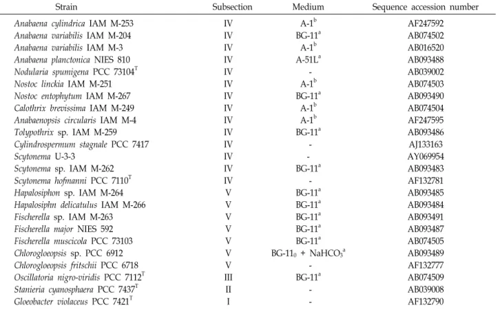
Table 1. Cyanobacterial strains used in this study
Strain Subsection Medium Sequence accession number
Anabaena cylindrica IAM M-253 Anabaena variabilis IAM M-204 Anabaena variabilis IAM M-3 Anabaena planctonica NIES 810 Nodularia spumigena PCC 73104T Nostoc linckia IAM M-251 Nostoc entophytum IAM M-267 Calothrix brevissima IAM M-249 Anabaenopsis circularis IAM M-4 Tolypothrix sp. IAM M-259 Cylindrospermum stagnale PCC 7417 Scytonema U-3-3
Scytonema sp. IAM M-262 Scytonema hofmanni PCC 7110T Hapalosiphon sp. IAM M-264 Hapalosiphn delicatulus IAM M-266 Fischerella sp. IAM M-263 Fischerella major NIES 592 Fischerella muscicola PCC 73103 Chlorogloeopsis sp. PCC 6912 Chlorogloeopsis fritschii PCC 6718 Oscillatoria nigro-viridis PCC 7112T Stanieria cyanosphaera PCC 7437T Gloeobacter violaceus PCC 7421T
IV IV IV IV IV IV IV IV IV IV IV IV IV IV V V V V V V V III II I
A-1b BG-11a
A-1b A-51La
- A-1b BG-11a
A-1b A-1b BG-11a
- - BG-11a
- BG-11a BG-11a BG-11a BG-11a BG-11a BG-110 + NaHCO3a
- BG-11a
- -
AF247592 AB074502 AB016520 AB093488 AB039002 AB074503 AB093490 AB074504 AF247595 AB093486 AJ133163 AY069954 AB093483 AF132781 AB093485 AB093484 AB093491 AB093487 AB074505 AB093489 AF132777 AB074509 AB039008 AF132790 Sequences in bold were determined in this study. T: type strain.
aRippka and Herdman, 1992 [6]; b IAM Catalogue of strains, 1998 [26].
the heterocytous members of cyanobacterial Subsection IV and V are monophyletic [8, 11, 21, 23, 24].
In this study, we investigated the phylogenetic relation- ships of the cyanobacterial strains of the Subsection IV and V based on 16S rRNA and compared the phylogenetic posi- tion of cyanobacterial strain with each of them.
Materials and Methods
Cyanobacterial strains and growth conditions The cyanobacterial strains investigated and media are list- ed in Table 1. All were cultured at 20℃, 12:12 hr L: D (light : dark) with fluorescent lamp illumination of 500 lux.
The preparation of cell lysate
Cells were harvested from 1.5 ml culture broth by cen- trifugation, washed with 1 ml of deionized water, and sus- pended in 100 μl of 20 mM Tris (pH 8.0) containing 0.1 mM EDTA, 0.5% Tween 20, and 0.1% Non-iodet P-40 (Boehringer Mannheim, Germany). Final lysis was achieved by the addi- tion of 10 μg of Proteinase K and incubation for 20 min at
60℃.
PCR amplification
The almost complete 16S rDNA from the genomic DNA of the respective strains was amplified by PCR using oligo- nucleotide primers of 1R (forward primer: 5’-AGAGTTTGA TCCTGGCTCAG-3’) and 16C (reverse primer: 5’-AAGGAG GTGATCCAGCCGCA-3’) [27]. PCR was performed at 3 min at 94℃ and then at 30 cycles with the following features:
1 min at 94℃, 1 min at 55℃, and 2 min at 72℃, followed by a final elongation step for 10 min at 72℃.
Sequencing of 16S rRNA
The refined PCR products were directly sequenced with
the use of a BigDye Terminator Cycle Sequencing Ready
Reaction Kit (Applied Biosystems, USA). Primers used for
the cycle sequencing were 1R, 320R (5’-CTGCTGCCTCCC
GATA-3’), 520F (5’-CAGCAGCCGCGGTAATAC-3’), 704R
(5’-TCTACGCATTTCACCGCTAC-3’), 926F (5’-AAACTCAA
AGGAATTGACGG-3’), 1100R (5’-GGGTTGCGCTVGTTG-
3’) (V, G or C or A), and 16C. The cycle sequencing reaction
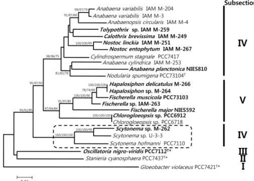
Fig. 1. Phylogenetic tree of cyanobacterial 16S rRNA sequences. Numbers at nodes are bootstrap values. NJ, ML, and MP bootstrap values are indicated as NJ/ML/MP. A total of 1335 unambiguously aligned positions were used. Local bootstrap probabilities are indicated at nodes if larger than 60. The strains of which the 16S rRNA gene sequences were determined in this study are indicated in bold. Starred strains were used as outgroups. T: type strain. Bar, 0.01 substitution per nucleotide position.
was performed with a PCR Thermal Cycler
MP(Takara, TP3080, Japan). The sequencing reaction was performed for 5 min at 96℃ at first and consisted of 25 cycles of the follow- ing: 10 sec at 96℃, 10 sec at 52℃, and 2 min at 60℃. The cycle sequencing products were purified with use of CENTRI-SEP Spin Columns (Applied Biosystems, USA).
DNA sequences were analyzed using an ABI PRISM 310 Genetic Analyzer (Applied Biosystems, USA).
Alignment and phylogenetic analysis
The nucleotide sequences of 16S rRNA were aligned using the CLUSTAL W computer program, version 1.81 [20]. 16S rRNA sequences were aligned based on their secondary structures with a selection of cyanobacterial reference se- quences obtained from the DNA Data Bank of Japan (DDBJ), and the alignment was manually corrected.
Neighbour-Joining (NJ)
The phylogenetic tree was constructed from the evolu- tionary distance matrix calculated by the neighbour-joining method [19] with Kimura`s two-parameter method [12].
Neighbour-joining analysis was performed using the MEGA2 program [13]. All gaps in the alignment were ex- cluded in order to draw the tree. Bootstrap analyses were
performed with 1,000 replicates.
Maximum-likelihood (ML) and Maximum-parsimony (MP)
A total of 100 bootstrap samples for alignment was pro- duced by using the program SEQBOOT from PHYLIP ver- sion 3.6 [6, 7], and phylogenetic trees were inferred from each bootstrap sample by using maximum-likelihood (DNAMLK software in PHYLIP version 3.6) and maximum- parsimony (DNAPARS software in PHYLIP). The resulting trees were combined to yield a consensus tree (CONSENSE software in PHYLIP). A matrix of evolutionary distances was also derived from bootstrap alignment by using the DNADIST software in PHYLIP. Trees were inferred from the matrices by using the FITCH software in PHYLIP. The resulting trees were visualized to yield consensus trees (CONSENSE software in PHYLIP) using the TREEVIEW ver- sion 1.6.6 software [14].
Results and Discussion
The new 16S rDNA sequences were deposited in the
DDBJ; accession numbers for each cyanobacterial strain used
in this study and reference strains are given in Table 1.
Positions with gaps and undetermined and ambiguous se- quences were removed. A total of 1335 sites were used for the phylogenetic analysis.
Constructed phylogenetic trees, the NJ, the ML, and MP trees, reveal that all the numbers of cyanobacterial Subsec- tion V were monophyletic (Fig. 1). However, as shown in Figure 1, the cyanobacterial strains of Subsection IV are clearly separated into two clusters. All members of cyano- bacterial Subsection IV, except the genus Scytonema, consist of a major cluster on phylogentic trees, supported by boot- strap values of 99% of NJ, 95% of ML, and 75% of MP (Fig.
1). The cluster of the genus Scytonema is placed externally to those of Subsections IV and V, forming a well-supported cluster with bootstrap values of 95% of NJ, 87% of ML, and 88% of MP (Fig. 1). In some reports published earlier, al- though unicellular cyanobacterial strains of Subsections I and II, and filamentous cyanobacterial strains of Subsection III were shown to be polyphyletic, heterocystous strains of Subsections IV and V were shown to be monophyletic [8, 10, 11, 21, 23, 24]. However, interestingly, our phylogenetic analyses based on sequence determination of the 16S rDNA indicate Subsection IV was not monophyletic.
Our phylogenetic analyses indicate that the genus Scytone- ma could be a common ancestor of cyanobacterial Subsec- tions IV and V. The results reveal that the divergence of the genus Scytonema is earlier than any other genus of Subsections IV and V. The results of analyses based on NJ, ML, and MP strongly support this (Fig. 1). Actually, in Bergey’s Manual of Systematic Bacteriology, the Scytonema hofmanni PCC 7110 was described as the following: “Scytone- ma hofmanni PCC 7110 has no close relatives in the phyloge- netic trees with the recent addition of new sequences, how- ever, some interesting relationships are emerging” [25]. Our results may constitute evidence to help explain the evolu- tional relationships of cyanobacterial Subsection IV and V.
References
1. Bryant, D. A. 1994. The molecular biology of cyanobacteria.
pp. 881. Kluwer Academic: Dordrecht, The Netherlands.
2. Castenholz, R. 1992. Species usage, concept, and evolution in cyanobacteria (blue-green algae). J. Phycol. 28, 737-745.
3. Castenholz, R. 2001. Phylum BX. Cyanobacteria oxygenic photosynthetic bacteria, Vol. 3, pp. 473-599. In: Boone, D.
and Castenholz, R. (eds.), Bergey`s Manual of Systematic Bacteriology 2nd edn. Springer-Verlag: New York, USA.
4. Castenholz, R. and Waterbury, J. 1989. Group I cyanobac- teria, Vol. 3. pp. 1710-1728. In:Krieg, N. R. and Holt, J. G.
(eds), Bergey`s Manual of Systematic Bacteriology. Williams and Wilkins: Baltimore, USA.
5. Douglas, S. E. and Turner, S. 1991. Molecular evidence for the origin of plastids from a cyanobacterium-like ancestor.
J. Mol. Evol. 33, 267-273.
6. Felsenstein, J. 1989. PHYLIP-Phylogeny inference package (version 3.2). Cladisticts 5, 164-166.
7. Felsenstein, J. and Churchill, G. A. 1996. A hidden Markov model approach to variation among sites in rate of evolution. Mol. Biol. Evol. 13, 93-104.
8. Giovannoni, S., Turner, S., Olsen, G. J., Barns, S., Lane, D.
J. and Pace, N. R. 1988. Evolutionary relationships among cyanobacteria and green chloroplasts. J. Bacteriol. 170, 3584- 3592.
9. Henson, B. J., Watson, L. E. and Barnum, S. R. 2002.
Molecular differentiation of hetero cyanobacteria, Nostoc and Anabaena, based on complete nifD sequences. Curr.
Microbiol. 45, 161-164.
10. IAM Catalogue of Strains. 1998. pp. 291-294. Editorial Board of IAM Catalogue of Strains. (eds.), Center for Cellular and Molecular Research. Institute of Molecular and Cellular Biosciences, The University of Tokyo.
11. Ishida, T., Watanabe, M. M., Sugiyama, J. and Yokota, A.
2001. Evidence for polyphyletic origin of the members of the orders of Oscillatoriales and Pleurocapsales as de- termined by 16S rDNA analysis. FEMS Microbiol. Lett. 201, 79-82.
12. Kimura, M. 1980. A simple method for estimating evolu- tionary rate of base substitution through comparative stud- ies of nucleotide sequence. J. Mol. Evol. 16, 111-120.
13. Kumar, S., Tamura, K., Jacobsen, I. B. and Nei, M. 2001.
Mega2: molecular evolutionary genetics analysis software.
Bioinformatics 17, 1244-1245.
14. Page, R. D. M. 1996. TREEVIEW: an application to display phylogenetic trees on personal computers. Comput. Appl.
Biosci. 12, 357-358.
15. Rippka, R. 1988. Recognition and identification of cyanobac- teria. Methods Enzymol. 167, 28-67.
16. Rippka, R., De Reuelles, J., Waterbury, J., Herdman, H. and Stanier, R. 1979. Generic assignments, strain histories, and properties of pure cultures of cyanobacteria. J. Gen. Microbiol.
111, 1-61.
17. Rippka, R. and Herdman, H. 1992. Pasteur culture collection of cyanobacteria catalogue and taxonomic handbook I.
Catalogue of strains. Institute Pasteur: Paris, France.
18. Robertson, B. R., Tezuka, N., and Watanabe, M. M. 2001.
Phylogenetic analyses of Synechococcus strains (cyanobacter- ia) using sequences of 16S rDNA and part of the phycocya- nin operon reveal multiple evolutionary lines and reflect phycobilin content. Int. J. Syst. Bacteriol. 51, 861-871.
19. Saitou, N. and Nei, M. 1987. The neighbor-joining method:
A new method for reconstructing phylogenetic tree. Mol.
Biol. Evol. 4, 406-425.
20. Thompson, J. D., Higgins, D. G. and Gibson, T. J. 1994.
CLUSTAL W: Improving the sensitivity of progressive mul- tiple sequence alignment through sequence weighting, posi-
초록:16S rRNA 분석에 의한 Subsection IV cyanobacteria 균주들의 다계통성 기원의 증거
신용국
1․서필수
2*
(1세명대학교 한방바이오융합과학부, 2순창농업기술센터 친환경농업과)
Subsection I과 II의 시아노박테리아 균주들은 단세포성이며, Subsection III의 시아노박테리아 균주들은 섬유상 의 다계통성, 이형 사이토시스 형성성 균주들인 반면, Subsections IV와 V는 단일계통성으로 보고되어있다. 본 연 구에서 13 균주의 시아노박테리아의 the small subunit rRNA (16S rRNA) 염기서열들이 - Subsection III의 Oscillatoria nigro-viridis PCC7112, Subsection IV에 속하는 Anabaena, Nostoc, Tolypothrix, Calothrix 및 Scytonema 속을 포함한 6 균주, Subsection V에 속하는 Hapalosiphon, Fischerella and Chlorogloeopsis 속의 6 균주 - 결정되었 다. 결정된 16S rRNA 염기서열을 이용하여 시아노박테리아의 분자계통분석을 수행하였다. 그러나, 16S rRNA의 염기서열 결정을 근거로 한 본 연구의 계통분석결과 Subsection IV는 단일 계통성이 아닌 다계통성이며, 반면 Subsection V는 이전에 보고되어진 것처럼 단일 계통성임을 나타내었다. 또한, 본 연구 결과는 Scytonema속이 이 형 사이토시스 형성성 시아노박테리아인 Subsection IV 및 V의 공통 조상일 수 있음을 강력하게 나타낸다. 부가적 으로, 본 연구의 분자계통 분석을 통해 Anabaena속은 다계통성으로 계통학적으로 다양한 종들로 구성되어 있음을 나타내고 있다. 본 연구 결과는 Anabaena속이 좀 더 세밀하게 재분류 되어져야 함을 나타낸다.
tion-specific gap penalties and weight matrix choice. Nucleic Acids Res. 22, 4673-4680.
21. Turner, S. 1997. Molecural systematics of oxygenic photo- synthetic bacteria. Plant Syst. Evol. 11, 13-52.
22. Turner, S., Burger-Wiersma, T., Giovannoni, S., Mur, L. R.
and Pace, N. R. 1989. The relationship of a prochlorophyte Prochlorothrix hollandica to green chloroplasts. Nature 337, 380-382.
23. Turner, S., Pryer, K. M., Miao, V. P. W. and Palmer, J. D.
1999. Investigating deep phylogenetic relationships among cyanobacteria and plastids by small subunit rRNA sequence analysis. J. Eukaryot. Microbiol. 46, 327-338.
24. Wilmote, A. 1994. Molecural evolution and taxonomy of cy- anobacteria, pp. 1-25. In: Bryant, D. (eds.), The molecural biology of cyanobacteria. Kluwer Academic Publisher, The Netherlands.
25. Wilmotte, A. and Herdman, M. 2001. Phylogenetic relation- ships among the cyanobacteria based on 16S rRNA se- quences. The Archaea and deeply Braching and Phototropic Bacteria, Vol. 1, pp. 487-493. In: Boone, D. R., Castenholz, R. W. and Garrity, G. M. (eds.), Bergey`s Manual of Syste- matic Bacteriology, 2nd edn, Springler-Verlag, New York.
26. Wilmotte, A., Turner, S., Van de Peer, Y. and Pace, N. R.
1992. Taxonomic study of marine Oscillatoriacean strains (cyanobacteria) with narrow trichomes. II. Nucleotide se- quence analysis of the 16S ribosomal RNA. J. Phycol. 28, 828-838.
27. Wilmote A., Van der Auwera, G. and De Watcher, R. 1993.
Structure of 16S ribosomal RNA of the thermophilic cyano- bacterium Chlorogloepsis HTF (`Mastigocladus laminosus HTF`) strains PCC 7518, and phylogenetic analysis. FEBS Lett. 371, 96-100.