In vivo Micronucleus Test of Cyclohexanone and Mutagenicity Classification According to a Globally Harmonized System
Soo-Jin KIM, Kyung-Taek RIM* and Cheol-Hong LIM
Chemicals Safety and Health Center, Occupational Safety and Health Research Institute, Korea Occupational Safety and Health Agency, Daejeon 305-380, Korea
Received May 16, 2014 /Revised July 02, 2014 /Accepted July 22, 2014
A micronucleus test of cyclohexanone has not yet been conducted. To classify the chemical hazard posed by cyclohexanone according to a globally harmonized system of classification and labeling of chemicals (GHS), we investigated its mutagenicity by micronucleus induction in ICR bone marrow cells of 7-weeek-old male mice. The mice were administered three dosages of the chemical for 24 hr via the oral route. After 24 hr, the mice were sacrificed, and their bone marrow cells were prepared for smearing slides. Based on counts of micronucleated polychromatic erythrocytes (MNPCEs) of 2,000 polychromatic erythrocytes, cyclohexanone did not inhibit bone marrow cell proliferation in any of the treated groups, but it resulted in micronucleus induction. According to the results of the mammalian bone marrow micronucleus test, this chemical is mutagenic and classified as category 2 in the GHS.
Key words : Cyclohexanone, GHS (globally harmonized system of classification and labeling of chemicals), micronucleus, mutagenicity
*Corresponding author
*Tel : +82-42-869-0345, Fax : +82-42-863-9001
*E-mail : [email protected]
This is an Open-Access article distributed under the terms of the Creative Commons Attribution Non-Commercial License (http://creativecommons.org/licenses/by-nc/3.0) which permits unrestricted non-commercial use, distribution, and reproduction in any medium, provided the original work is properly cited.
Journal of Life Science 2014 Vol. 24. No. 7. 804~811 DOI : http://dx.doi.org/10.5352/JLS.2014.24.7.804
Introduction
Chemical substances may have various hazardous effects on human health or the environment. Some of them may be carcinogenic, mutagenic or reproductive toxins, which are categorized as CMRs [1]. In view of the hazards they present, these classified substances and their mixtures are subject to restrictive regulations, particularly in the workplace. The ne- cessity for chemical hazard assessments has increased be- cause the number of workers exposed to chemicals has risen with the development of many industries, and it is necessary to determine what these substances are and how they are regulated [7].
Carcinogens are substances and preparations that, if in- haled, ingested, or penetrated into the skin, may induce can- cer or increase its incidence. Mutagens are substances that may induce heritable genetic defects or increase their incidence. They trigger permanent changes in the amount or structure of the genetic material in a cell, and these chem- icals, in accordance with the UN globally harmonized sys-
tem of the classification and labeling of chemicals (GHS), shall be considered mutagens for the purposes of this article [15].
To classify a chemical’s mutagenicity according to GHS, an in vivo micronucleus (MN) test was performed on mam- malian bone marrow cells treated with cyclohexanone (CAS No. 108-94-1), for which the definitive information is in- sufficient for its mutagenicity. Although many toxicological studies have been conducted other than the MN test, the available genotoxic data on cyclohexanone are still con- troversial with and without mammalian metabolic activation (S9). Therefore, it was necessary for further study, according to the good laboratory practice (GLP) guidelines to secure a quality assurance of the test [9]. The purpose of this MN test is to screen the cytogenetic damage that results in the formation of micronuclei containing lagging chromosome fragments or whole chromosomes. Micronuclei were first used to quantify chromosomal damage and are now recog- nized as one of the most successful and reliable assays for genotoxic carcinogens [12].
Cyclohexanone is used as a metal-degreasing solvent for
lacquers, resins, and insecticides [13]. It is also produced for
use as a raw material in the production of the precursors
to Nylon-6 and Nylon-6,6. Other important applications in-
clude its use as a cleaning agent and as a solvent in the
production of pesticides, magnetic tapes, and paint [8]. The
physicochemical and toxicological information regarding cy-
- Note -
Table 1. Physicochemical and toxicological information of cyclohexanone Chemical name Cyclohexanone
CAS No. 108-94-1
Synonyms
AnonCicloesanone Cyclohexyl ketone Hytrol O
Ketohexamethylene Nadone
Molecular formula
C6H10O
Molecular weight 98.14 Partition coefficient 0.81
Melting point -47℃ Boiling point 155℃
Forms Clear colorless liquid Water solubility 86 g/l at 20°C (68°F)
Stability and reactivity
OSHA Hazards: Combustible Liquid, Harmful by ingestion, Irritant Chemical stability: Stable under recommended storage conditions Conditions to avoid: Heat, flames and sparks
Materials to avoid: Oxidizing agents, Plastics
Hazardous decomposition products formed under fire conditions. - Carbon oxides
Toxicity
Target Organs: Liver, Kidney, Central nervous system, Lungs LD50 Oral - rat - 1,534 mg/kg
LC50 Inhalation - rat - 4 hr - > 6.2 mg/l LD50 Dermal - rabbit - 794 - 3,160 mg/kg Skin - rabbit - Irritating to skin.
Eyes - rabbit - Risk of serious damage to eyes. - 24 hr
Genotoxicity
in vitro
- Ames test -S. typhimurium
- with or without metabolic activation - negative Genotoxicityin vitro
- Human - fibroblast - with or without metabolic activation - Laboratory experiments have shown mutagenic effects.It is not classifiable as to its carcinogenicity based on its IARC, ACGIH, NTP, or EPA classification.
(IARC Group 3: Not classifiable as to its carcinogenicity to humans)
Overexposure may cause reproductive disorder (s) based on tests with laboratory animals.
GHS classification
Flammable liquids (Category 3) Acute toxicity, Oral (Category 4) Acute toxicity, Inhalation (Category 4) Acute toxicity, Dermal (Category 4) Skin irritation (Category 2) Serious eye damage (Category 1)
Sourced by searching in ChemIDplus Advanced, US National Library of Medicine (http://chem.sis.nlm.nih.gov/chemidplus/rn/
108-94-1). Rockville Pike, Bethesda, MD 20894, and Material Safety Data Sheet, Sigma-Aldrich (http://www.sigmaaldrich.com/MSDS/
MSDS/DisplayMSDSPage.do?country=US&language=en&productNumber=C102180&brand=SIAL&PageToGoToURL=
http%3A%2F%2Fwww.sigmaaldrich.com%2Fcatalog%2Fproduct%2Fsial%2Fc102180%3Flang%3Den), MO, USA. Searches were con- ducted using keywords chemical name AND/OR CAS number.
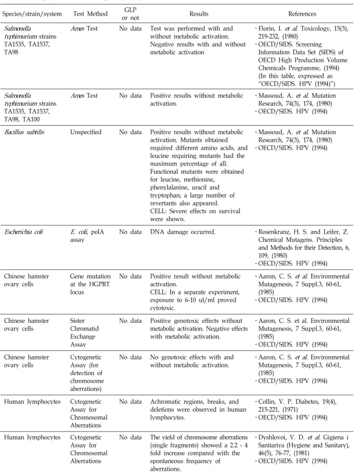
clohexanone is shown in Table 1. In addition, the major find- ings related to the mutagenicity of cyclohexanone are shown in Table 2.
Materials and Methods Chemicals and animal feeding conditions
In this in vivo MN test, cyclohexanone (Sigma-Aldrich,
99.8%, MO, USA, Cat. No. C8930) was used to test the
Table 2. Findings related to the mutagenicity of cyclohexanone Species/strain/system Test Method GLP
or not Results References
Salmonella
typhimurium
strains TA1535, TA1537, TA98Ames
Test No data Test was performed with and without metabolic activation.Negative results with and without metabolic activation
- Florin, I.
et al
. Toxicology, 15(3), 219-232, (1980)- OECD/SIDS. Screening Information Data Set (SIDS) of OECD High Production Volume Chemicals Programme, (1994) (In this table, expressed as
“OECD/SIDS. HPV (1994)”)
Salmonella
typhimurium
strains TA1535, TA1537, TA98, TA100Ames
Test No data Positive results without metabolicactivation. - Massoud, A.
et al
. Mutation Research, 74(3), 174, (1980) - OECD/SIDS. HPV (1994)Bacillus subtilis
Unspecified No data Positive results without metabolic activation. Mutants obtained required different amino acids, and leucine requiring mutants had the maximum percentage of all.Functional mutants were obtained for leucine, methionine,
phenylalanine, uracil and tryptophan; a large number of revertants also appeared.
CELL: Severe effects on survival were shown.
- Massoud, A.
et al
. Mutation Research, 74(3), 174, (1980) - OECD/SIDS. HPV (1994)Escherichia coli E. coli
, polAassay No data DNA damage occurred. - Rosenkranz, H. S. and Leifer, Z.
Chemical Mutagens. Principles and Methods for their Detection, 6, 109, (1980)
- OECD/SIDS. HPV (1994) Chinese hamster
ovary cells Gene mutation at the HGPRT locus
No data Positive result without metabolic activation.
CELL: In a separate experiment, exposure to 6-10 ul/ml proved cytotoxic.
- Aaron, C. S.
et al
. Environmental Mutagenesis, 7 Suppl.3, 60-61, (1985)- OECD/SIDS. HPV (1994)
Chinese hamster
ovary cells Sister
Chromatid Exchange Assay
No data Positive genotoxic effects without metabolic activation. Negative effects with metabolic activation.
- Aaron, C. S. et al. Environmental Mutagenesis, 7 Suppl.3, 60-61, (1985)
- OECD/SIDS. HPV (1994) Chinese hamster
ovary cells Cytogenetic Assay (for detection of chromosome aberrations)
No data No genotoxic effects with and
without metabolic activation. - Aaron, C. S.
et al
. Environmental Mutagenesis, 7 Suppl.3, 60-61, (1985)- OECD/SIDS. HPV (1994)
Human lymphocytes Cytogenetic Assay for Chromosomal Aberrations
No data Achromatic regions, breaks, and deletions were observed in human lymphocytes.
- Collin, V. P. Diabetes, 19(4), 215-221, (1971)
- OECD/SIDS. HPV (1994)
Human lymphocytes Cytogenetic Assay for Chromosomal Aberrations
No data The yield of chromosome aberrations (single fragments) showed a 2.2 - 4 fold increase compared with the spontaneous frequency of aberrations.
- Dyshlovoi, V. D.
et al.
Gigiena i Sanitariya (Hygiene and Sanitary), 46(5), 76-77, (1981)- OECD/SIDS. HPV (1994)
Table 2. Continued
Species/strain/system Test Method GLP
or not Results References
Human cells Unscheduled
DNA synthesis No data Negative results for genotoxicity with and without metabolic activation.
- National Toxicology Program.
Technical Report Series, (1983) - OECD/SIDS. HPV (1994) Human fibroblasts Unscheduled
DNA synthesis No data Negative results for genotoxicity with and without metabolic activation.
- Pevocco, P.
et al
. Toxicology Letters, 16(1-2), 69-76, (1983) - OECD/SIDS. HPV (1994)Drosophila
melanogaster
Sex-LinkedRecessive Lethal AssayNo data Negative effects of genotoxicity.Signs of toxicity were evident at 400 ppm.
- McGregor, D. B. National Technical Information Service (PB number), PB-83-127571, (1980) - OECD/SIDS. HPV (1994)
Drosophila
melanogaster
Phenocopies of tumor mutationsNo data A study of larvae reported the production of phenocopies of tumor mutations; however, other studies observed no effects.
- Goncharova, R. I. Tsitologiya i Genetika (Cytology and Genetics), 137-142, (1970)
- OECD/SIDS. HPV (1994)
Drosophila
melanogaster
Sex-LinkedRecessive Lethal AssayYes Negative result. Male sterility was only slightly increased over the controls.
- OECD/SIDS. HPV (1994)
Celltransformation No data Cyclohexanone did not cause behavior of cells to move closely resemble the cancerous state. (It is not clear if metabolic activation was used).
- Lijinsky, W. and Kovatch, R. M.
Journal of the National Cancer Institute (United States), 77(4), 941-949, (1986)
- OECD/SIDS. HPV (1994) L51784 th+/tk- mouse
lymphoma cells Forward
mutation assay No data No significant reductions in survival or increases in mutant fractions occurred at concentration up to 5000 ug/mL with and without metabolic activation.
- McGregor, D. B. et al.
Environmental and Molecular Mutagenesis, 12, 85-154, (1988) - OECD/SIDS. HPV (1994)
Dominant
Lethal Assay No data No effects on pregnancy frequency, numbers of corpora lutea and implantations, or the frequency of early deaths.
- McGregor, D. B. National Technical Information Service (PB number), PB83-127571, (1980) - OECD/SIDS. HPV (1994) Sperm
morphology No data Negative result. Abnormal sperm
frequency was not affected. - McGregor, D. B. National Technical Information Service (PB number), PB83-127571, (1980) - OECD/SIDS. HPV (1994)
In vivo
Cytogenetic Assay;
Chromosome Aberrations in Bone Marrow
No data Negative result. The frequency of chromosome aberrations were not increased.
- McGregor, D. B. National Technical Information Service (PB number), PB83-127571, (1980) - OECD/SIDS. HPV (1994)
Rat Cytogenetic
Assay
In vivo
; Chromosome Aberrations in Bone MarrowNo data Chromosome aberrations were induced at all doses and time intervals. Incidence of abnormalities increased with dose and decreased with time. They consisted of chromatid gaps and break, centric fusions, centrometric attenuation, chromatid exchanges and polyploidy.
- De Hondt, H. A. et al. Egyptian Journal of Genetics and Cytology, 12(1), 31-40, (1983)
- OECD/SIDS. HPV (1994)
Table 3. Animal body weight in micronucleus tests with oral exposure to cyclohexanone
Exposure method Concentration No. of animals Average body weight (mean ± SD)
Orally exposed to cyclohexanone for 24 hr
Negative control (Olive oil) 300 mg/kg b.w.a)
600 mg/kg b.w.
1,200 mg/kg b.w.
Positive control (MMC, 0.5 mg/kg b.w.)
6 6 6 6 6
37.30±1.72 g 37.16±1.76 g 37.07±1.74 g 37.28±1.58 g 37.16±1.49 g
a)b.w.: body weight
chemicals. Olive oil (Sigma-Aldrich, MO, USA, Cat. No.
C1514) was used as a solvent according to the results of the solubility test. The positive control used mitomycin C (MMC) (Sigma-Aldrich, MO, USA, Cat. No. M4287).
Animals and experimental design
The mouse (Mus musculus) bone marrow MN test was carried out according to the Organisation for Economic Co-operation and Development (OECD) test guideline (TG) 474 [9] as well as Hayashi [2] and Heddle [3]. Groups of specific pathogen-free male Institute for Cancer Research (ICR) mice were treated with the test substance at three dos- age levels, the highest dosage level being the estimated max- imum tolerated dose or the standard limit dose for the MN test, whichever is least. Concurrent negative and positive control groups were also treated. In this study, it was per- formed using 7-week-old male ICR mice at 300, 600, and 1,200 mg/kg dosages. At 24 hr post-treatment, with cyclo- hexanone administered orally, there were normally six male animals per group. An animal ethics committee approved the animal studies to ensure appropriate care before the ani- mals were obtained for research (Approval No.
IACUC-12-03).
Bone marrow preparation and MN test
Bone marrow cells were obtained from the femurs imme- diately following euthanization. Immature erythrocytes could be differentiated using a variety of staining techniques that rely on their relatively high content of residual DNA.
Mature erythrocytes with low nucleic acid content appeared pinkish-orange, while 5% Giemsa was used for mouse bone marrow, for peripheral blood, and for staining immature er- ythrocytes blue. Based on the cell cycles and maturation times of the erythrocytes, the bone marrow was harvested after 24 hr. The bone marrow was flushed from the femurs and spread onto slides. The slides were air-dried, fixed, and stained with a fluorescent DNA-specific stain that easily illu-
minates any micronuclei that may be present. The 2,000 pol- ychromatic erythrocytes (PCEs, reticulocytes; immature er- ythrocytes) were scored per animal regarding the frequency of micronucleated cells in each of the six animals per dosage group. In addition, the percentage of PCEs among the 500 erythrocytes in the bone marrow was recorded for each dos- age group as an indicator of chemically induced toxicity.
The presence of micronucleated PCEs was visually scored (at least 2,000 cells per mouse) by optical microscopy using a fluorescence microscope (Optiphot-2, Nikon, Tokyo, Japan) with a BA-2 filter. Cells were considered micronucleated when they neatly contained defined chromatin corpuscles with a diameter of less than one-third the diameter of the cell nucleus as well as when they were stained an equal or lighter shade than the nucleus of the cell from which the micronucleated cell was developed.
Statistical analysis
Data were presented as the mean number of micro- nucleated cells per 2,000 cells for each treatment group. The conclusion of the MN test was determined in consideration of the results of the statistical analyses. The experimental and control MN frequency for each specimen within and between the different mice strains were compared with a one-way ANOVA test and a Kruskal-Wallis test (α=0.05) us- ing SigmaStat v. 3.11.
Results and Discussion
There were no specific symptoms among the animals or-
ally exposed to cyclohexanone. The ranges of the body
weights of the animals exposed to this chemical was 35.33
to 39.02 g (Table 3). The preliminary tests were performed
to determine the maximum dosage. The inhibition of the
proliferation in the bone marrow cells was not observed in
this test. The frequencies of erythrocytes with MN in-
ductions were 0.18±0.03%, 0.21±0.09%, 0.22±0.05%, and 0.35
Table 4. Results of the main micronucleus test with cyclohexanone (for 24 hr)
Groups PCEa) observed MNPCEb)
observed MNPCE
frequency (%) (PCE+NCEc))
counted PCE
counted PCE/(PCE+
NCE) (%) Negative control
300 mg/kg b.w.d) 600 mg/kg b.w.
1,200 mg/kg b.w.
Positive control (MMC, 0.5 mg/kg b.w.)
2,022±10.81 2,029.67±10.80 2,022.83±17.52 2,017±12.59 2,018.33±14.99
3.67±0.52 4.17±1.94 4.33±1.03 7.00±1.41* 27.83±7.91
0.18±0.03 0.21±0.10 0.22±0.05 0.35±0.07* 1.38±0.40
517.83±18.63 514.17±9.75 510.67±7.23 511.00±12.79 513.33±16.94
269.00±74.40 267.00±47.72 254.50±30.04 278.00±67.36 247.17±71.22
52.21±15.05 51.84±8.61 49.86±6.04 54.29±12.47 48.09±13.69
a)PCE: polychromatic erythrocyte. b)MNPCE: micronucleated polychromatic erythrocyte.c)NCE: normochromatic erythrocyte.d)b.w.:
body weight.
All values are expressed as mean ± SD.
*: compare with negative control (0 mg/kg b.w.),
p
<0.05.A B
Fig 1. The presence of micronucleated polychromatic erythrocytes. Micronucleated mouse bone marrow cells with Giemsa staining by optical microscopy. Arrows show true MN: (A) Magnification 400×, (B) Magnification 1,000×.
±0.07% in the negative control group and 300, 600, and 1,200 mg/kg in the cyclohexanone-treated group, respectively.
The ratios of PCEs within the total number of erythrocytes were 52.21±15.05%, 51.84±8.61%, 49.86±6.04%, and 54.29±
12.47% in the negative control group and 300, 600, and 1,200 mg/kg in the cyclohexanone-treated group, respectively (Table 4).
The presence of micronucleated PCEs was visually scored by optical microscopy using a fluorescence microscope. Cells were considered micronucleated when they neatly contained defined chromatin corpuscles with a diameter of less than one-third the diameter of the cell nucleus as well as when they were stained an equal or lighter shade than the nucleus of the cell from which the micronucleated cell was devel- oped (Fig. 1).
Cyclohexanone did not inhibit bone marrow cell pro- liferation in all the treated groups, though it did initiate MN induction. Based on the results of these tests, it is concluded that this chemical is mutagenic, as demonstrated by the mammalian bone marrow MN test, and it can be classified
as a category 2 in mutagen, according to the GHS [14, 15].
In short, according to OECD test guideline [9], we used MMC as a positive control, and we determined as a positive result which is said that “There are several criteria for de- termining a positive result, such as a dose-related increase in the number of micronucleated cells or a clear increase in the number of micronucleated cells in a single dose group at a single sampling time. Biological relevance of the results should be considered first. Statistical methods may be used as an aid in evaluating the test results.”. Also we classified the test chemical as a category 2 in mutagen according to the UN GHS which is said that “Positive evidence obtained from experiments in mammals and/or in some cases from in vitro experiments, obtained from: (a) Somatic cell muta- genicity tests in vivo, in mammals; or (b) Other in vivo so- matic cell genotoxicity tests which are supported by positive results from in vitro mutagenicity assays.” [15].
There are three tests typically used to classify the the mu-
tagenicity, with the GHS category used for industrial chem-
icals, reverse mutation (Ames) tests, in vitro chromosomal
aberration tests, and in vivo MN tests. The objective of an in vivo MN test is to evaluate the test article for in vivo clasto- genic activity and/or disruptions of the mitotic apparatus by detecting micronuclei in the PCE of mouse bone marrow [3, 4, 11]. A MN assay is now recognized as one of the most successful and reliable assays for genotoxic carcinogens due to the MN formation results, which are from either chromo- some breakage (clastogenicity) or aneuploidy. By using pan- centromeric probes, it is possible to draw conclusions if the MN is formed because of chromosomal breakage (clastoge- nicity) or aneuploidy [5].
In this study, we performed in vivo MN tests based on the results of a dose range-finding assay; the maximum dose was estimated to be 1,200 mg/kg, as based on regulatory guidelines. Bone marrow was extracted, and at least 2,000 PCEs per animal were analyzed for the frequency of micronuclei. Cytotoxicity was assessed by scoring the num- ber of PCEs and normochromatic erythrocytes (NCEs) in at least the first 500 erythrocytes for each animal. Cyclohex- anone did not induce signs of clinical toxicity in the animals treated at the highest dose level (as based on regulatory guidelines). Cyclohexanone did induce statistically sig- nificant increases in the micronucleated PCEs at all doses.
In addition, it was not cytotoxic to the bone marrow (i.e., it did not produce statistically significant decreases in the PCE:NCE ratio) at any dose.
When a bone marrow erythroblast develops into a PCE, the main nucleus is extruded. Any MN that has been formed may remain behind in the otherwise anucleated cytoplasm.
Visualization of the micronuclei is facilitated in these cells because they lack a main nucleus. An increase in the fre- quency of MNPCEs in treated animals is an indication of induced chromosomal damage.
Rodent bone marrow is routinely used in this test since PCEs are produced in that tissue. The measurement of mi- cronucleated, immature (polychromatic) erythrocytes in pe- ripheral blood is equally acceptable in any species in which the inability of the spleen to remove micronucleated eryth- rocytes has been demonstrated, or which has shown an ad- equate sensitivity to detect agents that cause structural or numerical chromosomal aberrations. There are several cri- teria for determining a positive result, such as a dose-related increase in the number of micronucleated cells or a clear increase in the number of micronucleated cells in a single dose group at a single sampling time. The biological rele- vance of the results should be considered first. Statistical
methods may be used as aids in evaluating the test results [6, 10]. Statistical significance should not be the only de- termining factor for a positive response; positive results in a MN test indicate that a substance induces micronuclei that are the result of chromosomal damage or damage to the mi- totic apparatus in the erythroblasts of the test species.
We recommend that additional studies focusing on lung exposure and the long-term effects of these low-level con- taminants are also needed to improve the assessment of haz- ardous effects to predict risks for human health.
Acknowledgments
This work was supported by Korea Occupational Safety and Health Agency, Ministry of Employment and Labor, Republic of Korea, and a Grant-in-Aid for chemical hazard evaluation, 2012.
References
1. Combes, R., Grindon, C., Cronin, M. T., Roberts, D. W. and Garrod, J. 2007. Proposed integrated decision-tree testing strategies for mutagenicity and carcinogenicity in relation to the EU REACH legislation.
Altern Lab Anim
35, 267-287.2. Hayashi, M. 1991.
The micronucleus test
, Monograph series No. 2. Scientist Press Co., Ltd., Japan.3. Heddle, J. A., Cimino, M. C., Hayashi, M., Romagna, F., Shelby, M. D., Tucker J. D., Vanparys, P. and MacGrefor, J. T. 1991. Micronuclei as an index of cytogenetic damage:
past, present, and future.
Environ Mol Mutagen
18, 277-291.4. Heddle, J. A., Hite, M., Kirkhart, B., Larson, K., MacGregor, J. T., Newell, G. W. and Salamone, M. F. 1983. The induction of micronuclei as a measure of genotoxicity. A report of the US Environmental Protection Agency Gene-Tox Pro- gram.
Mutat Res
123, 61-118.5. Kim, S. J., Rim, K. T., Kang, M. G., Kim, J. K., Chung, Y.
H. and Yang, J. S. 2010. A Study of Micronucleus Induction with Methyl Formate and 2-Methylbutane in Bone Marrow Cells of Male ICR Mice.
Saf Health Work
1, 80-86.6. Lovell, D. P., Anderson, D., Albanese, R., Amphlett, G. E., Clare, G., Ferguson, R., Richold, M., Papworth, D. G. and Savage, J. R. K. 1989. Statistical Analysis of
In Vivo
Cytogenetic Assays In: Kirkland DJ (Ed.)Statistical Evaluation of Mutagenicity Test Data
. UKEMS Sub-Committee on Guidelines for Mutagenicity Testing, Report, Part III.Cambridge University Press, New York, USA.
7. Morita, T., Hayashi, M., Nakajima, M., Tanaka, N., Tweats, D. J., Morikawa, K. and Sofuni, T. 2009. Practical issues on the application of the GHS classification criteria for germ cell Mutagens.
Regul Toxicol Pharmacol
55, 52-68.8. Organisation for Economic Co-operation and Development.
1994. Screening Information Data Set (SIDS) of OECD High
초록:Cyclohexanone의 in vivo 소핵시험을 통한 GHS 변이원성 구분 김수진․임경택*․임철홍
(한국산업안전보건공단 산업안전보건연구원 화학물질센터)
Cyclohexanone의 GHS 분류 기준에 따른 화학물질의 변이원성 구분을 위해, 다른 연구에서는 아직까지 수행된
바 없는 ICR계 마우스의 골수세포를 이용하는 in vivo 소핵시험을 수행하였다. 7주령의 수컷 ICR계 마우스에 동
시험물질의 3가지 농도를 투여하였으며, 경구투여 24시간 후에 도살, 골수세포를 채취하여 슬라이드 표본을 제작
하였고 , 2,000개의 다염성적혈구 중 소핵을 갖는 다염성 적혈구(MNPCE)를 계수하였다. 모든 투여군에서 cyclo-
hexanone은 골수세포의 증식을 억제하지 않았으며, 소핵을 유발하였다. 이 골수세포를 이용한 소핵시험의 결과로 동 시험물질 (cyclohexanone)은 GHS 분류기준에 의거, 변이원성 구분2로 분류하였다.
Production Volume Chemicals Programme: Paris, France.
9. Organisation for Economic Co-operation and Development.
1997. OECD Guidelines for the Testing of Chemicals. TG 474: Paris, France.
10. Richold, M., Ashby, J., Bootman, J., Chandley, A., Gatehouse, D.G. and Henderson, L. 1990.
In Vivo
Cytogenetics Assays, In:.Kirkland DJ (Ed.) Basic Mutagenicity Tests, UKEMS Recommended Procedures. UKEMS Subcommittee on Guidelines for Mutagenicity Testing. Report. Part I revised.Cambridge University Press: NY, USA.
11. Schmid, W. 1975. The micronucleus test.
Mutat Res
31, 9-15.12. Scott, D. and Evans, H. J. 1967. X-ray-induced chromosomal aberrations in
vicia faba
: changes in response during the cellcycle.
Mutat Res
4, 579-599.13. Sittig, M. 1985. Handbook of Toxic and Hazardous Chemicals and Carcinogens, 2nd ed. Park Ridge, NJ (2nded.), pp. 280:
Noyes Data Corporation, USA.
14. UK government. 2012. Health and Safety Executive. United Nations Globally Harmonised System of Classification and Labelling of Chemicals (GHS). HSE: Merseyside, UK.
15. United Nations Economic Commission for Europe (UNECE).
2013. Globally Harmonized System of classification and la- beling of chemicals (GHS). 5th revised edition., In Part 3.
Health hazards. Chapter 3.5. Germ cell mutagenicity. pp.
159-65, Geneva, Switzerland.