Received: 15 May, 2017 Revised: 22 May, 2017 Accepted: 23 May, 2017
Ⓒ The Korean Society of Mycology
This is an Open Access article distributed under the terms of the Creative Commons Attrib- ution Non-Commercial License (http://creative- commons.org/licenses/by-nc/4.0/) which permits unrestricted non-commercial use, distribution, and reproduction in any medium, provided the original work is properly cited.
Kor. J. Mycol. 2017 June, 45(2): 102-106 https://doi.org/10.4489/KJM.20170012
pISSN : 0253-651X eISSN : 2383-5249
OPEN ACCESS
RESEARCH ARTICLE
Characterization of Rhizodermea veluwensis Isolated from the Roots of Rhododendron mucronulatum in Korea
Hyeok Park, Ahn-Heum Eom
*Department of Biology Education, Korea National University of Education, Cheongju 28173, Korea
*Corresponding author: [email protected]
Abstract
A fungal strain was isolated from surface-sterilized roots of Rhododendron mucronulatum, a plant species belonging to the Ericaceae family, collected from Mt. Minjujisan, Korea. This fungal strain was identified as Rhizodermea veluwensis based on its morphological characteristics and based on phylogenetic analysis of its internal transcribed spacer regions and large-subunit rDNA. R. veluwensis has not been previously reported in Korea, and for the first time, we report and describe it herein.
Keywords: Endophytic fungi, Ericaceae, Korean rosebay, Rhizodermea veluwensis
Introduction
The genus Rhizodermea Verkley & Zijlstra is a new taxon recently discovered by Verkley et al. [1] during evaluation of the unidentified fungi recorded as Helotiales sp.
Currently, only one species, Rhizodermea veluwensis Verkley & Zijlstra, belongs to this genus. The genus Rhizodermea belongs to the family Dermateaceae, order Helotiales, class Leotiomycetes, and phylum Ascomycota. Through phylogenetic analysis of DNA sequences, this genus was clustered within the family Dermateaceae as a distinct clade basal to the genus Pezicula [2]. The teleomorph has not been found in R. veluwensis. It was known that the fungi produce only chlamydospore-like structures in culture and no other reproductive structure was found to date [1, 2].
R. veluwensis has been mainly isolated from ericaceous plants. It was first reported from
surface-sterilized roots of Erica tetralix in Netherlands [1] and isolated from roots of
Empetrum nigrum and Vaccinium spp. In Morocco, this fungus was isolated from roots of
Calluna vulgaris and Vaccinium myrtillus [3]. However, the host range of the endophytic
fungus could be plants other than those belonging to the family Ericaceae. It was isolated
from roots of Larix decidua belonging to the family Pinaceae and Clethra barbinervis
belonging to the family Clethraceae [4]. In addition, the internal transcribed spacer (ITS) sequence of fungi isolated from roots of Banksia spinulosa belonging to the family Proteaceae in Australia showed 100% identity to R. veluwensis [1].
In this study, a fungal strain was isolated from the surface-sterilized roots of Rhododendron mucronulatum, a plant belonging to the family Ericaceae, and the strain was confirmed as R. veluwensis based on its morphological characteristics and phylogenetic analysis. To the best of our knowledge, this species has not been previously reported in Korea. Here, we describe the morphological characteristics and phylogenetic analysis of the strain.
Materials and Methods
Root sampling and isolation of the fungal strain.
Roots of R. mucronulatum were collected from Mt. Minjuji located in Chungbuk, Korea (N 36° 2 ˊ 49.88˝ , E 127° 46 ˊ 36.7 ˝ ). These samples were packed in a polyethylene bag and transported to the laboratory. The roots were washed with sterilized distilled water and then treated with 70% ethanol and 3% NaClO solution [5]. The surface-sterilized roots were cut into 0.5 cm length segments. The root segments were placed on water agar (WA) medium and the plates were incubated under dark conditions at 25°C. Mycelia growing out from the root segments were transferred to potato dextrose agar (PDA) and incubated at 25°C for 7 days. The pure isolate, 16E003, was stored in 20% glycerol at ‒80°C at the Mycology laboratory of Korea National University of Education, Cheongju, Korea, and deposited as glycerol stock at the Culture Collection of National Institute of Biological Resources (NIBR), Incheon, Korea, with an accession number NIBRFG0000499917.
Morphological characterization
PDA and malt extract agar (MEA) were used for morphological characterization of the fungal strain. Morphological characteristics of the isolate 16E003 were determined after incubation on both the media under dark conditions at 25°C for 7 days. Fungal isolates were mounted using lactophenol solution and observed under a light microscope (AXIO imager A1; Carl Zeiss, Oberkochen, Germany).
Phylogenetic analysis
Genomic DNA was extracted from the isolate 16E003 using Exgene Plant SV mini kit
(GeneAll, Seoul, Korea) according to the manufacturer’s protocol. The ITS including 5.8S
and the large subunit (LSU) regions of ribosomal DNA were amplified using ITS1F/ITS4
[6] and LR0R/LR16 primers [7], respectively. The amplified PCR product was sequenced
by SolGent Co. (Daejeon, Korea). The sequence was deposited in NCBI GenBank (accession
number MF042207). Phylogenetic analysis was conducted using neighbor-joining methods
in MEGA 6 software [8]. Support for specific nodes on the tree was estimated by bootstrapping 1,000 replications. The sequence for Phialocephala fortinii was used as an outgroup.
Results and Discussion
Taxonomy of isolate 16E003
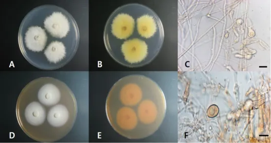
Rhizodermea veluwensis Verkley & Zijlstra, Persoonia 24: 131. 2010 (Fig. 1, Table 1) Diameters of the colonies on PDA grown for 7 days were 36~40 mm, and obverse of the colony was observed to be ivory and the reverse was observed to be pale yellow in color.
The colonies were flattened and serrated at the margin. Diameters of the colonies on MEA grown for 7 days were 34~37 mm, and obverse of the colony was observed to be beige and reverse was observed to be reddish brown in color. Colonies were flat and blunt-serrated at the margin. At the margin of the colonies, aerial mycelium was formed and its color was hyaline or vinaceous. The hyphae were 1~2 µm wide initially and 3~6 µm wide later, and were isodiametric inflated cells, rarely big gourd-shaped cells about 15 µm wide.
Chlamydospores were globose to limoniform, hyaline to yellowish, 1-septate, smooth- walled or warted, 15~20 µm in diameter.
Specimen examined: KOREA; Mt. Minjuji, N 36° 2 ˊ 49.88 ˝ , E 127° 46 ˊ 36.7 ˝ ; isolated from roots of R. mucronulatum; September 21, 2016; Culture 16E003 (NIBRFG 0000499917, GenBank MF042207).
Fig. 1. Morphological characteristics of Rhizodermea veluwensis 16E003. A, obverse colonies on potato dextrose agar (PDA); B, reverse colonies on PDA; C, mycelium; D, obverse colonies on malt extract agar (MEA); E, reverse colonies on MEA; F, chlamydospore (scale bars: C, F
=10 µm).
Phylogenetic analysis
BLAST results in NCBI showed that the ITS sequence of this isolate was closely related to that of R. veluwensis KR859283.1 with 99% similarity and the LSU region of this isolate was closely related to that of R. veluwensis KR859076.1 with 100% similarity. Both the sequences were obtained from fungal strains isolated from roots of species belonging to the family Ericaceae (Vaccinium myrtillus and Erica tetralix, respectively) inhabiting the Netherlands. Phylogenetic tree using the combined sequence of both ITS and LSU regions in Fig. 2 showed that the sequence from this study formed a monophyletic group with the strain R. veluwensis CBS 110605 isolated from roots of Erica tetralix in Netherlands, supported by 99% bootstrap value.
The morphological characteristics of fungal strain 16E003 isolated from the roots of R.
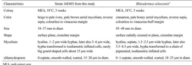
mucronulatum, in this study, were generally consistent with the original description of the species [1]. Additionally, phylogenetic analysis using ITS and LSU sequences strongly Table 1. Morphological characteristics of fungal strain 16E003 isolated from this study
Characteristics Strain 16E003 from this study Rhizodermea veluwensis
aColony MEA, 18°C, 3 weeks MEA, 18°C, 3 weeks
Color beige to pale ivory, pale brown aerial mycelium, reverse sepia, colourless to vinaceous margin
cinnamon, pale honey aerial mycelium, reverse sepia, colourless to vinaceous-buff margin
Size 34~37 mm in diam 45~48 mm in diam
Shape surface plane, crenulate margin surface radially creased or plane, crenulate margin Mycelium hyaline, 1~2 µm wide hyphae, later also 3~6 µm wide,
hypha transformed to isodiametric inflated cells, rarely big gourd-shaped cells about 15 µm wide
hyaline, septate, 1.5~2.5 µm wide hyphae, later also 5.5~8.5 µm wide, hypha transformed to a chain of pigmented, isodiametric inflated cells
chlamydospores 0-septate, smooth-walled, warted, 15~20 µm in diam. 0~1-septate, smooth-walled, warted, 18~25 µm in diam.
MEA, malt extract agar.
a