⋅교신저자 : 이용흠, 강원도 원주시 연세대길 1
연세대학교 보건과학대학 의공학과 백운관 418호 Tel. 033-760-2863, E-mail: [email protected]
⋅투고 : 2011/08/25 심사 : 2011/09/06 채택 : 2011/09/10
음포혈(陰包, LR9)에서
펄스형 전자기장 및 가시광 LED 복합 자극이 대퇴부 근피로 회복에 미치는 영향 분석
이나라1⋅김정윤2⋅박선우2⋅김수병1⋅이희영2⋅안순재2⋅김영호2⋅이용흠1
1연세대학교 보건과학대학 의공학과 한의공학연구실
2연세대학교 보건과학대학 의공학과 생체역학연구실
The Analysis of the Muscle Fatigue Recovery Effect on LR9 by Compound Stimulation of the PEMFs and LED
Na-Ra Lee1, Jung-Yoon Kim2, Sun-Woo Park2, Soo-Byeong Kim1, Hee-Young Lee2, Soon-Jae Ahn2, Young-Ho Kim2, Yong-Heum Lee1
1Dept. of Biomedical Engineering, College of Health Science Oriental medical Engineering Lab, Yon-Sei University
2Dept. of Biomedical Engineering, Institute of Medical Engineering, Yon-Sei University Abstract
Objectives : The aim of this study was to develop the combined medical stimulus system consisted of the PEMFs (Pulsed electromagnetic fields) and LED which are able to stimulate local point such as acupoints and trigger points.
Methods : To evaluate the therapeutic effect on the musculoskeletal disorders and the possibility of alternative method on manual acupuncture, we compared the fatigue recovery of two groups by analyzing the EMG and peak torque (non-stimulation and, stimulation group) after strenuous knee exercise. We chose the LR9 as a stimulation point.
Results : The median frequency (MF) and fatigue index (F.I) of the stimulation group were recovered faster than those of the non-stimulation group. Also the peak torques of both groups were not restored until after 20 minutes. However, the peak torque of the stimulation group was regained higher than that of the non-stimulation group.
Conclusions : We confirmed that the proposed combined stimulus system had useful effects as treatment instrument of musculoskeletal disorder using non-invasive method of PEMFs and LED.
Key words : Pulsed Electromagnetic Fields (PEMFs), Light Emitting Diode Therapy (LEDT), Muscle Fatigue, Acupoint, trigger point, EMG
Ⅰ. 서 론
근육은 격렬한 운동 중에는 활동성이 점진적으 로 감소하며 일정 휴식 시간 이후 활동성이 정상
상태로 회복된다. 이러한 운동 중에 유발된 근육의 활동성 감소는 근피로를 의미하는 것이며 이는 운 동의 형태 및 강도, 사용된 근육의 종류, 생화학적 인 환경 등에 따라 정도가 다르다1). 근피로 유발 시, 생체조직 내 Oxidative-stress와 Reactive Oxygen Species(ROS)가 발생하여 미토콘드리아의 기능을 억제시키며 근육세포의 탈분극화와 힘의 감소를 유발하는 요인으로 작용한다2). 또한 근피로와 근조 직의 손상이 동반될 수 있으며3), 이는 근조직의 구 조적이상, 세포막 손상, 근섬유분절 장애, 염증성 반응 등 다양한 근골격계 질환의 원인을 제공하는 요인이다1).
이러한 실정에 따라 근피로 및 근조직의 손상을 회복시키기 위한 다양한 연구가 보고되었으며, 대 표적인 사례로 Active recovery4), 냉동요법5), 마사 지6), 전기 자극7)을 일컬을 수 있다. 또한 한의학에 서는 근피로 및 근조직의 손상이 유발된 지점에 근접한 경혈 및 아시혈에 수기자극을 이행함으로 써 치료하고 있다8). 한의학적 치료방법으로써 수기 자극이 근피로 및 근조직 손상에 미치는 치료효과 를 규명하고자 다수 연구가 진행되었다. 이침(耳 鍼)은 운동 중 산소의 공급을 원활하게 하며 젖산 의 생성을 지연시키는 효과가 있다고 보고되었다9). 이와 유사하게 근피로가 유발된 근육에 수기자극 을 적용할 시, 근육의 수축 정도를 활성화되도록 유도하며10) 또한 Muscle force capacity의 회복에 유의한 효과가 있음을 보고하였다11). 그러나 이러 한 수기자극은 근육의 상태를 회복시키는 탁월한 치료 효과에도 불구하고, 침습적인 치료방법으로 인하여 대상 환자들로부터 기피되어지고 있는 실 정이다.
이러한 기피현상에 따라 국소적 부위인 경혈만 을 자극 가능하며 비침습적인 방법으로 경피 자극 방식이 아닌 피하조직을 자극하는 자극방법이 다 양하게 제시되었으며12-14), 이 중 자극원을 자기장 으로 선정한 연구가 다수 보고되었다15-17). 자기장 은 근골격계 질환뿐만 아니라 골절, 통증완화, 동
맥경화, 파킨슨병 등과 같은 다양한 질환에 적용되 고 있다15). 자기장의 근골격계 관련 질환에 대한 치 료 효과는 다수의 연구결과를 통하여 검증되었으며 또한 다양한 치료기기들의 개발이 이루어짐으로써 근 통증 치료에 다양하게 이용되고 있다15-18).
최근 근 통증 치료에 관련된 연구 동향을 살펴 보면 인공광을 이용한 광선요법(phototherapy)이 활발히 연구되고 있다. 이 중 가시광선 대역에서의 적색 파장 대역과 근적외선 파장 대역이 근 통증 완화 효과가 있다고 보고되었다19,20). 근 통증 완화 효과를 보고한 다수 연구 결과는 레이저를 이용하 여 좁은 파장대역에서의 고출력의 광을 조사한 방 식이다. 이러한 고출력 형태의 레이저는 침투 깊이 가 깊으나 다수 경혈을 동시 자극이 가능하지 않 으며, 피부에 직접 부착하기 어려운 단점을 내포하 고 있다. 이와 달리 Light Emitting Diode Therapy (LEDT)는 저출력으로써 파장대역이 넓고 레이저 에 비하여 보다 넓은 부위를 자극 할 수 있으며 Laser에 비하여 시스템의 구현이 편리할 뿐만 아니 라 가격이 저렴한 장점이 있다. 또한 LEDT를 이 용한 근 통증완화와 관련된 질환의 치료효과를 규 명한 연구가 다수 보고되었다21-23).
이러한 기존연구 결과를 기반으로 하여, 본 연구 에서는 수기자극을 대신하기 위한 자극원으로 자 기장과 LEDT를 선정하였다. 두 개의 자극원은 비 침습적인 방법으로 국소적 부위인 경혈을 자극하 며, 경피 자극 방식이 아닌 피하조직까지 자극 가 능하나 각 자극원마다 유도하는 생체 내 반응이 다르며 이에 따른 치료효과가 상이하므로 근피로 및 근조직 손상의 치료효과를 극대화하기 위하여 LEDT와 자기장의 교번자극이 가능한 시스템을 제 작하였다. 기존의 근피로 및 근조직 손상에 효과가 있는 파장 및 자기장 자극 방법을 조사해본 결과 Light Emitting Diode(LED)는 642~645nm의 파장 대역의 적색광원을21,23), 자기장 자극 방법 중 Pulsed electromagnetic fields(PEMFs)를17,18) 선택하였다.
이에 대퇴부 사두근에서 본 시스템의 근피로 회복
효과를 평가하였으며 그 결과 PEMFs와 LED의 교번 자극이 근피로 회복에 유의한 치료 효과를 가짐을 확인하였기에 이를 보고하는 바이다.
Ⅱ. 재료 및 방법 1. 피험자 선정
근골격계 및 신체에 특정한 질병이 없는 남자 대학생 20명이 실험에 참가하였으며, 피험자들은 무자극군(10명), 자극군(10명)으로 배정하여 실험 을 진행하였다. 임상실험을 진행하기 전 피험자들 에게 연구의 목적과 방법을 상세히 설명하였으며, 자발적 참여자에 한하여 실험 참여 동의를 받은 인원에 한하여 선정하였다. 모든 피험자는 근피로 (muscle fatigue)로 인한 EMG의 오차를 고려하여 실험 참여 2주 전부터 일상생활의 일반적 수행 외 운동을 금지하였다. 또한 측정 전극의 부착지점에 화상 및 피부 상처가 존재하지 않은지 확인하였다.
2. 자극 위치 선정
대퇴부 사두근을 근피로 유발 부위로 선정하였 으며, 족궐음간경(LR)에 속한 경혈 중 대퇴부에 위치하고 있는 음포혈(LR9)을 자극부위로 선정하 였다. 음포혈은 슬개골 내측상 4촌처, 봉공근과 반 막양근간의 요함처에 위치하고 있는데 봉공근하부 의 TP(Trigger Point)와 해부학적 위치가 100% 일 치하는 부위이다27).
3. 자극주파수 선정
PEMFs에 사용된 주파수는 기존 연구 결과에서 근 골격계 치료에 효과가 있음이 밝혀진 Extremely Low Freuquency(ELF≤300Hz) 대역을 선정하였다
15-17). Transcutaneous electrical acupoint stimulation (TEAS)를 이용하여 경혈만의 경피 신경 전기 자
극을 통한 근피로 회복에 관한 기존의 연구 결과 에 의하면 2Hz이상의 자극 주파수에서는 피험자가 통증을 자각하는 사례가 보고되었다24,25). 이에 본 연구에서는 PEMFs와 LEDT의 교번 자극 주파수 를 2Hz로 선정하였다.
4. 전극부 설계
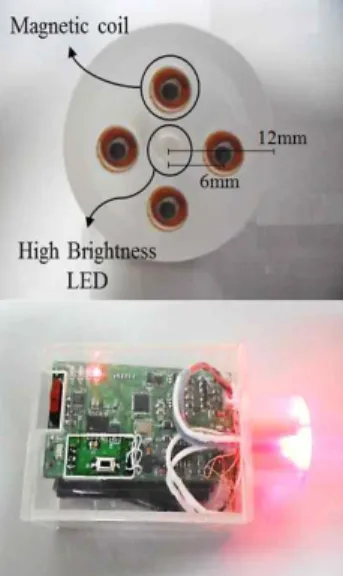
Fig. 1과 같이 PEMFs와 LED의 광 조사의 교 번 방식이 국소적 부위인 경혈을 중심으로 반경 1cm 외 다른 경피 조직을 자극하지 못하도록 최소 화하였다. 자속밀도의 발생을 높이기 위하여 SM45C 재질을 사용하여 코어를 제작하였으며, 본 코어를 중심으로 두께 0.2㎜의 코일을 이용하여 총 400회 감았다.
LEDT에 사용된 LED는 근 통증에 유의한 효과 가 있음이 밝혀진 642~645nm의 파장 대역을 선정
하였으며21,23) 광도(Luminous Intensity) 50~70mcd
를 출력하는 고휘로 LED를 사용하였다. 코일과 LED를 포함하는 케이스의 직경은 12mm이며, 코 일과 LED의 중심 간의 거리는 6mm 간격으로 이 루어져 있다.
Fig. 1. Electrode for operating magnetic flux &
LED and designed system.
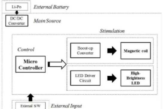
본 시스템은 PEMFs와 LED의 광 조사를 교번 자극하기 위한 시스템으로 Fig 2와 같이 구성되어 있다. 3.7V의 Li-Polymer battery의 전원부로 구성 되어 있으며, a constant voltage regulator에 의하 여 시스템 동작전압은 3.3V이다. 코일에 흐르는 전 류를 통하여 강한 전자기장(electromagnetic fields) 을 형성하므로, boost-up converter를 자체 설계하 여 16V의 PEMFs의 source voltage를 별도로 생성 하여 높은 자속밀도(magnetic flux)를 유도하였다.
자극 주파수에 따른 LED의 광량을 일정하게 하기 위하여 constant current circuit를 설계하였다.
Fig. 2. Block diagram of the PEMFs and LEDT system.
PEMFs 방법으로 신경의 전도성을 10% 줄이기 위해서는 24 Tesla 이상의 자속밀도가 요구되지만
26), 통증 완화를 위하여서는 대부분 300~500 Gauss 가 사용되어진다. 이에 source voltage인 16V와 자 극 주파수 2Hz에서 통증 완화에 사용되는 300~500 Gauss의 범위 내의 자속밀도가 발생하도록 코일을 자체 제작하였으며 제작된 전극의 자속밀도를 측 정한 결과, 347.84Gauss가 발생하였다.
X-axis Y-axis Z-axis Vector Magnitude Magnetic
flux density 153.90 147.37 275.07 347.84 Table 1. Magnetic Flux of the PEMFs Coil (unit:
Gauss)
5. EMG 측정
근피로 회복을 평가하기 위하여 본 실험에서는 대퇴직근(Rectus femoris)에서 근전도를 측정하였 다. 근전도 측정을 위하여 Ag/AgCl 표면 전극 (Noraxons, USA)을 이용하였다. 표본주파수(sampling frequency)는 1kHz로 하였으며, 10Hz와 500Hz의 구간만 사용하도록 filtering 하였다. 근전도는 Noraxon Myoresearch XP software에서 제공하는 GUI를 통 하여 실시간 확인하였으며 측정된 근전도의 power spectrum을 분석하였다.
근피로를 평가하기 위하여 본 연구에서는 Median Frequency(MF)를 분석하였다. 기존의 연구결과에 따르면 등척성 운동으로 인하여 근피로가 유발될 시, MF가 낮아지게 되며 근피로가 회복됨에 따라 낮아진 주파수가 초기 값으로 회복된다. 따라서 MF의 변화 추이를 통하여 근피로의 회복 여부를 판단할 수 있으며, 이를 보다 쉽게 확인하기 위하 여 Fatigue Index(F.I)를 추가적으로 계산하였다.
Equation 1. Fatigue Index
6. 실험방법
본 연구에서 Maximal Voluntary Contraction (MVC) 측정 및 근피로 유발을 시키기 위하여 Biodex System 3(Biodex Medical Inc., Shirley, NY, USA)을 사용하였다. 실험절차는 Fig. 3과 같 이 구성되었다. 첫 번째로 모든 피험자는 MVC를 측정하였다. 그 후 근피로 유발에 앞서 Biodex의 운동에 익숙해지기 위하여 측정된 MVC의 30%의 힘으로 knee extension/flexion을 10회씩 3회 실시한 다. 근피로 유발 전 근육의 충분한 휴식과 피험자 의 지침을 방지하기 위하여 3회 warm up 사이마 다 2분간의 휴식시간을 적용하였다. MVC를 재측 정한 후 근피로를 유발하기 위하여 측정된 MVC의
65%의 힘으로 knee extension/flexion을 10회씩 5회 실시하였다. 10회 운동이 끝날 때 마다 2분간의 휴 식시간을 적용시켰다. 운동이 끝난 직후 MVC를 측정하며, 그 뒤 2분 간격으로 MVC를 측정하고
측정 사이 시간에는 자극군은 PEMFs와 LED 광 조사를 교번 자극하였으며, 무자극 군의 경우 휴식 시간을 유지하도록 하였다.
Fig. 3. Procedure of experiment.
7. 통계처리
자극군과 무자극군의 근피로 회복을 평가하기 위하여 one-way repeated measures analysis of variance를 이용하여 대비검정을 실시하였다. 모든 변인에 대한 통계적 유의수준은 P<0.05로 설정하였 다. 측정된 모든 데이터는 초기 값을 1로 적용하여 normalization을 하였으며, 백분위 그래프로 표현하 였다.
Ⅲ. 결 과
1. 무자극군과 자극군의 MF 결과 비교
⋅분석
Fig. 4와 같이 모든 피험자에게서 운동직후 감소 한 MF가 시간의 흐름에 따라 운동전의 MF 값으 로 회복되는 동일한 현상을 확인하였다. 그러나 운 동 후에 감소한 MF가 운동전의 정상상태의 MF로 도달하는 시간은 각 군마다 차이가 관찰되었다. 자 극군은 약 3분 정도에 운동전의 근피로 유발 전의 MF로 회복되었으며, 무자극군의 경우 8분과 10분 의 회복 구간사이에 회복되었다.
Fig. 4. The mean and standard error of the MF for non-stimulation group and stimulation group as time passed.
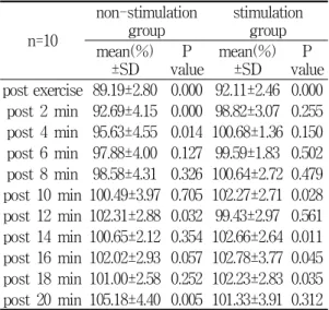
이에 one-way repeated measures analysis of variance를 이용하여 대비검정을 통하여 분석해본 결과, 자극군은 운동 후 2분 후에 98.82±3.07(p=0.255) 로 무자극군은 6분 후에 97.88±4.00(p=0.127)로 확 인되었다. 즉 무자극군에 비하여 PEMFs와 LED 광의 교번 조사를 한 자극군의 MF의 자극 전 정 상상태로 4분 먼저 회복되었음을 확인하였다.
n=10
non-stimulation
group stimulation group mean(%)
±SD P
value mean(%)
±SD P
value post exercise 89.19±2.80 0.000 92.11±2.46 0.000 post 2 min 92.69±4.15 0.000 98.82±3.07 0.255 post 4 min 95.63±4.55 0.014 100.68±1.36 0.150 post 6 min 97.88±4.00 0.127 99.59±1.83 0.502 post 8 min 98.58±4.31 0.326 100.64±2.72 0.479 post 10 min 100.49±3.97 0.705 102.27±2.71 0.028 post 12 min 102.31±2.88 0.032 99.43±2.97 0.561 post 14 min 100.65±2.12 0.354 102.66±2.64 0.011 post 16 min 102.02±2.93 0.057 102.78±3.77 0.045 post 18 min 101.00±2.58 0.252 102.23±2.83 0.035 post 20 min 105.18±4.40 0.005 101.33±3.91 0.312 Table 2. The Analysis of the MF in the Non-stimulation Group and Stimulation Group as Time Passed
2. 무자극군과 자극군의 F.I 결과 비교
⋅분석
MF를 비교할 시, 운동 직후 근피로가 유발되는 정도가 상이하다는 문제점을 해결하기 위하여 F.I 를 분석한 결과이다. Fig. 5와 같이, 무자극군은 회 복시간 8분과 10분 사이 구간에서 근피로 유발 전 의 상태로 회복되었으며, 자극군은 회복시간 4분과 6분 사이 구간에서 회복되었다. MF의 결과와 동일 하도록 무자극군보다 자극군이 운동 전의 정상상 태로 대략 4분정도 먼저 신속히 회복되고 있음을 확인하였다.
Fig. 5. The mean and standard error of the F.I for non-stimulation group and stimulation group as time passed.
Table 3은 각 군에 대한 F.I를 one-way repeated measures analysis of variance를 이용하여 대비검정 한 결과로 무자극군은 6분이 경과한 후에 72.68±
52.91(p=0.138)로 자극군은 2분이 지난 후에 67.39
±25.36(p=0.271)로 무자극군에 비하여 자극군의 회 복속도가 빠른 것을 확인하였다.
n=10
non-stimulation
group stimulation group mean(%)
±SD P
value mean(%)
±SD P
value post 2 min 25.95±48.41 0.001 67.39±25.36 0.271 post 4 min 56.98±44.35 0.013 90.84±20.17 0.198 post 6 min 72.68±52.91 0.138 105.57±22.94 0.782 post 8 min 87.88±35.40 0.310 123.53±31.09 0.527 post 10 min 109.25±41.56 0.497 115.40±18.65 0.031 post 12 min 126.12±32.12 0.030 138.04±30.36 0.428 post 14 min 104.64±24.35 0.558 134.77±36.36 0.009 post 16 min 117.36±23.09 0.042 117.08±33.22 0.045 post 18 min 107.31±22.45 0.329 137.72±46.26 0.080 post 20 min 149.14±43.21 0.006 139.21±50.23 0.447 Table 3. The Analysis of the F.I in the Non-stimulation Group and Stimulation Group as Time Passed
3. 무자극군과 자극군의 peak torque 결과 비교⋅분석
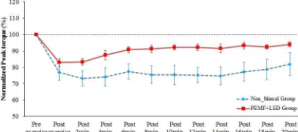
Fig. 6은 무자극군과 자극군의 peak torque의 변 화추이를 나타낸 것으로 대비검정 결과와 동일하 게 두 군 모두 초기의 peak torque로 회복하지 못 하는 것을 확인하였다. 두 군 모두 운동전의 peak torque를 회복하지 못하였으나 무자극군은 운동직 후 77.11±15.62로 떨어진 peak torque가 81.94±21.82 로, 자극군은 운동직후 83.11±7.72에서 운동 후 20 분 후에 93.95±4.67로 자극군이 무자극군에 비하여 회복되는 정도가 더 높음을 확인하였다.
Fig. 6. The mean and standard error of the peak torque for non-stimulation group and stimulation group as time passed.
Table 4는 무자극군과 자극군의 peak torque의 데이터를 시간의 흐름에 따라 one-way repeated measures analysis of variance를 이용하여 대비검정 한 결과이다. 20분이 지난 후까지 두 군 모두 초기 peak torque를 회복하지 못하는 동일한 결과를 확 인하였다(p<0.05).
n=10
non-stimulation
group stimulation group mean(%)
±SD P
value mean(%)
±SD P
value Post exercise 77.11±15.62 0.001 83.11±7.72 0.000 post 2 min 73.21±14.52 0.000 83.33±6.65 0.000 post 4 min 74.15±17.69 0.001 87.52±9.10 0.002 post 6 min 77.46±15.10 0.001 90.84±5.40 0.000 post 8 min 75.43±16.85 0.001 91.35±6.93 0.003 post 10 min 75.37±18.82 0.003 92.20±5.11 0.001 post 12 min 75.15±14.73 0.000 92.18±6.46 0.004 post 14 min 74.86±17.32 0.001 91.52±8.27 0.010 post 16 min 77.27±17.95 0.003 93.24±6.44 0.009 post 18 min 78.74±19.53 0.007 92.40±4.36 0.000 post 20 min 81.94±21.82 0.028 93.95±4.67 0.003 Table 4. The Analysis of the Peak Torque in the Non-stimulation Group and Stimulation Group as Time Passed
Ⅳ. 고 찰
본 연구에서는 수기자극이 분명한 치료효과8-11) 를 가지고 있음에도 불구하고 침습적인 방법으로
인하여 환자들로부터 기피되고 있으므로 이를 대 안할 수 있는 비침습적인 치료 방법을 제시하고자 하였다. 이에 비침습적 방식으로 국소적 부위인 경 혈만을 자극 가능하며 피하 아래의 조직까지 영향 을 미칠 수 있는 자극원을 조사하였으며 그 결과 PEMFs와 LEDT가 최적이라 판단하였다. 이에 본 연구에서 두 자극원의 생체 내 치료기전이 서로 상이하므로 치료효과의 극대화를 위하여 두 자극 원을 교번 자극 가능한 시스템을 제작하였으며, 대 퇴사근에서의 근피로를 유발한 후, 제시된 자극원 이 경혈만을 자극하였을 때의 치료효과를 관찰하 였다. 그 결과, 무자극군에 비하여 근피로 회복에 탁월한 효과가 있음이 확인되었다. 이는 PEMFs와 LEDT의 광 조사의 교번 방식이 다음과 같은 생체 내 반응을 유도하여 이와 같은 결과가 도출되었다 고 사료된다.
체외에서 PEMFs의 자극은 생체 내에 Faraday current28)를 유도하며 이는 해당 부위에서 통증을 감소시키고 염증을 완화시키며 세포의 재생을 활 성화를 유도 한다29,30). 통증은 Na+ 이온의 증가와 세포막의 감극 현상, action potential의 발생에 의 해 유발 된다31). 이에 PEMFs 자극은 세포막의 삼 투성과 action potential을 감소시켜 통증 완화에 효과가 있음이 제시되고 있다. 특히 PEMFs는 혈 관 이완과 혈류 증가에 효과가 있으며 주변 신경 의 탈 분극화와 근조직을 자극함으로써 해당 근육 을 강화시키고 통증완화에 효과가 있음이 발표되 었다32).
LED의 642~645nm대역에 대한 기존의 연구결 과에 따르면, creatine kinase(CK) 효소의 활성 지 표를 통하여 근조직의 손상 정도33,34)를 판별한 결 과, CK효소가 활성화됨을 보고하였다. 또한 젖산 의 농도는 혈액과 세포내의 PH 감소 정도와 H+이 온의 증가를 확인하는 2차적인 지표35)로써 혈중 젖 산 농도를 측정을 통하여 근육의 회복 평가를 한 결과36), 혈중 젖산 농도 감소 효과가 있음이 밝혀
졌다22,23). 즉 CK와 혈중 젖산 농도의 감소는 LEDT
가 근조직의 손상 및 근육의 회복 평가에 효과가 있음을 의미하는 것이며, 이는 LEDT의 치료 효과 중 미세순환(microcirculation)의 증가로 인한 것으 로 보고되었다23). 또한 높은 강도와 반복적인 근 수축은 근 조직에 염증성 반응을 일으키며37), 보편 적으로 사용되어 온 염증의 정도를 판단하는 기준 인 C-reative protein(CRP)38)로 LEDT의 치료 효 과를 판별한 결과, 염증을 감소시킨다고 보고되었 다21). 더욱이 LEDT는 신경 전도성을 통하여 진통 의 효과와 일정 시간동안의 sensory impulse의 발 생 수를 줄임으로써 통증완화의 효과를 가지고 있 다고 보고되었다.39)
이와 같은 PEMFs와 LEDT가 생체에 미치는 영향으로 인하여 비침습적 방식으로 경혈을 자극 하였을 시, 탁월한 근피로 회복 효과를 유도한 것 으로 판단하였다.
Ⅴ. 결 론
본 연구에서는 국소적 부위인 경혈만을 비침습 적 방식으로 자극 가능한 PEMFs와 LED 광 조사 교번 자극기기를 개발하였다. 이에 따른 근골격계 질환 중 근피로 회복을 평가하고자 대퇴직근에 반 복적으로 격렬하게 수축/이완을 시킴으로써 근피 로를 유발하였다. 시간에 따라 회복되는 정도를 비 교함으로써 PEMFs와 LED의 교번 자극이 근피로 회복에 미치는 영향을 확인하였다. 실험 결과, 근 피로 유발 후 MF, F.I 와 peak torque가 감소되고 회복됨에 따라 다시 증가한다는 기존의 연구결과 와 동일한 변화 추이를 확인하였으며, MF, F.I와 peak torque 모두 무자극군에 비하여 자극군이 운 동전의 정상 상태로 신속히 회복되는 추이를 확인 하였다. 이에 향후 수기자극을 대신하여 경혈만을 자극하는 근골격계 질환 관련 치료방식으로서의 가능성을 확인하였다. 그러나 이는 PEMFs와 LED 의 교번자극과 무자극간의 비교만을 통하여 시스 템을 평가한 것으로 교번자극 방식의 치료효과를
보다 객관적으로 확인하기 위하여 PEMFs, LED의 단일 자극과의 비교실험이 필요하다. 이에 추후 단 일 자극과의 비교 실험 및 무자극군의 객관적 비 교를 위해 double blind, placebo-controlled 실험을 진행함으로써 새로운 한의학적 자극 시스템으로써 의 가능성을 확인하고자 한다.
감사의 글
본 연구는 지식경제부 지역산업기술개발사업의 연구비를 지원 받았음[과제고유번호 R0000129].
참고문헌
1. Allen DG, Lamb GD, Westerblad H. Skeletal muscle fatigue: cellular mechanisms. Physiol Rev. 2008 ; 88(1) : 287-332.
2. Nethery D, Callahan LA, Stofan D, Mattera R, DiMarco A, Supinski G. PLA(2) dependence of diaphragm mitochondrial formation of reactive oxygen species. J Appl Physiol. 2000 ; 89(1) : 72-80.
3. Cheung K, Hume P, Maxwell L. Delayed onset muscle soreness: treatment strategies and performance factors. Sports Med. 2003 ; 33(2) : 145-64.
4. Ahmaidi S, Granier P, Taoutaou Z, Mercier J, Dubouchaud H, Prefaut C. Effects of active recovery on plasma lactate and anaerobic power following repeated intensive exercise. Med Sci Sports Exerc. 1996 ; 28(4) : 450-6.
5. Cheung K, Hume P, Maxwell L. Delayed onset muscle soreness: treatment strategies and performance factors. Sports Med. 2003 ; 33(2) : 145-64.
6. Martin NA, Zoeller RF, Robertson RJ, Lephart SM. The comparative effects of sports massage,
active recovery, and rest in promoting blood lactate clearance after supramaximal leg exercise.
J Athl Train. 1998 ; 33(1) : 30-5.
7. Lattier G, Millet GY, Martin A, Martin V.
Fatigue and recovery after high-intensity exercise. Part II: Recovery interventions. Int J Sports Med. 2004 ; 25(7) : 509-15.
8. Ehrlich D, Haber P. Influence of acupuncture on physical performance capacity and haemodynamic parameters. Int J Sports Med. 1992 ; 13(6) : 486-91.
9. Lin JG, Salahin HS, Lin JC. Investigation on the effects of ear acupressure on exercise -induced lactic acid levels and the implications for athletic training. Am J Acupunct. 1995 ; 23(4) : 309-13.
10. Qu ZQ, Lu DH, Wang YR. Mechanism of acupuncture and effects on recovery of contractile force and ultrastructure in the exhausted muscles. J Beijing Inst Phy Educ.
1993 ; 16(2) : 35-44.
11. Wang WY, Niu D, Wang H, Chiu DH, Hu YS. A clinical study on treating exercise- induced fatigue with acupuncture. Chin Acup Moxi. 1999 ; 1 : 13-5.
12. So RC, Nq JK, Nq GY. Effect of transcutaneous electrical acupoint stimulation on fatigue recovery of the quadriceps. Eur J Appl Physiol.
2007 ; 100(6) : 693-700.
13. Baxter GD, Bleakley C, McDonough S. Clinical effectiveness of laser acupuncture: a systematic review. J Acupunct Meridian Stud. 2008 ; 1(2) : 65-82.
14. 이나라, 이승욱, 김영대, 김수병, 이경중, 이용 흠. 지연성 근육통증 회복 평가를 위한 경혈 부위에서의 자기장자극에 대한 체열변화 분석.
한국해양정보통신학회논문지. 2011 ; 15(3) :
645-53.
15. Rosch P. Markov M. Bioelectromagnetic Medicine.
New York: Marcel Dekker. 2004.
16. Smania N, Corato E, Fiaschi A, Pietropoli P, Aglioti SM, Tinazzi M. Therapeutic effects of peripheral repetitive magnetic stimulation on myofascial pain syndrome. Clin Neurophysiol.
2003 ; 114(2) : 350-8.
17. Smania N, Corato E, Fiaschi A, Pietropoli P, Aglioti SM, Tinazzi M. Repetitive magnetic stimulation: a novel therapeutic approach for myofascial pain syndrome. J Neurol. 2005 ; 252(3) : 307-14.
18. Nuruki A, Tsujumura S, Furuich H, Takenaka T, Yunokuchi K. Application of Pulsed magnetic stimulation for recovery from muscle fatigue in hypokalemic and normal wistar rats. Trans Jpn Soc Med Biol Eng. 2009 ; 47(1) : 57-63.
19. Camara K, Danao-Camara T. Awareness of, use and perception of efficacy of alternative therapies by patients with inflammatory arthropathies. Hawaii Med J. 1999 ; 58(12) : 329-32.
20. Leal Junior EC, Lopes-Martins RA, Dalan F, Ferrari M, Sbabo FM, Generosi RA et al.
Effect of 655-nm low level laser therapy on exercise-induced skeletal muscle fatigue in humans. Photomed Laser Surg. 2008 ; 26(5) : 419-24.
21. Leal Junior EC, Lopes-Martins RA, Rossi RP, De Marchi T, Baroni BM, de Godoi V et al.
Effect of cluster multi-diode light emitting diode therapy (LEDT) on exercise-induced skeletal muscle fatigue and skeletal muscle recovery in humans. Lasers Surg Med. 2009 ; 41(8) : 572-7.
22. Vinck E, Cagnie B, Coorevits P, Vanderstraeten
G, Cambier D. Pain reduction by infrared light-emitting diode irradiation: a pilot study on experimentally induced delayed-onset muscle soreness in humans. Lasers Med Sci. 2006 ; 21(1) : 11-8.
23. Leal Junior EC, de Godoi V, Mancalossi JL, Rossi RP, De Marchi T, Parente M et al.
Comparison between cold water immersion therapy (CWIT) and light emitting diode therapy (LEDT) in short-term skeletal muscle recovery after high-intensity exercise in athletes -preliminary results. Lasers Med Sci. 2011 ; 26(4) : 493-501.
24. Melzack R. Prolonged relief of pain by brief, intense transcutaneous somatic stimulation.
Pain. 1975 ; 1(4) : 357-73.
25. Fields HL, Basbaum AI. Endogenous pain control mechanisms. Edinburgh: Churchill Livingstone. 1989.
26. Wikswo JP, Barach JP. An estimate of the steady magnetic field strength required to infl uence nerve conduction. IEEE Trans Biomed Eng. 1980 ; 27(12) : 722-3.
27. 김형준, 허동석, 오만석. 근육의 Trigger Points 와 경혈(經穴)의 연관성에 관한 연구 (II) - 요방형근에서 발의 심층 내재근들까지. 한의학 논문집. 2007 ; 16(1) : 127-32.
28. Ramey DW. Magnetic and electromagnetic therapy. Sci Rev Altern Med. 1998 ; 2 : 13-9.
29. Bassett CA. Beneficial effects of electromagnetic fields. J Cell Biochem. 1993 ; 51(4) : 387-93.
30. Basset CA. Fundamental and practical aspects of therapeutic uses of pulsed electromagnetic fields. Crit Rev Biomed Eng. 1989 ; 17(5) : 451-529.
31. Markov MS, Colbert A. Magnetic and electromagnetic field therapy. J Back Musculoskelet Rehabil.
2000 ; 15(1) : 17-29.
32. Graven-Nielsen T, McArdle A, Phoenix J, Arendt -Nielsen L, Jensen TS, Jackson MJ, et al. In vivo model of muscle pain: quantification of intramuscular chemical, electrical, and pressure changes associated with saline-induced muscle pain in humans. Pain. 1997 ; 69(1-2) : 137-43.
33. Smith TL, Wong-Gibbons D, Maultsby J. Micro circulatory effects of pulsed electromagnetic fields. J Orthop Res. 2004 ; 22(1) : 80-4.
34. MacDougall JD, Hicks AL, MacDonald JR, McKelvie RS, Green HJ, Smith KM. Muscle performance and enzymatic adaptations to sprint interval training. J Appl Physiol. 1998
; 84(6) : 2138-42.
35. Cairns SP. Lactic acid and exercise performance:
culpid or friend?. Sports Med. 2006 ; 36(4) : 279-91.
36. Baldari C, Videira M, Madeira F, Sergio J, Guidetti L. Lactate removal during active recovery related to the individual anaerobic and ventilatory thresholds in soccer players.
Eur J Appl Physiol. 2004 ; 93(1-2) : 224-30.
37. Milias GA, Nomikos T, Fragopoulou E, Athanasopoulos S, Antonopoulou S. Effects of eccentric exercise-induced muscle injury on blood levels of platelet activating factor (PAF) and other inflammatory markers. Eur J Appl Physiol. 2005 ; 95(5-6) : 504-13.
38. Malm C, Sjödin TL, Sjöberg B, Lenkei R, Renström P, Lundberg IE, Ekblom B.
Leukocytes, cytokines, growth factors and hormones in human skeletal muscle and blood after uphill or downhill running. J Physiol.
2004 ; 556(3) : 983-1000.
39. Vinck E, Coorevits P, Cagnie B, Muynck MD, Vanderstraeten G, Cambier D. Evidence of
changes in sural nerve conduction mediated by light emitting diode irradiation. Lasers Med
Sci. 2005 ; 20(1) : 35-40.