• 검색 결과가 없습니다.

Fatal Interstitial Pneumonitis Rapidly Developed after the First Cycle of CHOP with Etoposide Combination Chemotherapy in a Patient with Lymphoma

N/A
N/A
Protected

Academic year: 2021

Share "Fatal Interstitial Pneumonitis Rapidly Developed after the First Cycle of CHOP with Etoposide Combination Chemotherapy in a Patient with Lymphoma"

Copied!
5
0
0

로드 중.... (전체 텍스트 보기)

전체 글

(1)

Tuberc Respir Dis 2013;74:235-239

CopyrightⒸ2013. The Korean Academy of Tuberculosis and Respiratory Diseases. All rights reserved.

Fatal Interstitial Pneumonitis Rapidly Developed after the First Cycle of CHOP with Etoposide Combination Chemotherapy in a Patient with Lymphoma

Hyung Chul Park, M.D.1, Jae-Sook Ahn, M.D., Ph.D.1, Deok-Hwan Yang, M.D., Ph.D.1, Sung-Hoon Jung, M.D.1, In-Jae Oh, M.D., Ph.D.2, Song Choi, M.D., Ph.D.3, Seung-Shin Lee, M.D.1, Mi-Young Kim, M.D.1, Yeo-Kyeoung Kim, M.D., Ph.D.1, Hyeoung-Joon Kim, M.D., Ph.D.1, Je-Jung Lee, M.D., Ph.D.1,4

Departments of 1Hematology-Oncology, 2Pulmonology, and 3Radiology, Chonnam National University Hwasun Hospital, Chonnam National University Medical School, Hwasun, 4The Brain Korea 21 Project, Center for Biomedical Human Resources at Chonnam National University, Gwangju, Korea

Several chemotherapeutic agents are known to develop pulmonary toxicities in cancer patients, although the frequency of incidence varies. Cyclophosphamide is a commonly encountered agent that is toxic to the lung.

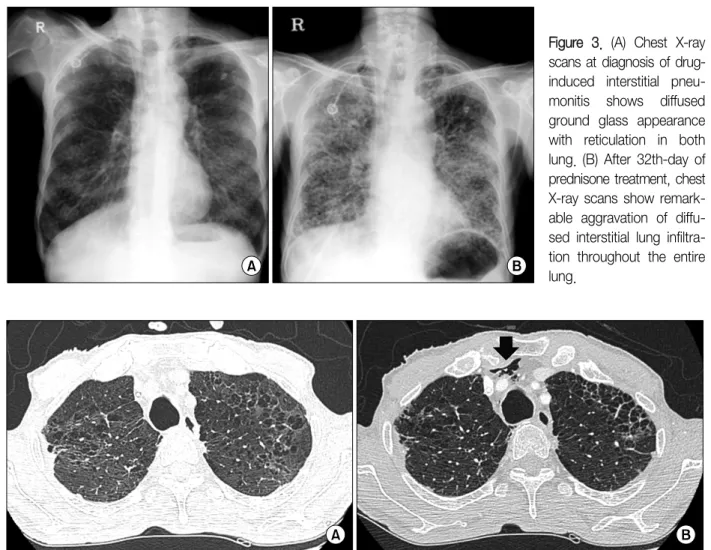
Additionally, granulocyte colony-stimulating factor (G-CSF) being used for the recovery from neutropenia can exacerbate lung injury. However, most of the patients reported previously that the drug-induced interstitial pneumonitis were developed after three to four cycles of chemotherapy. Hereby, we report a case of peripheral T cell lymphoma which rapidly developed a fatal interstitial pneumonitis after the first cycle of combined chemotherapy with cyclophosphamide, adriamycin, vincristine, prednisolone, and etoposide and the patient had also treated with G-CSF during neutropenic period.

Key Words: Lymphoma; Lung Diseases, Interstitial; Granulocyte Colony-Stimulating Factor; Drug Therapy

Address for correspondence: Je-Jung Lee, M.D., Ph.D.

Department of Hematology-Oncology, Chonnam National University Hwasun Hospital, Chonnam National University Medical School, 322 Seoyang-ro, Hwasun 519-763, Korea Phone: 82-61-379-7638, Fax: 82-61-379-7628

E-mail: [email protected] Received: Jun. 4, 2012

Revised: Jul. 23, 2012 Accepted: Oct. 5, 2012

CCIt is identical to the Creative Commons Attribution Non-Commercial License (http://creativecommons.org/licenses/by-nc/3.0/).

Introduction

Patients with hematological malignancies can develop to various acute pulmonary diseases during the course of chemotherapy, and interstitial pneumonitis induced by chemotherapeutic agents is commonly seen as a pul- monary complication

1,2

. In addition, the use of gran- ulocyte colony-stimulating factor (G-CSF) during the treatment of chemotherapy-induced neutropenia en- hanced the lung injury

2-5

. However, in most reported cases, the patients developed chemotherapeutic drug-in-

duced interstitial lung disease after several cycles of che- motherapy

1

.

Here, we report a rare case of peripheral T cell lym- phoma, not otherwise specified (PTCL, NOS), with rap- idly deteriorating fatal interstitial lung disease that devel- oped after the first cycle of cyclophosphamide, adriamy- cin, vincristine, and prednisolone (CHOP) and etopo- side combination chemotherapy along with G-CSF treat- ment.

Case Report

A 69-year-old man was admitted to the hospital pre-

senting with multiple palpable masses in the neck and

groin area. He had hypertension and a 30-pack year

smoking history. The Eastern Cooperative Oncology

Group performance status was grade 2, and lactate de-

hydrogenase level was 824 IU/L (normal, 218–472 IU/L)

at diagnosis. A lymph node biopsy was performed in

the right groin, and the patient was diagnosed with

(2)

Figure 1. Positron emission tomography scan showing disseminated lymphomatous involvements in the tonsils, spleen, left anterior chest wall, and multiple lymph nodes on both sides of the diaphragm.

Figure 2. Follow-up chest computed tomography scan during chemotherapy. (A) At diagnosis of peripheral T cell lympho- ma, not otherwise specified, multiple lymph node enlargement and diffuse centrilobular emphysema were observed.

(B) After the first cycle of chemotherapy with cyclophosphamide, adriamycin, vincristine, and prednisolone along with etoposide, the scan shows newly-developed diffused ground glass opacities with reticulation and peripheral predom- inance in both lungs.

PTCL, NOS. Further, neck, chest, and abdominal com- puted tomography (CT), positron emission tomography/

computed tomography, and bone marrow examination were conducted for the staging work-up. Lymphoma- tous involvements were found in the tonsil, spleen, left anterior chest wall, ileocecal area, and multiple lymph nodes on both sides of the diaphragm (Figure 1).

However, no involvement in the bone marrow was

noted. The initial chest CT scan showed multiple lym- phadenopathies and, incidentally, diffuse centrilobular emphysema (Figure 2). Finally, the patient was diag- nosed with Ann Arbor stage IV PTCL, NOS and he was considered to be at high risk according to the interna- tional prognostic index. He received the first cycle of CHOP with etoposide combination chemotherapy (750 mg/m

2

cyclophosphamide i.v. on day 1 [D1], 1.4 mg/m

2

vincristine i.v. on D1, 50 mg/m

2

doxorubicin i.v. on D1, 60 mg/m

2

prednisolone p.o. on D1–5, and etoposide 100 mg/m

2

i.v. on D1). Eight days after the chemo- therapy, he developed a neutropenia, and, then, lenog- rastim (5 μg/kg, i.v.) was administered for 7 consec- utive days. The second cycle of chemotherapy was de- layed for 1 week because he had developed a moderate degree of small bowel bleeding while recovering from the neutropenia; the bleeding was controlled by suppor- tive care only.

After 30 days of the first cycle of combination chemo-

therapy, the patient was hospitalized for the second cy-

cle of CHOP with etoposide chemotherapy. Unfortuna-

tely, he complained of exertional dyspnea and shortness

of breath without fever or purulent sputum. Crackles

were noted in both lower lung fields on chest auscul-

tation. The initial laboratory examination revealed a

white blood cell count of 12,900/μL, hemoglobin level

(3)

Figure 3. (A) Chest X-ray scans at diagnosis of drug- induced interstitial pneu- monitis shows diffused ground glass appearance with reticulation in both lung. (B) After 32th-day of prednisone treatment, chest X-ray scans show remark- able aggravation of diffu- sed interstitial lung infiltra- tion throughout the entire lung.

Figure 4. Chest computed tomography (CT) scan showing small amounts of newly appearing pneumomediastinum (black arrow), without evidence of aero-digestive tract injury (B) comparison with the follow-up chest CT on 30th days after the first cycle of chemotherapy with cyclophosphamide, adriamycin, vincristine, and prednisolone along with etoposide (A).

of 12.9 g/dL, and platelet count of 328,000/μL. The level of C-reactive protein was 14.31 mg/dL. Arterial blood gas analysis (ABGA) with 2 L/min oxygen ad- ministered via nasal prongs showed that the pH was 7.489, PaO

2

75.9 mm Hg, PaCO

2

28.4 mm Hg, bicar- bonate level 21.1 mmol/L, and SaO

2

96.3%, showing respiratory alkalosis. A chest X-ray showed diffuse ground glass appearances with reticulation in both lungs (Figure 3). A chest CT scan showed to the mark- edly improvements of previous multiple lymphomatous involvements in the thorax, but it also revealed that both lungs had a newly developed peripherally pre- dominant diffuse ground glass appearance with retic-

ulation (Figure 2).

For evaluation of associated pulmonary infection, the

patient was performed the rapid influenza antigen im-

munoassays that can identify influenza A and B viral nu-

cleoprotein antigens, sputum culture for the evaluation

of bacterial pneumonia, acid-fast bacillus (AFB) smear,

Mycobacterium tuberculosis polymerase chain reaction

(PCR) test and Pneumocystis PCR in the sputum. Addi-

tionally, we checked the serum galactomannan assay for

screening of aspergillosis infection before treatment of

antibiotics. The influenza A or B antigen immunoassay,

AFB stain, M. tuberculosis PCR test and Pneumocystis

PCR were all negative and bacteria was not cultured in

(4)

the respiratory specimen. The galactomannan assay showed the negative result (0.1 optical density index) and there was no objective evidence about pulmonary infection. Further diagnostic studies such as bron- choalveolar lavage (BAL) or lung biopsy were not per- formed because the symptom of dyspnea was deteriorated. Based on the clinical status and the radio- logical finding, the patient was diagnosed with drug-in- duced interstitial pneumonitis, and 125 mg/day pre- dnisone was administered immediately with the empiri- cal antibiotics such as tazobactam and clarithromycin.

After the 15th day of prednisone therapy, a chest CT scan showed no changes, and a small amount of pneu- momediastinum had newly developed (Figure 4). After the 32th day of prednisone treatment, the symptoms of dyspnea were aggravated up to grade 4 according to the Medical Research Council Dyspnea Scale, and a chest X-ray showed diffuse interstitial lung infiltration throughout the entire lung (Figure 2). At that time, we did not check the additional evaluations, such as Pneumocystis PCR or BAL study, because of the wors- ened his condition. ABGA showed that the patient was experiencing respiratory distress as his PaO

2

was 59.6 mm Hg with 10 L/min oxygen supply via mask. There- fore, 375 mg/day prednisone was administered, but the patient died of respiratory failure on the 38th day after the treatment of prednisone.

Discussion

The previously reported chemotherapeutic drugs that induce interstitial pneumonitis include bleomycin, me- thotrexate, cyclophosphamide, and busulfan

2

. In fact, the mechanisms by which bleomycin and cyclopho- sphamide induce pulmonary damages have been stud- ied in detail

6

. These drugs induce the production of O

2

radicals by neutrophils (sometimes in the presence of Cu

2+

or Fe

2+

), which result in the lipid peroxidation and DNA damage

7

. It follows that interstitial pneumo- nitis induced by these drugs is exacerbated under con- ditions of high O

2

pressure. Thus, O

2

radicals produced by neutrophils possibly contribute to the development

of drug-induced interstitial pneumonitis, particularly in lungs already damaged by certain chemotherapeutics

8

. G-CSF has been reported to stimulate superoxide pro- duction in neutrophils when administered with N-for- mylmethionyl-leucyl-phenylalanine or ionomycin in vi- tro

9

, G-CSF probably induces interstitial pneumonitis through neutrophil elastase production

10

. G-CSF is usu- ally produced by fibroblasts, and it in turn enhances their proliferation

11

. Thus, these four mechanisms, namely, 1) superoxide production by cytotoxic drugs, 2) oxidant production by G-CSF, 3) elastase production by G-CSF, and 4) fibroblast proliferation by G-CSF, are potentially involved in the development of interstitial pneumonitis. In our case, chemotherapeutic agents, in- cluding cyclophosphamide, and G-CSF administration, can be considered the contributing factors in the devel- opment of interstitial pneumonitis.

The interval between the administration of chemo- therapeutic agents and the onset of respiratory symp- toms varies from weeks to years

12

. However, in many cases reported around the world, interstitial develop- ments were observed in the third or subsequent chemo- therapeutic cycles

13,14

. However, in the present case, the patient showed clinical signs of acute interstitial pneu- monitis only after the first cycle of chemotherapy. He had a 30-pack year smoking history, and the initial chest CT scan had shown emphysematous changes. Previous studies established that pneumonitis associated with chemotherapy occurred significantly and more fre- quently in patients with preexisting interstitial lung dis- ease

15

. Therefore, the cyclophosphamide and G-CSF may have accelerated the lung damage in this patient who had an existing lung disease.

This case has several limitations because we did not perform the lung biopsy for tissue confirmation and the BAL to identify the bacteriologic pathogen also was not checked for reason of deteriorated condition. However, the laboratory findings, the typical pulmonary fibrosis on the imaging study, and rapid clinical courses strongly indicated the diagnosis of drug-induced interstitial pneumonitis.

This patient illustrates the PTCL, NOS, with rapidly

(5)

deteriorating, fatal interstitial lung disease that devel- oped after the first cycle of chemotherapy along with G-CSF therapy. Modification of the G-CSF dose may be useful in preventing pneumotoxicity, and early applica- tion of a high-dose corticosteroid for patients with inter- stitial pneumonitis would be considered in addition to the discontinuation of G-CSF

1

. Therefore, clinicians should be alert to the signs and symptoms of lung injury during chemotherapy, especially in patients with preex- isting interstitial lung disease.

References

1. Nakase K, Tsuji K, Nagaya S, Tamaki S, Tanigawa M, Ikeda T, et al. Acute interstitial pneumonitis during chemotherapy for haematological malignancy. Eur J Cancer Care (Engl) 2005;14:336-41.

2. Meadors M, Floyd J, Perry MC. Pulmonary toxicity of chemotherapy. Semin Oncol 2006;33:98-105.

3. Azoulay E, Attalah H, Yang K, Herigault S, Jouault H, Brun-Buisson C, et al. Exacerbation with granulocyte colony-stimulating factor of prior acute lung injury dur- ing neutropenia recovery in rats. Crit Care Med 2003;

31:157-65.

4. Aggarwal A, Baker CS, Evans TW, Haslam PL. G-CSF and IL-8 but not GM-CSF correlate with severity of pul- monary neutrophilia in acute respiratory distress syndrome. Eur Respir J 2000;15:895-901.

5. Wiedermann FJ, Mayr AJ, Hobisch-Hagen P, Fuchs D, Schobersberger W. Association of endogenous G-CSF with anti-inflammatory mediators in patients with acute respiratory distress syndrome. J Interferon Cytokine Res 2003;23:729-36.

6. Cooper JA Jr, White DA, Matthay RA. Drug-induced pulmonary disease. Part 1: Cytotoxic drugs. Am Rev Respir Dis 1986;133:321-40.

7. Burger RM, Peisach J, Horwitz SB. Activated bleomy-

cin: a transient complex of drug, iron, and oxygen that degrades DNA. J Biol Chem 1981;256: 11636-44.

8. Lindemann A, Herrmann F, Oster W, Haffner G, Meyenburg W, Souza LM, et al. Hematologic effects of recombinant human granulocyte colony-stimulating factor in patients with malignancy. Blood 1989;74:

2644-51.

9. Yuan L, Inoue S, Saito Y, Nakajima O. An evaluation of the effects of cytokines on intracellular oxidative production in normal neutrophils by flow cytometry.

Exp Cell Res 1993;209:375-81.

10. Iijima S, Tsuji T, Sugita M, Yamada N, Satoh T, Kubo T. Leukocyte activity and occurrence of tissue injury by G-CSF. Nihon Sanka Fujinka Gakkai Zasshi 1993;45:

213-9.

11. Mendoza JF, Caceres JR, Santiago E, Mora LM, Sanchez L, Corona TM, et al. Evidence that G-CSF is a fibroblast growth factor that induces granulocytes to increase phagocytosis and to present a mature morphology, and that macrophages secrete 45-kd molecules with these activities as well as with G-CSF-like activity. Exp Hematol 1990;18:903-10.

12. Brieva J. Cyclophosphamide-induced acute respiratory distress syndrome. Respirology 2007;12:769-73.

13. Katsuya H, Suzumiya J, Sasaki H, Ishitsuka K, Shibata T, Takamatsu Y, et al. Addition of rituximab to cyclo- phosphamide, doxorubicin, vincristine, and predni- solone therapy has a high risk of developing interstitial pneumonia in patients with non-Hodgkin lymphoma.

Leuk Lymphoma 2009;50:1818-23.

14. Ennishi D, Terui Y, Yokoyama M, Mishima Y, Takahashi S, Takeuchi K, et al. Increased incidence of interstitial pneumonia by CHOP combined with ritu- ximab. Int J Hematol 2008;87:393-7.

15. Togashi Y, Masago K, Handa T, Tanizawa K, Okuda C, Sakamori Y, et al. Prognostic significance of preex- isting interstitial lung disease in Japanese patients with small-cell lung cancer. Clin Lung Cancer 2012;13:

304-11.

수치

Figure 2. Follow-up chest computed tomography scan during chemotherapy.  (A) At diagnosis of peripheral T cell lympho- lympho-ma,  not  otherwise  specified,  multiple  lymph  node  enlargement  and  diffuse  centrilobular  emphysema  were  observed.
Figure  3.  (A)  Chest  X-ray  scans at diagnosis of  drug-induced  interstitial   pneu-monitis  shows  diffused  ground  glass  appearance  with  reticulation  in  both  lung

참조

관련 문서