계명대학교 의과대학 내과학교실1, 예방의학교실2, 핵의학교실3 이정은1, 민보람1, 박재석1, 박훈표1, 전미정2, 원경숙3, 최원일1
Right Ventricle Ejection Fraction Contributes Severity of Dyspnea in Chronic Obstructive Pulmonary Disease (COPD)
Jung Eun Lee, M.D.1, Bo Ram Min, M.D.1, Jae Seok Park, M.D.1, Hun Pyo Park, M.D.1, Mi Jung Jun, M.S.2, Kyung Sook Won, M.D.3, Won Il Choi, M.D.1
Departments of Medicine1, Preventive Medicine2, and Nuclear Medicine3, Keimyung University School of Medicine, Daegu, Korea
Background: Patients with COPD generally complain of very different degrees of dyspnea regardless of their pulmonary function. The study, we assessed the right ventricular ejection fraction in relation to dyspnea in COPD patient.
Methods: The pulmonary function including the diffusion capacity was measured. The right ventricle ejection fraction (RVEF) was measured using a first-pass radionuclide scan by multigated acquisition (MUGA). Forty patients with chronic obstructive pulmonary disease (COPD) were stratified for dyspnea according to the Medical Research Council (MRC) scale. Moderate dyspnea and severe dyspnea is defined as MRC 2/3 (n = 16) and MRC 4/5 (n = 24) respectively.
Results: The baseline pulmonary function tests including DLCO and the resting arterial blood gas were similar in the moderate and severe dyspnea group, with the exception of the residual volume (% predicted) (moderate 160 ± 27, severe 210 ± 87, p < 0.03). The right ventricle ejection fraction was significantly (p < 0.001) lower in the severe dyspnea group (25 ± 8) than in the moderate group (35 ± 6). The independent factor assessed by multiple logistic regression revealed only the severity of dyspnea to be significantly associated with RVEF (p < 0.02).
Conclusion: This study showed that the right ventricle ejection fraction would contributes to severity of dyspnea in patients with a similar pulmonary function (Tuberc Respir Dis 2006; 60: 631-637)
Key Words: COPD, Ejection fraction, Dyspnea.
Address for correspondence : Won-Il Choi, M.D.
Department of Medicine,
Keimyung University School of Medicine 194 Dongsan-Dong, Jung-Gu,
Daegu, 700-712, Korea
Telephone : +82-53-250-7405 Fax : +82-53-250-7434 E-mail : [email protected]
Received : Apr. 18. 2006 Accepted : Jun. . 2006
서 론
만성폐쇄성폐질환에서 호흡곤란은 매우 중요한 증 상 중의 하나이며 호흡곤란의 정도는 기류폐색의 정 도보다 더 의미 있는 생존율 예측인자로 알려져 있다
1,2. 일반적으로 폐기능이 감소할수록 호흡곤란이 심 해지지만, 비슷한 정도로 폐기능이 감소한 환자에서 도 호흡곤란의 정도는 다양하다3,4.
만성폐쇄성폐질환에서 호흡곤란을 만드는 중요한 이유로는 중추계에서 일어나는 호흡근육 자극의 증 가에 비해 흡기근육의 부적절한 반응5-8과 동적인 폐
과팽창에 의해 발생하는 것으로 알려져 있다9,10. 그러 나 임상에서는 폐활량 및 폐용적 그리고 일산화탄소 확산능 등이 매우 유사함에도 불구하고 다양한 정도 의 호흡곤란을 호소하는 환자를 볼 수 있다.
만성폐쇄성폐질환은 폐순환의 혈류역학을 변화시 켜 폐혈관저항을 증가시키고 궁극적으로는 우심실 기능부전을 일으킬 수 있다. FEV1 (forced expiratory volume in one second)예측치의 평균이 40% 이하인 만성폐쇄성폐질환 환자의 혈류역학을 조사한 연구에 서 폐동맥압은 정상범위에 있었지만 폐혈관저항은 증가하였고 폐혈관저항의 증가는 생존률과 역상관 관계가 있음을 보고한 바 있다.11 우심실박출계수는 우심실 수축기능을 반영하며 우심실 후부하, 즉 폐혈 관저항에 의해서 영향을 받는다12-14. 그러므로 폐혈관 저항이 증가하는 만성폐쇄성폐질환에서는 우심실 박 출계수가 감소할 수 있다.
종합하면, 호흡곤란은 만성폐쇄성폐질환의 예후의 중요한 예측인자 중의 하나이며, 예후는 폐혈관저항
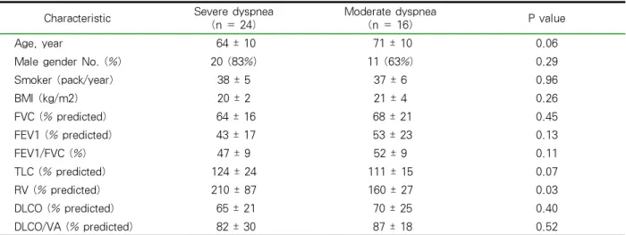
Characteristic Severe dyspnea (n = 24)
Moderate dyspnea
(n = 16) P value
Age, year 64 ± 10 71 ± 10 0.06
Male gender No. (%) 20 (83%) 11 (63%) 0.29
Smoker (pack/year) 38 ± 5 37 ± 6 0.96
BMI (kg/m2) 20 ± 2 21 ± 4 0.26
FVC (% predicted) 64 ± 16 68 ± 21 0.45
FEV1 (% predicted) 43 ± 17 53 ± 23 0.13
FEV1/FVC (%) 47 ± 9 52 ± 9 0.11
TLC (% predicted) 124 ± 24 111 ± 15 0.07
RV (% predicted) 210 ± 87 160 ± 27 0.03
DLCO (% predicted) 65 ± 21 70 ± 25 0.40
DLCO/VA (% predicted) 82 ± 30 87 ± 18 0.52
Values are patient number or means with standard deviation (percentage) BMI = body mass index
FEV1 = forced expiratory volume in one second FVC = forced vital capacity
TLC = total lung capacity RV = residual volume
DLCO = carbon monoxide diffusion capacity VA = alveolar volume
Table 1. Characteristics and Lung function of the study subjects 과도 밀접한 연관이 있으므로, 호흡곤란의 정도와 폐
혈관저항을 반영하는 우심실 박출계수와의 관계를 알아보고자 본 연구를 계획하였다. 외래 환자들을 대 상으로 비침습적인 방법을 통해 우심실 기능을 측정 하고, 이것과 호흡곤란과의 연관이 있는지를 확인하 고자 한다.
대상 및 방법 1. 대상환자
본 연구는 호흡곤란, 만성기침 및 객담을 주소로 내원한 환자에서 20 갑-년 이상의 흡연력이 있으면 서, FEV1/FVC비가 70미만인 만성폐쇄성폐질환 환자 중에서 3개월 이상 호흡곤란의 증상의 변화가 없고, 과거 심근경색의 병력이 없는 자들을 대상으로 하였 고, 2003년 12월부터 2005년 5월까지 계명의대 동산 병원을 방문한 자를 대상으로 조사하였다. 흉부방사 선에서 간질성폐질환의 음영이 의심되는 경우, 근육 질환, 또는 흉벽변형이 있는 경우에는 대상자에서 제
외하였다.
2. 검사장비 및 방법
폐활량은 미국 SensorMedics사의 6200 Autobox DL Pulmonary Function Laboratory를 이용하였다.
Plethysmographic 방법으로 기능적잔기량(functional residual capacity; FRC)을 측정하였다15. 폐활량과 폐 용적 및 폐확산능의 추정정상치는 유럽흉부학회에서 제시한 식으로 계산하였다16. 폐확산능은 전술한 바와 같이 측정하였다17.
3. 우심실 박출계수의 측정
우심실 박출계수의 측정은 게이티드(gated) 심장 혈액풀 스캔을 이용하였다. 먼저 Tc-99m에 환자의 적혈구를 표지하였는데 이는 간접법을 이용하였고 방법은 아래와 같다. 적혈구표지 바이알(RBC kit)에 생리식염수 3 ml를 넣어 희석시킨 다음, 5분 후에 희 석액 1 ml를 뽑아 환자에게 정맥주사하였다. 다시 20
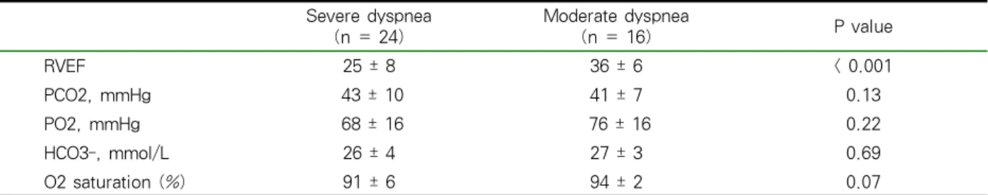
Severe dyspnea (n = 24)
Moderate dyspnea
(n = 16) P value
RVEF 25 ± 8 36 ± 6 < 0.001
PCO2, mmHg 43 ± 10 41 ± 7 0.13
PO2, mmHg 68 ± 16 76 ± 16 0.22
HCO3-, mmol/L 26 ± 4 27 ± 3 0.69
O2 saturation (%) 91 ± 6 94 ± 2 0.07
Values are means with standard deviation
Table 2. Right ventricle ejection fraction (RVEF) and resting blood gas of the study subjects 분을 기다려 Tc-99m 20 mCi를 환자에게 정맥주사하
고, 5-10분 후 촬영을 시작하였다. 스캔은 환자를 앙 와위로 놓고 안정시킨 후 좌전사위로 검출기가 환자 의 심장부위에 가깝게 위치하도록 한 후 전체 600개 의 심박수를 얻었다. 저에너지 일반목적용 조준기를 장착한 감마카메라(Vertex, ADAC Co., USA)를 사 용하였으며 매트릭스 크기는 64×64, 줌(zoom)은 2.19, 심박주기당 16개의 frame을 얻었으며 허용변이범위 는 20%로 얻었다. 얻어진 영상에서 우심실에 반자동 방법으로 이완기말(end diastolic)과 수축기말(end systolic)의 관심영역을 그려 우심실 박출계수를 측정 하였다
4. 통계처리
폐기능 검사치 및 우심실 박출계수의 결과는 평균
± 표준편차로 표현하였다. 두 집단의 연관성 분석은 Fisher’s exact test, 두 군 사이의 평균치 비교는 Wilcoxon signed rank test, 그리고 호흡곤란에 미치 는 인자는 다중회귀분석법을 이용하였다. 통계패키지 는 spss 11.0 version을 사용하였고 유의수준 5% 이 하로 검증하였다.
결 과 1. 대상군 특성
조사 대상자는 모두 40명으로, Medical Research Council (MRC) 호흡곤란 등급을 기준으로 4도와 5도 의 호흡곤란을 호소하는 경우 중증군으로 분류하였
고, MRC 2도와 3도의 호흡곤란을 호소하는 경우를 중등도군으로 분류하였다. 중증군은 24명이었고 중등 도군은 16명이었다. 중증군과 중등도군의 기저 특성 의 비교에서 폐활량치 및 확산계수는 두 군 사이에 유의한 차이는 관찰되지 않았으나, 중증군에서 잔기 량이 중등도군에 비해 유의하게 증가하였다(Table 1).
2. 우심실 박출계수와 동맥혈가스 소견
우심실 박출계수는 중증군이 중등도군에 비해 유 의하게 낮았다(P < 0.001). 동맥혈가스 분석에서는 중증군이 중등도군보다 산소포화도가 낮은 경향을 보였으나 통계적으로 유의하지 않았다(P = 0.07). 이 산화탄소분압, 산소분압 등은 두 군 사이에 유의한 차 이가 관찰되지 않았다.
3. 호흡곤란 관련 인자
호흡곤란에 영향을 미칠 수 있는 변수들을 다중회 귀분석방법으로 분석한 결과에서 우심실 박출계수만 이(P = 0.02) 유의한 인자로 관찰되었다(Table 3).
고 찰
본 연구에서는 만성폐쇄성폐질환 환자를 중증 호 흡곤란군(MRC 4/5도)과 중등증 호흡곤란군(MRC 2/3도)으로 나누어 비교분석하였다. 우심실박출계수 는 중증군에서 중등증군에 비해 유의하게 낮았고, 잔 기량은 중증군에서 유의하게 증가 되었으나 폐활량 은 두 군 사이에 유의한 차이가 관찰되지 않았다
B S.E. Sig. OR 95% CI
Age 0.01 1.29 0.19 1.10 0.95 ~ 1.28
Smoking -0.04 -1.25 0.21 0.95 0.89 ~ 1.28
FEV1 (% predicted) 0.12 1.17 0.24 1.12 0.92 ~ 1.38
FEV1/FVC (%) -0.05 -0.58 0.24 0.94 0.77 ~ 1.15
TLC (% predicted) 0.11 1.10 0.26 1.12 0.91 ~ 1.35
RV (% predicted) -0.04 -1.17 0.24 0.95 0.88 ~ 1.03
DLCO (% predicted) -0.06 -0.06 0.37 0.94 0.82 ~ 1.07
O2 Saturation (%) 0.31 0.31 0.59 1.37 0.42 ~ 4.44
RVEF 0.27 2.26 0.02 1.30 1.03 ~ 1.63
Abbreviations: B., regression coefficient; S.E., standard error; Sig., significance; OR, odd ratio; CI, confidence interval Table 3. Factors associated with dyspnea in patients with COPD
(Table 1, 2). 다중회귀분석을 통해서 우심실박출계수 가 호흡곤란에 유의하게 영향을 미친 독립적인 인자 로 밝혀졌다(Table 3).
우심실 박출계수의 정상치는 43-65% 정도이며, 좌 심실 박출계수의 정상치는 60-70%이며, 우심실 박출 계수가 좌심실 박출계수 보다 낮다18-20. 일반적으로 우심실 박출계수가 40-45% 이하이면 감소된 것으로 평가한다. 만성폐쇄성폐질환에서 우심실 박출계수는 19-70%로 매우 다양한 정도로 변화가 있으며21, 이러 한 차이는 폐활량의 감소 정도와는 연관성이 적었다
22. 본 연구에서도 폐활량이 유사한 두 군에서 우심실 박출계수는 유의한 차이가 있었다. 중증 폐기종환자 의 우심실 박출계수의 평균이 32% 였으며23, 본 연구 에서도 이와 유사하게 우심실 박출계수의 평균은 31% 였고 범위는 11-63%였다.
만성폐쇄성폐질환의 폐혈관저항 증가는 혈류역학 에서 중요한 변화로 인식되고 있으나 폐혈관저항의 증가가 폐동맥 고혈압으로 바로 이어지지는 않는 것 으로 보인다. 만성폐쇄성폐질환 환자 1264명에서 폐 동맥압을 조사한 결과 이 중 단지 11명의 환자만이 폐동맥고혈압이 폐쇄성폐질환에 의해 발생한 것으로 추정되었다24. 이러한 결과 미루어 볼 때 만성폐쇄성 폐질환 환자에서 폐동맥고혈압이 있을 경우 다른 병 발 질환을 고려하여야 함을 알 수 있다. FEV1의 예측 치가 45% 정도인 환자 131명을 7년간 추적해서 관찰 한 폐동맥압의 변화를 보면 안정시 폐동맥 고혈압을 보인 환자가 없다가 7년 후 33명(25%)의 환자에서 경 증의 폐동맥 고혈압이 발생하였다25. 이러한 보고들로
판단해 보면, 만성폐쇄성폐질환에서 폐동맥 고혈압의 발생은 빈도가 낮고 발생한다 하더라도 폐동맥혈압 은 정상에 비해 조금 더 상승한 정도이다. 따라서 만 성폐쇄성폐질환에서는 다른 질환의 감별이 필요한 경우에 선택적으로 침습적 방법을 사용하여 폐동맥 압을 측정하는 것이 바람직 할 것으로 보인다. 폐동맥 압의 측정은 도플러심장초음파검사를 이용해서도 할 수 있는데, 이 방법으로는 이미 많이 진행한 폐질환에 서는 폐동맥압을 과대평가를 할 수 있다26. 이에 만성 폐쇄성폐질환에서 비 침습적인 방법이면서 우심실기 능 및 폐순환의 간접적인 평가를 할 수 있는 우심실 박출계수의 임상적인 유용성을 확인하는 노력이 있 어왔다23,27-30.
만성폐쇄성폐질환에서 호흡곤란의 기전으로는 동 적인 기도압박 (dynamic airway compression)에 의 한 기도의 수용체 자극에 의해 발생할 수 있으며31, 저 산소혈증, 그리고 고탄산혈증을 들 수 있다32. 만성폐 쇄성폐질환에서는 흔히 폐의 과다팽창소견이 관찰되 며, 폐의 과다팽창은 횡격막을 편평하게 만들어서 흡 기시 호흡일을 증가시키게 된다. 호흡일의 증가 또한 호흡곤란을 일으키는 이유이며, 이에 더해서 폐의 과 다팽창은 폐혈관저항을 증가시켜서33 호흡곤란을 더 악화시킬 수 있음을 예상할 수 있다. 본 연구에서는 폐의 동적 과다팽창을 조사하지 못했지만, 안정시의 폐활량과 폐용적이 유사하다 할지라도 동적과다팽창 정도에 따라 활동시 호흡곤란이 다르게 나타날 수 있 을 것임을 예측할 수 있다.
폐용적감소술을 통해서 얻은 결과에 따르면, 우심
실기능의 부전은 과팽창에 의해 주로 발생하는 것으 로 볼 수 있다23. 그러나 과팽창 이외에도 혈관내피세 포 손상에 의해서도 혈류역학에 문제를 초래할 수 있 음이 알려져 있다. 경증의 만성폐쇄성폐질환에서부터 중증의 만성폐쇄성폐질환에까지 모두 폐동맥의 혈관 내피세포의 기능부전이 있으며34,35, 이러한 혈관내피 세포의 기능부전은 endothelial nitric oxide synthase (eNOS)의 발현 감소와 연관되어 만성폐쇄성폐질환 환자에서 폐동맥고혈압을 유발한다36. 또한 흡연에 의 해서도 eNOS의 발현이 감소될 수 있다37. 결과에 기 술하지 않았지만, 본 연구에서는 폐용적 차이와 우심 실기능부전 사이의 유의한 상관관계가 관찰되지 않 았으며 이러한 점을 고려해 볼 때, 흡연에 대한 폐용 적 반응과 폐혈관 반응이 일치하지 않을 수 있으리라 추측할 수 있으며, 비슷한 폐기능에도 불구하고 우심 실 박출계수가 감소한 것은 혈관내피세포 손상의 정 도의 차이에 따라서 폐혈관저항이 증가하여 발생한 것으로 유추할 수 있겠다.
결론적으로, 만성폐쇄성폐질환에서 안정시 측정한 우심실 박출계수와 호흡곤란은 서로 연관성이 있으 며, 특히 폐기능에 비해 호흡곤란이 심한 환자에서 우 심실 박출계수를 측정하는 것은 호흡곤란의 인자를 감별하는데 도움이 되리라 사료된다.
요 약
배 경: 만성폐쇄성폐질환 환자의 호흡곤란은 일반 적으로 폐활량에 반비례하나 유사한 폐기능에서도 서로 다른 호흡곤란을 호소한다. 본 연구는 만성폐쇄 성폐질환 환자에서 우심실박출계수와 호흡곤란의 정 도와 연관관계가 있는지를 알아보고자 한다.
방 법: 호흡곤란의 정도는 Medical Research Council (MRC) 호흡곤란척도로 분석하였고, MRC 4/5도인 중증군 24명과, MRC 2/3도인 중등증군 16명 을 비교 분석하였다. 심전도게이트 일회통과법을 이 용한 방사성동위원소 심조영술을 이용하여 우심실 박출계수를 구했으며, 안정시 동맥가스분석 및 폐기 능검사를 시행하였다.
결 과: 기저 폐기능에서 잔기량의 예측치 평균이
(%) 중증군에서(210 ± 87) 중등증군(160 ± 27)에 비 해 유의하게 증가되었으나(P < 0.03), 폐활량 및 확산 계수 등에서는 유의한 차이가 관찰되지 않았다. 우심 실 박출계수(%)는 중증군에서(25 ± 8) 중등증군(35 ± 6)에 비해 유의하게 감소되었으나(P < 0.001), 동맥혈 가스는 두 군 사이에 유의한 차이가 관찰되지 않았다.
다중회귀분석을 통해 우심실 박출계수가 독립적으로 호흡곤란에 영향을 미치는 인자로 밝혀졌다.
결 론: 만성폐쇄성폐질환에서 우심실 박출계수가 호흡곤란의 정도에 영향을 미치는 것으로 보인다.
참 고 문 헌
1. Celli BR, Cote CG, Marin JM, Casanova C, Montes de Oca M, Mendez RA, et al.The body-mass index, airflow obstruction, dyspnea, and exercise capacity index in chronic obstructive pulmonary disease. N Engl J Med 2004;350:1005-12.
2. Nishimura K, Izumi T, Tsukino M, Oga T. Dyspnea is a better predictor of 5-year survival than airway obstruction in patients with COPD. Chest 2002;121:
1434-40.
3. Mahler DA, Harver A. A factor analysis of dyspnea ratings, respiratory muscle strength, and lung func- tion in patients with chronic obstructive pulmonary disease. Am Rev Respir Dis 1992;145:467-70.
4. Wegner RE, Jorres RA, Kirsten DK, Magnussen H.
Factor analysis of exercise of exercise capacity, dyspnoea ratings and lung function in patients with severe COPD. Eur Respir J 1994;7:725-9.
5. Dodd DS, Brancatisano T, Engel LA. Chest wall mechanics during exercise in patients with severe chronic air-flow obstruction. Am Rev Respir Dis 1984;129:33-8.
6. Hamilton AL, Killian KJ, Summers E, Jones NL.
Muscle strength, symptom intensity, and exercise capacity in patients with cardiorespiratory disorders.
Am J Respir Crit Care Med 1995;152:2021-31.
7. Montes de Oca M, Rassulo J, Celli BR. Respiratory muscle and cardiopulmonary function during exercise in very severe COPD. Am J Respir Crit Care Med 1996;154:1284-9.
8. Marin JM, Montes de Oca M, Rassulo J, Celli BR.
Ventilatory drive at rest and perception of exertional dyspnea in severe COPD. Chest 1999;115:1293-300.
9. Belman MJ, Botnick WC, Shin JW. Inhaledbron- chodilators reduce dynamic hyperinflation during
exercise in patients with chronic obstructive pulmonary disease. Am J Respir Crit Care Med 1996;153:967-75.
10. O'Donnell DE, Webb KA. Exertional breathlessness in patients with chronic airflow limitation: the role of lung hyperinflation. Am Rev Respir Dis 1993;148:
1351-7.
11. Burrows B, Kettel LJ, Niden AH, Rabinowitz M, Diener CF. Patterns of cardiovascular dysfunction in chronic obstructive lung disease. N Engl J Med 1972;286:912-8.
12. Brent BN, Berger HJ, Matthay RA, Mahler D, Pytlik L, Zaret BL. Physiologic correlates of right ventricular ejection fraction in chronic obstructive pulmonary disease: a combined radionuclide and hemodynamic study. Am J Cardiol 1982;50:255-62.
13. Burger W, Allroggen H, Kober G. Right ventricular volumes determined by computerized thermodilution in ischaemic heart disease: effect of exercise and nitroglycerin. Int J Cardiol 1991;33:33-41.
14. Dhainaut JF, Brunet F, Monsallier JF, Villemant D, Devaux JY, Konno M, et al. Bedside evaluation of right ventricular performance using a rapid compu- terized thermodilution method. Crit Care Med 1987;
15:148-52.
15. Park HP, Park SH, Lee SW, Seo YW, Lee JE, Seo CK, et al. Change of lung volumes in chronic obstructive pulmonary disease patients with improve- ment of airflow limitation after treatment. Tuberc Respir Dis 2004;57:143-7.
16. Quanjer PH, Tammeling GF, Cotes JE, Pedersen OF, Peslin R, Yernault JC. Lung volumes and forced ventilatory flows: official statement of the European Respiratory Society. Eur Respir J 1993;6 (Suppl 16):5-40.
17. Seo YW, Choi WI, Lee JE, Park HP, Ko SM, Won KS, et al. Importance of carbon monoxide transfer coefficient (KCO) interpretation in patients with airflow limitation. Tuberc Respir Dis 2005;59:374-9.
18. Sechtem U, Pflugfelder PW, Gould RG, Cassidy MM, Higgins CB. Measurement of right and left ventricular volumes in healthy individuals with cine MR imaging. Radiology 1987;163:697-702.
19. Brynjolf I, Kelbaek H, Munck O, Godtfredsen J, Larsen S, Eriksen J. Right and left ventricular ejection fraction and left ventricular volume changes at rest and during exercise in normal subjects. Eur Heart J 1984;5:756-61.
20. Kumar A, Anel R, Bunnell E, Habet K, Zanotti S, Haery C, et al. Pulmonary artery occlusion pressure and central venous pressure fail to predict ventricular filling volume, cardiac performance, or the response
to volume infusion in normal subjects. Crit Care Med 2004;32:691-9.
21. Berger HJ, Matthay RA, Loke J, Marshall RC, Gottschalk A, Zaret BL. Assessment of cardiac performance with quantitative radionuclide angiocar- diography: right ventricular ejection fraction with reference to findings in chronic obstructive pulmonary disease. Am J Cardiol 1978;41:897-905.
22. Ellis JH Jr, Kirch D, Steele PP. Right ventricular ejection fraction in severe chronic airway obstruction.
Chest 1977;71:281-2.
23. Mineo TC, Pompeo E, Rogliani P, Dauri M, Turani F, Bollero P, et al. Effect of lung volume reduction surgery for severe emphysema on right ventricular function. Am J Respir Crit Care Med 2002;165:
489-94.
24. Chaouat A, Bugnet AS, Kadaoui N, Schott R, Enache I, Ducolone A, et al. Severe pulmonary hypertension and chronic obstructive pulmonary disease. Am J Respir Crit Care Med 2005;172:189-94.
25. Kessler R, Faller M, Weitzenblum E, Chaouat A, Aykut A, Ducolone A, et al."Natural history" of pulmonary hypertension in a series of 131 patients with chronic obstructive lung disease. Am J Respir Crit Care Med 2001;164:219-24.
26. Arcasoy SM, Christie JD, Ferrari VA, Sutton MS, Zisman DA, Blumenthal NP, et al.Echocardiographic assessment of pulmonary hypertension in patients with advanced lung disease. Am J Respir Crit Care Med 2003;167:735-40.
27. Keller CA, Ohar J, Ruppel G, Wittry MD, Goodgold HM. Right ventricular function in patients with severe COPD evaluated for lung transplantation.
Lung Transplant Group. Chest 1995;107:1510-6.
28. Mahler DA, Brent BN, Loke J, Zaret BL, Matthay RA. Right ventricular performance and central circulatory hemodynamics during upright exercise in patients with chronic obstructive pulmonary disease.
Am Rev Respir Dis 1984;130:722-9.
29. Matthay RA, Berger HJ, Davies RA, Loke J, Mahler DA, Gottschalk A, et al. Right and left ventricular exercise performance in chronic obstructive pulmon- ary disease: radionuclide assessment. Ann Intern Med 1980;93:234-9.
30. Olvey SK, Reduto LA, Stevens PM, Deaton WJ, Miller RR. First pass radionuclide assessment of right and left ventricular ejection fraction in chronic pulmonary disease: effect of oxygen upon exercise response. Chest 1980;78:4-9.
31. O'Donnell DE, Sanii R, Anthonisen NR, Younes M.
Effect of dynamic airway compression on breathing pattern and respiratory sensation in severe chronic
obstructive pulmonary disease. Am Rev Respir Dis 1987;135:912-8.
32. Manning HL, Schwartzstein RM. Pathophysiology of dyspnea. N Engl J Med 1995;333:1547-53.
33. Harris P, Segel N, Green I, Housley E. The influence of the airways resistance and alveolar pressure on the pulmonary vascular resistance in chronic bronhcitis.
Cardiovasc Res 1968;2:84-92.
34. Dinh-Xuan AT, Higenbottam TW, Clelland CA, Pepke-Zaba J, Cremona G, Butt AY, et al.
Impairment of endothelium-dependent pulmonary- artery relaxation in chronic obstructive lung disease.
N Engl J Med 1991;324:1539-47.
35. Peinado VI, Barbera JA, Ramirez J, Gomez FP, Roca J, Jover L, et al. Endothelial dysfunction in pulmonary arteries of patients with mild COPD. Am J Physiol 1998;274:L908-13.
36. Giaid A, Saleh D. Reduced expression of endothelial nitric oxide synthase in the lungs of patients with pulmonary hypertension. N Engl J Med 1995;333:
214-21.
37. Barbera JA, Peinado VI, Santos S, Ramirez J, Roca J, Rodriguez-Roisin R. Reduced expression of endothelial nitric oxide synthase in pulmonary arteries of smokers. Am J Respir Crit Care Med 2001;164:709-13.