Journal of Minimally Invasive Surgery Vol. 21. No. 2, 2018 https://doi.org/10.7602/jmis.2018.21.2.82
Laparoscopic Cholecystectomy in Two Patients with Situs Inversus Totalis: A Case Report
Jae Yool Jang, M.D., Woohyung Lee, M.D., Jinkyu Cho, M.D., Chi-Yeong Jeong, M.D., Ph.D., Soon-Chan Hong, M.D., Ph.D.
Department of Surgery, Gyeongsang National University Hospital, Jinju, Korea
Situs inversus totalis (SIT) is a rare condition in which the viscera are transposed in a mirror image reversal. We report two cases of laparoscopic cholecystectomy (LC) performed for SIT patients. A 63-year old male patient with SIT was diagnosed with symptomatic gallstones. We performed LC by 3-port method. The patient was discharged uneventfully on postoperative day 2. A 57-year old female patient with SIT underwent LC for acute cholecystitis. Due to severe inflammation an assistant was needed. The patient was discharged uneventfully on postoperative day 3. Over 80 cases of LCs in SIT patients have been reported so far and LC has become the standard treatment. The current report confirms the safety of laparoscopy in such cases. Laparoscopic cholecystectomy can be performed safely in SIT patients if care is taken. Surgeons need to be careful of reversed anatomy and unaccustomed working hand.
Keywords:
Laparoscopic cholecystectomy, Situs inversus totalis, Acute cholecystitisReceived August 16, 2017 Revised September 25, 2017 Accepted October 10, 2017
Corresponding author Soon-Chan Hong
Department of Surgery, Gyeongsang National University, College of Medicine, Gyeongsang National University Hospital, 79, Gangnam- ro, Jinju 52727, Korea
Tel: +82-55-780-8093 Fax: +82-55-757-5442 E-mail: [email protected] ORCID:
http://orcid.org/0000-0003-4499-8741
This is an Open Access article distributed under the terms of the Creative Commons Attribution Non-Commercial License (http://
creativecommons.org/licenses/by-nc/4.0/) which permits unrestricted non-commercial use, distribution, and reproduction in any medium, provided the original work is properly cited.
Copyright © 2018 The Journal of Minimally Invasive Surgery. All rights reserved.
Journal of Minimally Invasive Surgery
pISSN 2234-778X •eISSN 2234-5248 J Minim Invasive Surg 2018;21(2):82-85
CASE REPORT
INTRODUCTION
Situs inversus totalis (SIT) is a rare autosomal recessive anomaly in which thoracic and abdominal organs are trans- posed through the midline sagittal plane, resulting in a per- fect mirror image reversal of normal anatomy. This condition was first described by Fabricius in 1,600
1and its incidence is known to range from 1:10,000 to 1:20,000.
2Though there is no evidence that this condition is a risk factor for choleli- thiasis or acute/chronic cholecystitis,
3diagnosis can be dif- ficult when those diseases develop in SIT patients due to the confusing clinical presentations. Laparoscopic treatment is also challenging for surgeons because direction of approach is unfamiliar, and positioning of operator and ports placement should be planned in different way from those of conventional
method for patients with normal anatomy.
Herein we report our experience in two cases of laparo- scopic cholecystectomy performed for patients with SIT.
CASE REPORT
Case presentation 1
A 63-year old male patient was diagnosed with symptom-
atic gallbladder stones with SIT at a private clinic and referred
to our hospital for cholecystectomy. The patient complained of
recurrent colicky abdominal pain at left upper quadrant lasting
several months. On admission, the patient presented no symp-
toms of gallbladder stones. Laboratory results of liver function
tests and complete blood count were within normal range.
Laparoscopic Cholecystectomy in Situs Inversus Totalis
www.e-jmis.org
83
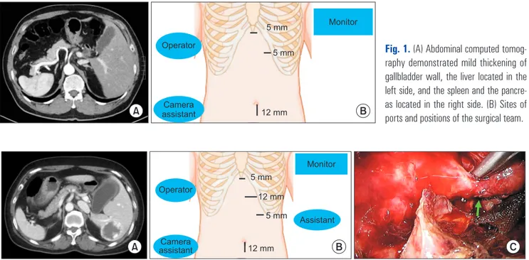
Abdominal computed tomography showed mild thickening of gallbladder wall and confirmed SIT (Fig. 1A). Abdominal ul- trasonography showed a few gallstones in the mildly collapsed gallbladder (Fig. 1B). No comorbid condition was identified from the evaluation for general anesthesia.
With the diagnosis of symptomatic gallbladder stones the patient underwent elective laparoscopic cholecystectomy. The patient was laid in supine position with his left side elevated, and the right-handed operating surgeon stood on the right side of the patient (Fig. 1B). The camera assistant stood on the right side of the patient and the laparoscopic stack was placed on the left side of the patient. A Veress needle was used for insufflation of the peritoneal cavity with carbon dioxide gas.
The port for a 12 mm flexible laparoscope was placed infra- umbilically, and two 5 mm ports were placed at the subxy- phoid midline and left subcostal margin of the midclavicular line respectively.
There was no sign of inflammation at the gallbladder and Calot’s triangle. Without traction of the Hartmann’s pouch by an assistant, dissection of the Calot ’s triangle seemed difficult and the operator decided to dissect the gallbladder from the liver first in a fundus-to-infundibulum direction. The 5 mm port placed at left subcostal midclavicular line was used as a working/dissecting port. After that the cystic duct and cystic artery were carefully isolated, clipped, and divided. Gallblad- der specimen was retrieved through the 12 mm port.
The operation took 60 minutes and estimated blood loss was
20 ml. There was no evidence of acute cholecystitis from the inspection of the specimen, and 5 black and round-shaped stones were detected. The patient was discharged uneventfully on the 2nd day after operation.
Case presentation 2
A 57-year old female patient with known SIT visited our emergency room with pain at left upper quadrant lasting for five days. The patient was afebrile, not jaundiced, and com- plained of tenderness and rebound tenderness at the left up- per quadrant. She had previously undergone total abdominal hysterectomy due to uterine myoma 5 years ago and that was when she was first informed to be diagnosed with SIT. Labo- ratory results were within normal range except for slightly increased white blood cell count (10,830 /mm
3) and serum C-reactive protein level (15.8 mg/dl). Abdominal computed tomography showed thickened gallbladder wall with pericho- lecystic fluid collection and confirmed SIT (Fig. 2A). She was a chronic hepatitis B virus carrier on antiviral medication.
With the diagnosis of acute cholecystitis the patient under- went emergency laparoscopic cholecystectomy. She was laid in supine position with her left side elevated, and the right- handed operating surgeon stood on the right side of the pa- tient (Fig. 2B). Position of surgical team was identical to that of case 1 with an additional assistant standing on the left side of the patient. Creation of pneumoperitoneum and port place-
Fig. 1. (A) Abdominal computed tomog- raphy demonstrated mild thickening of gallbladder wall, the liver located in the left side, and the spleen and the pancre- as located in the right side. (B) Sites of ports and positions of the surgical team.
A B
Operator
Camera assistant
Monitor 5 mm
12 mm 5 mm
Fig. 2. (A) Abdominal computed tomography demonstrated thickening of gallbladder wall with pericholecystic fluid collection, and confirmed SIT. (B) Sites of ports and positions of the surgical team. (C) Arrow indicates thickened cystic duct.
Assistant Monitor Operator
Camera
assistant 12 mm
5 mm
5 mm 12 mm
A B C
Jae Yool Jang et al.
Journal of Minimally Invasive Surgery Vol. 21. No. 2, 2018
84
ment were performed with the same method, with one ad- ditional 5 mm port placed at the left subcostal margin of an- terior axillary line. The port in the middle of the three 5 mm ports was used as a working/dissecting port, and was replaced by a 12 mm port later to apply large-sized surgical clips to the thickened cystic duct.
The gallbladder and hepatoduodenal ligament were covered by greater omentum which was detached easily. Wall of the gallbladder and Calot ’s triangle were severely inflamed and fi- brotic. To secure a clear view of Calot’s triangle, traction of the gallbladder was exerted by the assistant through the additional 5 mm port. Cystic artery was managed by ultrasonic scalpel, and the cystic duct was isolated from surrounding fibrotic tis- sues by blunt dissection using a suction device. After clipping and division of the cystic duct, the gallbladder was dissected from the liver using ultrasonic scalpel. After placement of a closed suction drain, hemostasis at the gallbladder bed by electrocautery was done. Gallbladder specimen was retrieved through the 12 mm port.
The operation took 90 minutes and estimated blood loss was 150 ml. Neck of the gallbladder was impacted by a 1.5 cm sized round pigment stone, and the pathologic result was gan- grenous cholecystitis with microabscess formation. The patient was discharged uneventfully on the 3rd day after operation
DISCUSSION
SIT is a genetic disorder of which the gene involved is lo- cated on the long arm of chromosome 14, and it is transmitted as an autosomal recessive nature with incomplete penetra- tion.
4Due to the mirror imaged transposition of thoracic and abdominal organs, diagnosis and surgical treatment of gall- bladder diseases developed in these patients may be more dif- ficult than that of orthotopic patients. The first laparoscopic cholecystectomy in a SIT patient was reported by Campos and Sipes in 1991,
5and since then over 80 other cases have been reported in literature, discussing the safety of laparoscopic cholecystectomy in such patients.
6Technical difficulties of laparoscopic cholecystectomy for SIT patients arise from the fact that everything in the surgi- cal field is transposed to mirror image except for operator ’s hands. Hence right-handed surgeons suffer from crossing of two devices when they use their right hand as a working hand in the setting of conventionally mirror imaged port placement, whereas left-handed or ambidextrous surgeons can perform the operation easily by simply changing the role of each hand.
To overcome this problem we used different methods for the two cases. In the case 1, we used 3 ports and approaching the Calot ’s triangle from the beginning of operation seemed diffi- cult without assisting traction. Hence the operator began dis-
section at the fundus first and to the infundibulum carefully.
This method was found to help isolating the cystic duct and cystic artery safely and easily later. In case 2, we added the 4th port at the costal margin of the left anterior axillary line for assisting traction of the distended and inflamed gallblad- der. By this method we could stretch the Calot’s triangle effi- ciently and isolate the cystic duct and the cystic artery without crossing two devices.
Other reports described variable methods to overcome the difficulties caused by the mirror-image anatomy. Ren JJ et al.
described that they used 4-port method with assisting trac- tion, and the right-handed operator used his left hand as a main operating hand while his right hand was holding the gallbladder.
7Iusco DR et al. laid the patient in French posi- tion with the operator standing between the patient ’s legs, and modified the placement of 3 ports - a camera port at the right upper quadrant, an infraumbilical port and a left flank port for the operator. The operator changed his working/dissecting hand as occasion demands.
8Alsabek MB et al. used conven- tional mirrored 4-port method, but in their case the operator was left-handed that dissection of the Calot ’s triangle was performed easily with his left hand throughout the operation.
Recently 7 cases of single incision laparoscopic cholecystec- tomy in patients with SIT have been reported.
9,10With this method the operators could perform dissection with their right hand more conveniently, without the necessity of crossing their hands at any point of time during the surgery.
As most of the case reports have recommended laparoscopic cholecystectomy as the standard treatment for benign gall- bladder diseases in SIT, we also treated two cases of symp- tomatic gallbladder stones and acute calculous cholecystitis by laparoscopic approach successfully. Laparoscopic cholecystec- tomy in SIT patients can be a technically challenging proce- dure especially for right-handed surgeons. However, it can be performed safely if care is taken throughout every step from the diagnosis to the port design and intraoperative procedures.
REFERENCES
1) Yaghan RJ, Gharaibeh KI, Hammori S. Feasibility of laparoscopic cholecystectomy in situs inversus. J Laparoendosc Adv Surg Tech A 2001;11:233-237.
2) Bopp P, Bussat P, Lemonnier J. Rheumatic heart disease and dex- trocardia. Arch Intern Med 1964;113:19-22.
3) Eisenberg D. Cholecystectomy in situs inversus totalis: a laparo- scopic approach. Int Med Case Rep J 2009;2:27-29.
4) Oh SP, Li E. Gene-dosage-sensitive genetic interactions between inversus viscerum (iv), nodal, and activin type IIB receptor (Ac- tRIIB) genes in asymmetrical patterning of the visceral organs along the left-right axis. Dev Dyn 2002;224:279-290.
Laparoscopic Cholecystectomy in Situs Inversus Totalis
www.e-jmis.org
85
5) Campos L, Sipes E. Laparoscopic cholecystectomy in a 39-year- old female with situs inversus. J Laparoendosc Surg 1991;1:123- 125; discussion 126.
6) Fanshawe AEE, Qurashi K. Laparoscopic cholecystectomy for gallstone pancreatitis in a patient with situs inversus totalis. J Surg Case Rep 2017;2017:rjx003.
7) Ren JJ, Li SD, Geng YJ, Xiao R. Modified laparoscopic cholecys- tectomy technique for treatment of situs inversus totalis: A case report. J Int Med Res 2017;45:1261-1267.
8) Iusco DR, Sacco S, Ismail I, Bonomi S, Virzi S. Three-trocar lapa-
roscopic cholecystectomy in patient with situs viscerum inversus totalis: case report and review of the literature. G Chir 2012;33:10- 13.
9) Deguchi Y, Mitamura K, Omotaka S, et al. Single-incision chole- cystectomy in a patient with situs inversus totalis presenting with cholelithiasis: A case report. Asian J Endosc Surg 2015;8:347-349.
10) Han HJ, Choi SB, Kim CY, Kim WB, Song TJ, Choi SY. Single- incision multiport laparoscopic cholecystectomy for a patient with situs inversus totalis: report of a case. Surg Today 2011;41:877- 880.