Copyrightⓒ 2009, The Korean Academy of Oral Biology
29
Journal of Oral Biology
Proteomic Analysis of Rat PC12 Cells Exposed to Cyclosporin A
Ji-Yeon Jung1, Kwang Seol1, Yeon-Jin Jeong1, Won-Jae Kim1**, and Sang-Jin Oh2*
1Department of Oral Physiology, Dental Science Research Institute, Brain Korea 21 Project, School of Dentistry Chonnam National University, 2School of Biological Sciences and Technology, Chonnam National University, Gwangju 500-757, Korea (received February 20, 2009 ; revised March 12, 2009 ; accepted March 16, 2009)
Cyclosporin A (CsA) has been used clinically as an immunosuppressive drug to prevent organ transplant rejection and in basic research as a mitochondrial permeability blocker. It has been reported that CsA has a protective role in severed neurons and a neurotrophic effect in neuronal cells. However, the molecular mechanisms underlying the stimulation of neuronal cell proliferation by CsA have not yet been elucidated. In our current study, we investigated CsA responsive proteins in PC12 cells using a systematic proteomic approach. The viability of these cells following CsA treatment increased in a dose- and time- dependent manner. Proteins in the CsA-treated PC12 cells were profiled by two-dimensional gel electrophoresis (2-DE) and identified by matrix-assisted laser desorption ionization time-of flight (MALDI-TOF) and electrospray ionization quadupole time-of-flight mass spectrometries (EIQ-TOF MS). This differential expression analysis showed significant changes for 10 proteins (6 up-regulated and 4 down-regulated) upon CsA treatment that were related to cell proliferation, metabolism and the stress response. These proteomics data further our understanding of the proliferation mechanisms of PC12 cells exposed to CsA and demonstrate that our methodology has potential to further elucidate the mechanisms and pathways involved.
Key words: Proteomics, PC12 cells, Cyclosporin A, Proliferation
Introduction
Cyclosporin A (CsA), a lipophilic cyclic polypeptide derived from the fungus tolypocladium inflatum, has been widely used as an immunosuppressant to prevent organ transplantation rejection (Coley et al., 1986; Faulds et al., 1993). At the cellular level, CsA binds to Cyp-M, a 17-kDa cyclophillin-family protein, forming a complex that interacts with the calcium-calmodulin dependent protein phosphatase activity of calcineurin associated with the mitochondrial permeability transition (MPT) pore. This reaction causes it to dissociate from the pore complex and increase the probability of pore closure, thereby disrupting the pathways that are controlled by various regulatory molecules, including interleukin-2 and tyrosine kinases.
CsA have been thought that it may exert a potent neuroprotective and neurotrophic action. Several previous studies have reported that CsA decreased the infarct size and the edema when administrated orally before or during a transient ischemia (Shiga et al., 1992). Surprisingly, CsA ameliorates CA1 hippocampal damage following transient forebrain ischemia in the rat (Uchino et al., 1995) and brain injury induced by cerebral ischemia-reperfusion (I/R) in vivo (Folbergrova et al., 1997). In addition, CsA promotes neurite outgrowth in both neuronal cells and sensory neuronal cultures (Lyons et al., 1994). In general, low concentrations of CsA are rather neuroprotective whereas CsA in high concentrations are rather neurotoxic.
Paradoxically, among the adverse effects of CsA occurring during long-term treatment as an immunosuppressive drug, a wide range of CsA-related neurological side effects has been associated with all types of organ transplantation, as well as with autoimmune disorders. Depending on the concentrations, CsA protects brain energy metabolism from I/R injury in the hypoxic condition and also result in significantly reduced brain energy metabolism under the
*Corresponding author: Sang-Jin Oh, School of Biological Sciences and Technology, Chonnam National University, Gwangju 500- 757, Korea. Tel.: +82-62-530-3413, E-mail: [email protected]
**Co-corresponding author: Won-Jae Kim, Department of Oral Physiology, School of Dentistry Chonnam National University, Gwangju 500-757, Korea. Tel.: +82-62-530-4881, Fax.: +82-62- 530-4885; E-mail: [email protected]
normoxic conditions. In contrast to the well-established immunosuppressive mechanisms of CsA action, biochemical aspects of CsA effects in the brain remain poorly understood.
Many hypotheses have been formed to explain the mechanisms underlying neuroprotective effect of CsA. It has been mainly demonstrated that CsA has neuroprotective effect through anti-imflammatory response by downregu- lation of cytokine expression or suppression of calcineurin (Folbergrova et al., 1997). The proposed mechanism of neuroprotection of CsA is related to the effect of drug on the MPT pore. CsA acts as the specific inhibitor of MPT pore, which directly inhibits formation of MPT pore. It prevents the changes in permeability of mitochondrial membranes and loss of transmembrane potential, and as a consequence, impedes subsequent events leading to apoptosis (Bernardi et al., 1994, Andre et al., 2004). Furthermore, recent reports have showed that pro-survival pathway activated by neurotrophic factor (NTF) participates in neuroprotective effect of CsA in the model of global ischemia (Miyata et al., 2001; Tanaka et al., 2004). The other mechanism of the neuroprotective effect of CsA has been explained in that CsA stimulates neuronal cell survival by mediating the antiapoptotic signals because the cell survival rate depends on the balance between cell proliferation and cell death.
However, the many pathways for neuroprotection by CsA have been discussed without distinguishing the principal molecular mechanism.
Recently, proteomic analysis has been highlighted as a technique that enables not only to find cellular proteins changed in response to internal stresses, external stimulations, or developmental changes, but also to assess the amount of the changes and posttranslational modification of proteins (Scheler et al., 1998). Developments in structural and functional proteomics have improved the understanding of molecular mechanism at the protein level in various cellular conditions and processes. The rat pheochromocytoma PC12 cells are derived from chromaffin cells of the adrenal medulla and are used widely as a model system for sympathetic ganglion-like neuron (Leprince et al., 1991). The present study was undertaken to investigate the different changes in the proteome of rat PC12 cells after the CsA treatment using proteomic analysis, providing advanced understanding on mechanisms of proliferation in neuronal cells by CsA.
Materials and Methods
Cell culture and cell viability assay
PC12 cells derived from rat adrenal pheochromocytoma were maintained in RPMI1640 media supplemented with 5 % fetal bovine serum (Gibco-BRL, Grand Island, USA) and 10 % horse serum (Gibco-BRL) under 5 % CO2 at 37oC. CsA (Sigma, St. Louis, USA) was dissolved in
distilled RPMI 1640 and sterilized through 0.2µm filter.
Cell viability was determined using 3-(4,5-dimethylthiazol- 2-yl)-2,5-diphenyltetra zolium bromide (MTT, Sigma) assay. PC12 cells were plated onto 96 well plates and exposed to CsA (Sigma)with different concentrations for 24 h or at 10µM for different time. After treatments, MTT was added to the culture medium in respective times at a final concentration of 0.1 mg/ml and incubated at 37oC for 4 h. The reaction product of MTT was extracted in dimethylsulfoxide (DMSO) and optical density (OD) was measured at 570 nm with DMSO as a blank using ELISA reader (ELx800uv, BIO Tek Instruments. Inc, Winooski, USA).
Proteomic analysis
Two-dimensional electrophoresis
After a lysis buffer containing 8 M urea, 4 % CHAPS, 40 mM Tris base and 1 % dithiothreitol (DTT) was added to 0.5 % immobilized pH gradient (IPG) buffer, the sample suspension was sonicated in short bursts on ice. The lysate was centrifuged in a microcentrifuge at 30,000×g for 15 min at 4oC. The supernatant was stored at -80oC until used. The first dimension of 2-dimensional electrophoresis (2-DE) was performed on an immobilized pH gradient (IPG) strip (Amersham Biosciences, Little Chalfont, UK). Linear pH 3- 10 IPG strips (24 cm) were rehydrated overnight at room temperature in rehydrating buffer (8 M urea, 1 % DTT, 2 % CHAPS, and 0.5 % IPG buffer). Sample of 500µg was applied during rehydration. The first dimension was run for 53,500 Vh at 20oC using the following conditions: 500 V for 1 h, 1,000 V for 1 h and 8,000 V for 6 h and 30 min. Next, gels were equilibrated for 30 min in a equilibration buffer I containing 50 mM Tris-Cl, 6 M urea, 30 % glycerol, 2 % sodium dodesyl sulfate (SDS) and 0.1 % DTT and subsequently equilibration buffer II containing 50 mM Tris- Cl, 6 M urea, 30 % glycerol, 2 % SDS and 0.25 % iodoacetic acid. The second dimension was run on Ettan DALT II system (Amersham Biosciences). A 12.5 % SDS- polyacrylamide slab gel was used for the second dimensional gel electrophoresis.
The IPG strips were placed on the surface of the second dimensional gel, and then were sealed with 0.5 % agarose in SDS electrophresis buffer containing 25 mM Tris base, 192 mM glycine and 0.1 % SDS. The gels were placed into Ettan DALT II system chamber containing 1 % SDS electrophoresis buffer. The gels were run overnight at 110 V until the dye front reached the bottom of the gel.
Silver staining
Silver staining of gels was performed using Silver Stain PlusOne kit (Amersham Biosciences). The use of glutaraldehyde in the sensitization step and formaldehyde in the silver impregnation step was omitted. After electrophoresis, the gels were fixed with 40 % methanol and
10 % acetic acid for 30 min. The gels were sensitized by incubating in sensitizing solution (0.2 % sodium thiosulfate, 30 % methanol, sodium acetate, 68 g/L), and rinsed with three changes of distilled water for 5 min each. They were then incubated in 0.25 % silver nitrate for 20 min, followed by rinsing, and the gels were rinsed twice with distilled water for 1 min and then developed in 0.15 % formaldehyde in 2.5 % sodium carbonate with intensive shaking. The development was terminated by incubating the gels with 1.46 % EDTA.
Image analysis
The silver-stained 2-DE gels were scanned with LabScan software on Imagescanner (Amersham Biosciences), digitized and analyzed using Image-Master 2D (Amersham Biosciences). Matching of the spots was performed by use of a reference gel prepared from five gels. Spot standardization was carried out for all matched spots.
Destaining
Silver-stained proteins were destained using chemical reducers to remove the silver as described previously with the following critical modifications (Scheler et al., 1998).
Two stock solutions of 30 mM potassium ferricyanide and 100 mM sodium thiosulfate were prepared in water. A working solution was prepared by mixing them at a 1:1 ratio prior to use. After interesting protein spots were excised from the gel, they were incubated in the working solution until the brownish color disappeared. The reaction was stopped by rinsing the gels in water and 200 mM ammonium bicarbonate. Subsequently, the gel was cut into small pieces, washed with water, and dehydrated repeatedly with changes of acetonitrile until the gel pieces turned into opaque white color. The gel pieces were dried in a vacuum centrifuge for 30 min.
Trypsin digestion of proteins in-gel enzymatic digestion was performed as previously described (Hellman et al., 1995). Briefly, the digestion was performed by incubating the proteins in 5-10 ng/µl of trypsin and 50 mM ammonium bicarbonate and incubated overnight at 37oC. The resultant peptides were extracted three times with 10-20µl of 5 % trifluoroacetic acid in 50 % acetonitrile and dried using a vacuum centrifuge for 30 min.
Identification of proteins
The dried samples were analyzed by matrix-assisted laser desorption/ionization-time-of flight (MALDI-TOF) mass spectrometer (Voyager-DE PRO) for peptide mass finger- printing, and by electrospray ionization quadupole time of flight (ESI-Q-TOF) mass spectrometry analysis for peptide sequencing. The database searches were carried out using MS-Fit, which has an access to world wide website at http://
kr.expasy.org or http://www.ncbi.nlm.nih.gov.
Statistical anaylsis
Statistics were performed using the unpaired Student's t- test, p<0.05 considered significant. Data were presented as a mean± standard deviations (SD).
Results
CsA promotes proliferation in PC12 cells
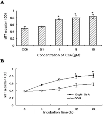
The effect of CsA on proliferation of PC12 cells was assessed by MTT assay. As shown in Fig. 1A, cell viability was gradually increased in a dose-dependent manner when PC12 cells were exposed to 0.1 ~ 10µM CsA for 24 h. At a dose of up to 0.1µM, CsA had no marked effect on cell proliferation. Treatment at a higher dose (>1µM) of CsA for 24 h induced the proliferation of PC12 cells to about 1.2- 1.5 folds of that in the control cells and showed maximal proliferation rate at 10µM. Similarly, cell viability of PC12 cells increased with time when cells were cultured with 10µM CsA (Fig. 1B).
Proteomic analysis in 10 µM CsA-treated PC12 cells for 12 h
Total proteins in CsA-treated PC12 cells were extracted and separated by 2-DE using pH 3-10 IPG strips in HEF, and
Fig. 1. Effects of CsA on proliferation in PC12 cells. Cell viability was determined by MTT assay as described in materials and meth- ods. PC12 cells were incubated with various concentrations of CsA for 24 h (A), or at 10µM for indicated times (B). CON denotes the control group. Data are mean± SD from 5 independent experi- ments (* p<0.05, compared with control).
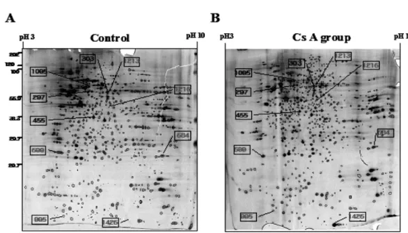
then the profiles were stained with sliver. A representation of one set among them is shown in Fig. 2. About 1,500 spots in approximately 500µg protein were observed on the gel.
The initial matching process was done by Labscan software program. Accuracy of the matches was confirmed by manually comparing each gel to the reference and other Fig. 2. A pair of representative 2-DE gels from CsA-treated group and CsA-untreated group (Control), which was stained with silver nitrate.
Ten spots that showed significantly and consistently different intensities between the proteom from CsA-treated group and that from the con- trol in PC12 cells were marked on 2-DE gel.
Fig. 3. Up-regulated 6 spots among 10 spots were significantly and consistently changed in CsA-treated PC12 cells compared to the CsA- untreated group. In each of 2-DE gels from the control and CsA-treated PC12 cells, the density of a spot in CsA-treated gel was compared to the density of the corresponding spot in the control gel. The averaged volumes of each spot were displayed as column bars from 5 pairs on the lower panel. CON denotes the control group (*p<0.05, compared with control group).
individual gels three times. The differential image analysis showed changed spots in CsA-treated PC12 cells. Spot numbers on 2-DE image indicate the protein spots, which shown significantly and consistantly different intensities between the proteom from CsA-treated group and the control in PC12 cells (Fig. 2). The numbers of changed spots showed above 1.5 folds increase or more than 50 % decrease in CsA-treated group compared to control. Ten proteins were significantly and consistently different between the CsA-treated group and the CsA-untreated group on 2-DE gels. Based on the cut-off value of significant difference of increase (over 1.5 folds) or decrease (over 50 %) in statistic analysis, 6 out of 10 spots were significantly increased (Fig. 3) as well as 4 out of 10 spots were significantly decreased (Fig. 4). The results of the MALDI-TOF MS and ESI Q-TOF MS on ten spots unequivocally indicated that the significantly up-regulated 6 proteins and down-regulated 4 proteins were identified.
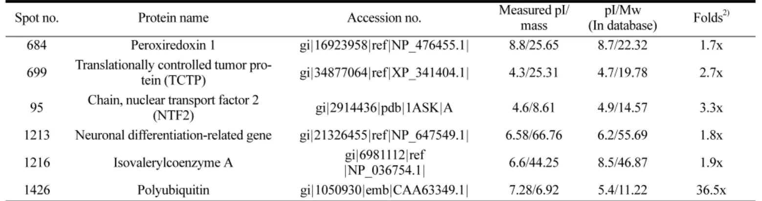
Table 1 and 2 summarized the results of protein identification showing protein definition. The identities of the 6 up-regulated proteins were peroxiredoxin 1 (Prx 1, sopt 684), translationally controlled tumor protein (TCTP, spot 699), nuclear transport factor 2 (NTF 2, spot 995), neuronal differentiation-related gene (spot 1213), isovaleryl
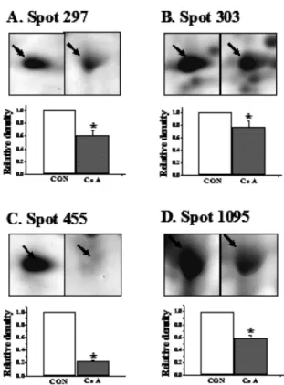
coenzyme A dehydrogenase (spot 1216) and polyubiquitin (spot 1426). In addition, the other significantly down- regulated 4 proteins were vacuolar H+-ATPase (V-ATPase, spot 297), chaperonin containing TCP-1 beta (CCT beta, spot 303), acidic ribosomal protein P0 (spot 455) and chaperonin containing TCP-1 epsilon subunit (CCT epsilon, spot 1095).
Discussion
Up to date, there has been much controversy about the effect of CsA with conflicting evidence as to which it acts as neuroprotection or neurotoxicity on neuronal cells. These adverse effects of CsA may be attributed to subpopulation of neuronal cells which have different characteristics in response to CsA. The previous reports have shown that CsA has different effects on cell proliferation according to cell types. CsA interfered with T cell proliferation whereas it promoted hair epithelial cell proliferation (Takahashi and Kamimura, 2001; Mascarell et al., 2004). We reported that CsA induced cell proliferation of human gingival fibroblast (Park et al., 2007). In the present study, CsA induced the proliferation of PC12 cells dose-dependently in the concentrations (0.1 ~ 10µM) of CsA which are similar to the plasma concentrations (100 ~ 200µg/ml) of patients undergoing CsA treatment. The present results indicated that CsA has a stimulating effect on the proliferation of PC12 cells.
Proteomic analysis is rapid and reproducible and it examines the expression of several different biomarkers simultaneously, providing a profile of protein expression that more accurately correspond to a particular type of cell proliferation (Scheler et al., 1998). In the present study, proteomic methods were employed to detect possible changes compared to control in the entire protein level after CsA treatment in PC12 cells. Proteins were identified to be significantly different expression compared to the control group (Table 1, 2). The present study is the first report identifying the significantly changed proteins in CsA- induced PC12 cells proliferation using proteomic analysis.
The analyzed protein spots were all those which displayed the differential changes above 1.5 folds increase or more than 50 % decrease of protein expression, suggesting that those proteins were closely related to proliferation response by CsA treatment at the concentrations inducing proliferation in PC12 cells. The identified proteins in the present study were fallen into four functional categories : (1) stress response associated proteins, including Prx 1 which was up-regulated; whereas acidic ribosomal protein P0 was down-regulated; (2) proliferation / differentiation associated proteins, including TCTP and neuronal differentiation- related gene, which were up-regulated; (3) the protein processing / transport / degradation / synthesis associated protein, NTF 2 and ubiquitin were up-regulated, whereas Fig. 4. Down-regulated 4 spots among 10 spots were significantly
and consistently changed in CsA-treated PC12 cells compared to the control. In each pair of 2-DE gels, the density of a spot in CsA- treated group gel was compared to the density of the corresponding spot in the control gel. The averaged volumes of each spot were displayed as colume bars from 5 pairs on the lower panel. CON denotes the control group (*p<0.05, compared with control group).
CCT beta, CCT epsilon and V-ATPase were down- regulated; (4) energy metabolism associated protein, including isoaleryl-coenzyme A dehydrogenase, which was up-regulated.
Polyubiquitin is a component of the ubiquitin system that selectively degrades cellular protein and proteins targeted to undergo proteolysis are initially covalently tagged with ubiquitin. The ubiquitin system contributes to a diversity of intracellular processes (intercellular stress, cell cycle, gene expression, apoptosis, and cancer) (Noga et al., 1997).
Ubiquitin has been described in neuroblastoma, a neuroendocine-related tumor, or in cell growth and/or cancer-promoting effects as secretory products. This protein may enhance the prognostic value related to proliferation in patients with neuroblastoma (Sandoval et al., 2006). In the present study, polyubiquitin was the most drastically up- regulated above 30 folds in CsA-treated PC12 cells. This finding represents that polyubiquitin may play an important role for proliferation process in neuronal cells, in consistent with those of previous reports.
Peroxiredoxins (Prxs) are a family of intracellular antioxidant proteins consisting of six members that protect cells against the damaging effects of reactive oxygen intermediates such as H2O2 (Hoffmann et al., 2002). In
addition to their antioxidant role, Prxs have been implicated in cellular functions such as regulation of gene expression, cell proliferation and differentiation (Kang et al., 1998). It was also reported that Prx proteins could protect cells from apoptosis through the inhibition of c-Abl, c-Myc, NF-κB and TNF-α, and with a mechanism similar to that of Bcl-2 (Zhang et al., 1997). In the present study, Prx protein was significantly up-regulated in CsA-treated PC12 cells proteomic analysis. Prosperi et al. (1998) showed that higher levels of pag gene (Prx 1) expression were observed following induction of proliferation and oxidative stress.
From the previous studies and the present results, it suggested the possibility that Prx may play a role in promoting proliferation in CsA-treated PC12 cells.
NTF 2, a small homodimeric protein, plays an important role in the trafficking of macromolecules, ions and small molecules between the cytoplasm and nucleus (Moore et al., 1994). Recently, it was reported that cells cease to be viable if the interaction between NTF 2 and nucleoporins is abolished, suggesting that NTF 2 may play a crucial role in process for cell survival (Quimby et al., 2001). NTF 2 was up-regulated in the present study, demonstrating that it is related to the proliferation in PC12 cells by CsA.
TCTP was up-regulated in CsA-treated PC12 cells in the Table 1. Identities of significantly up-regulated proteins in PC12 cells1)
Spot no. Protein name Accession no. Measured pI/
mass
pI/Mw
(In database) Folds2)
684 Peroxiredoxin 1 gi|16923958|ref |NP_476455.1| 8.8/25.65 8.7/22.32 1.7x
699 Translationally controlled tumor pro-
tein (TCTP) gi|34877064|ref |XP_341404.1| 4.3/25.31 4.7/19.78 2.7x 95 Chain, nuclear transport factor 2
(NTF2) gi|2914436|pdb|1ASK|A 4.6/8.61 4.9/14.57 3.3x
1213 Neuronal differentiation-related gene gi|21326455|ref |NP_647549.1| 6.58/66.76 6.2/55.69 1.8x
1216 Isovalerylcoenzyme A gi|6981112|ref
|NP_036754.1| 6.6/44.25 8.5/46.87 1.9x
1426 Polyubiquitin gi|1050930|emb|CAA63349.1| 7.28/6.92 5.4/11.22 36.5x
1)Six spots, which were upregulated over 1.5 folds difference in CsA-treated group compared to the control group, were identified by MALDI-TOF and ESI Q-TOF MS.
2)Increased folds in the normalized volume of a given protein spot indicated the difference between the CsA-treated group vs. the control.
Table 2. Identities of significantly down-regulated proteins in PC12 cells1).
Spot no. Protein name Accession no. Measured
pI/mass
pI/Mw
(In database) %2)
297 ATPase gi17105370|ref
|NP_476561.1| 5.8/63.05 5.6/56.88 40%
303 Chaperonin containing TCP-1 beta subunit
gi|34864883
|ref|XP_216891.2| 6.5/62.12 6.0/57.78 24%
455 Acidic ribosomal protein P0 gi|71138|pir|
|R5RT10 5.9/38..28 5.9/34.35 77%
1095 Chaperonin containing TCP-1 epsilon subunit
gi|34854926|ref
|XP_215516.2| 5.7/72.60 6.7/70.20 41%
1)Four spots, which were downregulated over 50 % decrease significantly in CsA-treated group compared to the control group, were identified by MALDI-TOF and ESI Q-TOF MS.
2)Decreased % in the normalized volume of a given protein spot indicated the differecne between the CsA-treated group vs. the control.
present study. TCTP was originally described in mouse tumor cells as a growth-related (Arcuri et al., 2004). A large amount of data has associated with the biological activity of TCTP to cell growth and differentiation (Yenofsky et al., 1983; Thomas, 1986; Sanchez et al., 1997). TCTP induction has been correlated with the mitogenic activity of non- neoplastic and cancer cell lines, suggesting an involvement of this protein in proliferation-related process. Furthermore, Li et al. (2001) have identified TCTP as an antiapoptotic protein in human cancer cells. It thus has the potential that one of the many functions of TCTP may be related to CsA- induced PC12 cells proliferation. However, to clarify the role of TCTP in CsA-induced PC12 cells proliferation, further studies are needed.
Molecular chaperons are proteins whose role is to mediate the folding of certain other polypeptides and their assembly into oligomeric structures (Hartl, 1996). In addition to protein folding, chaperone proteins exert protective actions by interfering with the stress-induced apoptotic pathway in the neuronal cells and are associated with neuritogenesis (Roobol et al., 1995). TCP-1, one of chaperonin family, is known to be involved in the folding of actin, and tubulin upon ATP hydrolysis. Six novel TCP-1-related polypeptides were isolated as subunit of the chaperonin containg TCP-1 (CCT): CCT beta, CCT gamma, CCT delta, CCT epsilon, CCT zeta and CCT eta. The fact that CCT is essential for its growth in budding yeast, suggests the possibility that CCT may play a role of proliferation in neuronal cells. The present study showed the down-regulated levels of CCT beta and CCT epsilon subunit in CsA-treated PC12 cells, demonstrating that CCT is associated with the proliferation in PC12 cells by CsA.
The neuroprotective effect could postulate to be mediated by stimulating the anti-apoptotic effect. Acidic ribosomal protein P0 containing 316 amino acids can be detected in the nucleus. Grabowski et al. (1991) suggested an extraribosomal function of P0 as a DNA repair protein. It was also identified as potential candidates for controlling apoptosis in B lymphocytes (Brockstedt et al., 1998). The present result showed the downregulation of P0 protein suggest that closely related to proliferation by CsA in PC12 cells may be mediated by regulating antiapoptosis effect.
In the present study, up-regulated neuronal differentiation- related gene, which may be expressed during transition between proliferation and differentiation, are not yet identified to play an exact role in the proliferation process of neuronal cells. In addition, the down-regulated isovalerylcoenzyme A, which is a mitochondrial enzyme that catalyzes a step in the catabolic pathway of the ketogenic branched chain amino acid leucine, is unexplainable at present.
Taken together, the identified ten proteins significantly different between CsA-treated group and the control in PC12 cells using proteomic analysis, which are mainly related with stress response, proliferation, metabolism and
protein processing, might be involved in the CsA-induced proliferation in PC12 cells, even if there needed direct evidences showing the relationship. Further quantitative confirmation of expression changes by Western blotting is needed to understand the proliferation effect of these proteins in neuronal cells and also comparative investigations of these proteins expression with in vivo study would be required. In conclusion, the data support the notion that down-regulated or up-regulated proteins play a relevant role in CsA-induced proliferation of PC12 cells.
The present information obtained with proteomic analysis may provide efficient approaches in understanding mechanisms of CsA-induced proliferation of PC12 cells.
Acknowledgement
This study was financially supported by Chonnam National University, 2005.
References
Andre N, Roquelaure B, Conrath J. Molecular effects of cyclosporin and oncogenesis: a new model. Med Hypotheses.
2004;63:647-52.
Arcuri F, Papa S, Carducci A, Romagnoli R, Liberatori S, Riparbelli MG, Sanchez JC, Tosi P, del Vecchio MT.
Translationally controlled tumor protein (TCTP) in the human prostate and prostate cancer cells: expression, distribution, and calcium binding activity. Prostate. 2004;60:
130-40.
Bernardi P, Vassanelli S, Veronese P, Colonna R, Szabo I, Zoratti M. Modulation of the mitochondrial permeability transition pore. Effect of protons and divalent cations. J Biol Chem. 1994;267:2934-9.
Brockstedt E, Rickers A, Kostka S, Laubersheimer A, Dorken B, Wittmann-Liebold B, Bommert K, Otto A. Identification of apoptosis-associated proteins in a human Burkitt lymphoma cell line. Cleavage of heterogeneous nuclear ribonucleoprotein A1 by caspase 3. J Biol Chem. 1998;273:
28057-64.
Coley C, Jarvis K, Hassell T. Effect of cyclosporin-A on human gingival fibroblasts in vitro. J Dent Res. 1986;65:
353-8.
Faulds D, Goa KL, Benfield P. Cyclosporin. A review of its pharmacodynamic and pharmacokinetic properties, and therapeutic use in immunoregulatory disorders. Drugs.
1993;45:953-1040.
Folbergrova J, Li PA, Uchino H, Smith ML, Siesjo BK.
Changes in the bioenergetic state of rat hippocampus during 2.5min of ischemia, and prevention of cell damage by cyclosporin A in hyperglycemic subjects. Exp Brain Res.
1997;114:44-50.
Grabowski DT, Deutsch WA, Derda D, Kelley MR.
Drosophila AP3, a presumptive DNA repair protein, is homologous to human ribosomal associated protein P0.
Nucleic Acids Res. 1991;19:4297.
Hartl FU. Molecular chaperones in cellular protein folding.
Nature. 1996;381:571-9.
Hellman U, Wernstedt C, Gonez J, Heldin CH. Improvement of an "In-Gel" disgestion procedure for the micropreparation of internal protein fragments for amino acid sequencing.
Anal Biochem. 1995;224:451-5.
Hoffmann B, Hecht HJ, Flohe L. Peroxiredoxins. Biol Chem.
2002;383:347-64.
Kang SW, Chae HZ, Seo MS, Kim K, Baines IC, Rhee SG.
Mammalian peroxiredoxin isoforms can reduce hydrogen peroxide generated in response to growth factors and tumor necrosis factor-α. J Biol Chem. 1998;273:6297-302.
Leprince P, Rogister B, Delre´e P, Rigo JM, Andre´ B, Moonen G. Modulation of proteolytic activity during neuritogenesis in the PC12 nerve cell: differential control of plasminogen activator and plasminogen activator inhibitor activities by nerve growth factor and dibutyryl-cyclic AMP. J Neurochem. 1991;57:665-74.
Li F, Zhang D, Fujise K. Characterization of fortilin, a novel antiapoptotic protein. J Biol Chem. 2001;276:47542-9.
Lyons WE, George EB, Dawson TM, Steiner JP, Snyder SH.
Immunosuppressant FK506 promotes neurite outgrowth in cultures of PC12 cells and sensory ganglia. Proc Natl Acad Sci USA. 1994;91:3191-5.
Mascarell L, Auger R, Alcover A, Ojcius DM, Jungas T, Cadet-Daniel V, Kanellopoulos JM, Truffa-Bachi P.
Characterization of a gene encoding two isoforms of a mitochondrial protein up-regulated by cyclosporin A in activated T cells. J Biol Chem. 2004;279:10556-63.
Miyata K, Omori N, Uchino H, Yamaguchi T, Isshiki A, Shibasaki F. Involvement of the brainderived neurotrophic factor/TrkB pathway in neuroprotective effect of cyclosporin A in forebrain ischemia. Neuroscience. 2001;105:571-8.
Moore MS, Blobel G. Purification of a Ran-interacting protein that is required for protein import into the nucleus. Proc Natl Acad Sci USA. 1994;91:10212-6.
Noga M, Hayashi T, Tanaka J. Gene expressions of ubiquitin and hsp70 following focal ischemia in rat brain. Neuroreport 1997;24:1239-41.
Park JI; Lee GS; Jeong YJ; Kim BK; Kim JH; Lim HS; Kim SH; Kim WJ; Jung JY. Involvement of Antiapoptotic Signals in Rat PC12 Cells Proliferation by Cyclosporin A Treatment. Int J of Oral Biol. 2007;32:51-7.
Prosperi MT, Ferbus D, Rouillard D, Goubin G. The pag gene product, a physiological inhibitor of c-abl tyrosine kinase, is overexpressed in cells entering S phase and by contact with agents inducing oxidative stress. FEBS Lett. 1998;423:39-44.
Quimby BB, Leung SW, Bayliss R, Harreman MT, Thirumala
G, Stewart M, Corbett AH. Functional analysis of the hydrophobic patch on nuclear transport factor 2 involved in interactions with the nuclear pore in vivo. J Biol Chem.
2001;276:38820-9.
Roobol A, Holmes FE, Hayes NV, Baines AJ, Carden MJ.
Cytoplasmic chaperonin complexes enter neurites developing in vitro and differ in subunit composition within single cells. J Cell Sci. 1995;108:1477-88.
Sanchez JC, Schaller D, Ravier F, Golaz O, Jaccoud S, Belet M, Wilkins MR, James R, Deshusses J, Hochstrasser D.
Translationally controlled tumor protein: A protein identified in several nontumoral cells including erythrocytes.
Electrophoresis. 1997;18:150-5.
Sandoval JA, Hoelz DJ, Woodruff HA, Powell RL, Jay CL, Grosfeld JL, Hickeyd RJ, Malkas LH. Novel peptides secreted from human neuroblastoma: useful clinical tools? J Pediatr Surg. 2006;41:45-51.
Scheler C, Lamer S, Pan Z, Li XP, Salinikow J, Jungblut P.
Peptide mass fingerprint sequence coverage from differently stained proteins on two-dimensional electrophoresis patterns by matrix assisted laser desorption/ionization-mass spectrometry (MALDI-MS). Electrophoresis. 1998;19:918- 27.
Shiga Y, Onodera H, Matsuo Y, Kogure K. Cyclosporin A protects against ischemia-reperfusion injury in the brain.
Brain Res. 1992;595:145-8.
Takahashi T, Kamimura A. Cyclosporin a promotes hair epithelial cell proliferation and modulates protein kinase C expression and translocation in hair epithelial cells. J Invest Dermatol. 2001;117:605-11.
Tanaka K, Ogawa N. Immunophilin ligands: from immuno- suppressants to neuroprotective drugs. Nihon Shinkei Seishin Yakurigaku Zasshi. 2004;24:71-4.
Thomas G. Translational control of mRNA expression during the early mitogenic response in Swiss mouse 3T3 cells:
Identification of specific proteins. J Cell Biol. 1986;103:
2137-44.
Uchino H, Elmer E, Uchino K, Lindvall O, Siesjo BK. A dramatically ameliorates CA1 hippocampal damage following transient forebrain ischemia in the rat. Acta Physiol Scand.
1995;155:469-71.
Yenofsky R, Cereghini S, Krowczynska A, Brawerman G.
Regulation of mRNA utilization in mouse erythroleukemia cells induced to differentiate by exposure to dimethyl sulfoxide. Mol Cell Biol. 1983;3:1197-203.
Zhang P, Liu B, Kang SW, Seo MS, Phee SG, Obeid LM.
Thioredoxin peroxidase is a novel inhibitor of apoptosis with a mechanism distinct from that of Bcl-2. J Biol Chem.
1997;272:30615-8.