155
산삼 배양액을 급여한 육계에서 근육의 프로테옴 분석
설재원·황인호
1·채준석·강형섭·류경선
2·강춘성
3·박상열*
전북대학교 수의과대학 생체안전성연구소
1
축산기술연구소
2
농업생명과학대학 동물자원과학부
3
㈜이앤티
(게재승인: 2005년 5월 24일)
Proteome analysis of chickens fed with tissue culture medium waste after harvest of Korean wild ginseng
Jae-Won Seol, In Ho Hwang
1, Joon-Seok Chae, Hyung-Sub Kang, Kyeong-Seon Ryu
2, Chun-Seong Kang
3, Sang-Youel Park*
Bio-Safty Research Institute, College of Veterinary Medicine, Chonbuk National University, Jeonju 561-756, Korea
1
National Livestock Research Institute, Suwon 441-350, Korea
2
Department of Animal Resources and Biotechnology, Chonbuk National University, Jeonju 561-756, Korea
3
E&T Co. Ltd., Nonsan 320-930, Korea
(Accepted: May 24, 2005)
Abstract : Proteomics is a useful approach to know protein expression, post-translational modification and protein function. We investigated the protein expression pattern and identity in chickens fed with the tissue culture medium waste after harvest of Korean wild ginseng (TCM-KWG) ( Panax ginseng ). Two groups (n=60/group) of day old broiler chickens were administered with 0 (control) and 0.8% (treatment) TCM- KWG through drinking water. After 5 weeks, we examined the protein expression pattern of fibularis longus and superficial pectoral muscle by Two-dimensional electrophoresis analysis. Interestingly, TCM-KWG treatment significantly increased five spot's density, and markedly reduced five spot's density in the muscles.
We identified 10 proteins (desmin, myosin light chain 1, heat shock 25 kDa protein, collapsin response mediator protein-2A, alpha enolase, vimentin, actin alpha 1, my023 protein, pyruvate kinase and troponin T) by the matrix-assisted laser desorption ionization time of flight (MALDI-TOF).
Key words : Proteomic analysis, Chicken, Gienseng, MALDI-TOF
서 론
프로테오믹스 (proteomics) 는 단백질체의 기능 이상과 구조변형 유무 등을 가려내는 분석기술을 뜻하는 것으 로 특정 단백질의 발현 양상의 변화 , post-translational
modification, 다른 단백질간의 상호작용과 단백질 전체
수준에서의 구성 및 기능 등을 연구하여 세포내 변형과
정과 네트웍 형성을 질병의 진행과정과 연계시켜 총괄
적으로 이해할 수 있는 연구 분야이다 [1, 17, 18]. 이차
원전기영동 (Two-dimensional electrophoresis; 2-DE) 은 한 개체내에서 발현되는 단백질의 발현 양상과 생리적 , 환
경적 변화에 따른 발현정도의 변화도 관찰할 수 있다
[12, 19]. 또한 분석 대상 단백질들을 세포의 특정 생리
조건에 따라 제조하여 특정한 spot 들의 양적인 변화를
본연구는농림기술개발사업
(
과제번호103145001)
에의해수행되었음.
*Corresponding author: Sang-Youel Park
College of Veterinary Medicine, Chonbuk National University, Jeonju 561-756, Korea
[Tel: +82-63-270-3886, Fax: +82-63-270-3780, E-mail: [email protected]]
보이는 단백질들을 적당한 크기로 잘라 The matrix- assisted laser desorption ionization time of fight(MALDI- TOF) mass spectrometry(MS) 등의 단백질 질량 분석기 로 분석하여 아미노산 서열을 결정하고 단백질의 정체 를 확인할 수 있다 .
산삼 (Korean wild ginseng, panax ginseng)은 오래전부 터 의학적으로 그 효용성이 증명되었으며 최근에는 첨 단 생명공학기술을 응용하여 산삼을 실험실에서 조직배 양시키는 기술이 국내 연구자들에 의해 개발되었다 . 산 삼배양액 (The tissue culture medium waste after harvest of Korean wild ginseng(TCM-KWG)) 이란 조직배양을 통 해 실험실에서 배양시킨 산삼을 분리하고 남은 배양액 을 의미한다 . 이 산삼배양액에는 사포닌 성분외에 많은
성분들이 다량 함유되어 있음이 발견되었다 . 산삼배양 액에서 가장 많은 성분으로 알려져 있는 사포닌 성분은 이미 인삼 등의 연구에서 잘 밝혀져 있는데 면역기능 증 강작용 , 암세포 증식 억제작용 , 항염증작용 그리고 단백 질 합성 촉진작용 등의 효능과 다양한 작용들을 보인다
[2, 4, 15, 21]. 그러나 아직까지 산삼 배양액을 이용한 연
구결과는 거의 보고되지 않은 상태이며 산업동물에 적 용하여 효능과 효과를 검증한 결과도 전무한 상태이다 .
따라서 본 연구에서는 지금까지 버려져 왔던 폐기물 로서의 산삼배양액을 산업동물에 적용하여 최근에 널리 연구되고 있는 프로테오믹스 기술을 이용하여 , 산삼배 양액을 투여한 육계와 투여하지 않은 육계의 긴비골근
(fibularis longus muscle) 과 얕은흉근 (superficial pectoral
muscle) 에서 단백질 발현의 변화를 조사하였다 . 또한 발
현양이 현저한 차이를 보이는 단백질들을 분리 , 동정함
으로써 육계에서의 산삼배양액의 효과 규명에 기초 자 료를 제공하고자 본 연구를 수행하였다 .
재료 및 방법
실험동물 및 시료
( 주 ) 하림으로부터 1 일령 Ross 수컷을 구입하여 5 주간 사양실험을 시행하였다 . 육계를 60 수씩 산삼배약액을 처
리하지 않는 그룹 ( 대조군 ) 과 산삼배양액을 0.8% 씩 처리 한 그룹 ( 실험군 ) 으로 나누었다 . 산삼배양액은 음수로 급 여하였다 . 대조군과 실험군에 5 주 동안 처리한 육계를
방혈 폐사시킨 후 대조군과 투여군의 긴비골근과 얕은
흉근 부위에서 1×2×1 cm 의 크기로 조직을 절제하여 시
료를 채취하였으며 실험에 사용전까지 액체질소에 보관 하였다 .
시료 준비와 2차원 전기영동
2-DE 는 Yan et al. [20] 에서 서술한 방법에 준하여 실
시하였다 . 시료에 lysis buffer(7M urea, 2M thiourea, 2%
CHAPS, 1% DTT 와 0.8% pharmalyte pH 3-10 nonlinear) 1 m l을 넣은 후에 1 분 동안 homogenizer 를 이용하여 균 질화시켰다 . 균질화된 용해물을 1 분 동안 잘 혼합해 주
고 30 분 동안 실온에서 rotational shaking 해 주었다 . 이 용해물을 20
oC 에서 1 시간동안 4000×g 로 centrifuge 하고
상층액의 2 번째 층을 취한 후에 단백질 농도를 Bradford
법을 이용하여 측정한 후 최종 단백질 농도가 3~10 mg/
m l이 되도록 희석시켰다 . 정량된 시료는 rehydration buffer(8 M urea, 0.5% CHAPS, 0.2% DTT 과 0.2%
pharmalyte pH 3-10 nonlinear) 와 혼합하여 IPG phore strip holder 에서 로딩하였다 . First-dimension Isoelectric focusing (IEF) 이 끝난 IPG strip 은 6M urea, 30% glycerol, 2% SDS, 0.002% bromophenol blue 와 1% DTT 가 포함된 50 mM Tris-HCl buffer(pH 8.8) 에 15 분간 반응시키고 다시 이
buffer 에 iodoacetamide 를 첨가하여 15 분간 더 반응시
킨 다음에 Ettan DALT system(Amersham Pharmacia Biotech, Amersham, UK) 을 이용하여 IPG strip 을 12.5%
acrylamide gel 의 상단부에서 로딩시켰다 .
염색
이차원 전기영동 (2-Dimensional Gel Electrophoresis; 2- DE) 을 한 후에 gel 에서 단백질 spot 을 검출하기 위하여 염색을 실시하였다 . Gel 을 2% phosphoric acid, 10%
ammonium sulphate 와 20% methanol 을 포함한 0.1%
colloidial Coomassie Briliant Blue G-250 을 이용하여 48
시간동안 염색하였다 [11].
이미지분석과 단백질 동정
가시화된 단백질은 2DE 이미지 분석용 프로그램인
PD Quest(Bio-Rad, USA) 를 이용하여 분석하였고 , 산삼
배양액 비투여군과 투여군간의 발현의 차이를 보이는 단백질들을 선발하였다 . 이 중에 관심있는 단백질을 동
정하기 위해서 관련 spot 을 1 mm
3으로 자르고 30 mM potassium ferricyanide 와 100 mM sodium thiosulfate(1:1)
을 혼합한 용액에 넣어 탈염색한 후에 세척하였다 . Gel
을 acetonitrile 100 µl을 넣어 탈수시키고 vacuum centrifuge 를 이용하여 완전히 건조시켰다 . 건조된 gel 에
50 mM ammonium bicarbonate(0.2 µ g modified trypsin 이
포함 ) 20 µl를 넣고 ice 상태에서 45 분 동안 반응시켰
다 . 남은 용액을 제거한 후에 50 mM ammonium
bicarbonate 30 µl를 더하고 37
oC 에서 overnight 함으로서
digestion 을 하였다 . Peptide 의 추출을 위해 acetonitrile
20 µl를 넣고 30 분간 vortex 시킨 후에 상층액만 수거하
여 vacuum centrifuge 를 이용하여 건조시켰다 . 다음에
MALDI-TOF 분석을 위하여 건조시킨 sample 과 matrix
solution(15-20 g/L α -cyanon-4-hydrosycinnamic acd;
CHCA in 70% acetonitrile) 을 혼합하고 , MALDI target 에
2 µl를 분주한 후 건조시켜서 MALDI-TOF 을 실시하였다
[16]. MALDI-MS spectra 는 delayed-extraction reflectron time-of-flight mass spectrometer(Model MALDI-R;
Micromass, UK) 를 이용하여 얻었다 . Peptide masses 는
MASCOT software 와 Profound software 를 사용하여 NCBI database 에서 단백질을 동정하였다 .
결 과
산삼배양액 투여로 인한 근육 내 단백질 발현 양상을
분석하기 위하여 대조군과 실험군으로 나누고 산삼배양 액을 음수로 급여하였다 . 산삼배양액 투여 5 주 후에 긴
비골근과 얕은 흉근 부위에서 1×2×1 cm 의 크기로 조직
을 절제하여 시료를 채취하였다 . 각 근육으로부터 추출
한 총 단백질을 Yan et al . [20] 의 방법에 준하여 2 차원
전기영동을 실시하였고 , gel 을 가시화 시키기 위하여
0.1% colloidial Coomassie Briliant Blue G-250 를 이용하
여 염색을 실시하였으며 가시화된 gel image 는 PD Quest
software 를 이용하여 분석하였다 , 그 결과 긴비골근과 얕
은 흉근의 gel image 에서 총 900 개의 spot 들을 관찰하였
다 (Fig. 1A, B). 이 spot 들 중 대조군에 비하여 산삼배양 액을 투여한 육계의 긴비골근과 얕은 흉근 모두에서 동
Fig. 1. 2-DE map of protein from control groups and TCM-KWG treatment groups in fibularis longus muscle (A) and
superficial pectoral muscle (B). Proteins were separated using an IPG 3-10 NL in the first dimension and 12.5% SDS
gel in the second dimension. The protein loading was 400 µ g and the gel was stained with coomassie blue G-250.
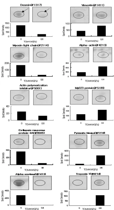
Fig. 2. Expression profiles of 10 spots between control groups and TCM-KWG treatment groups in fibularis longus muscle
and superficial pectoral muscle . An arrows show the relative spot density change in control group (0%) and TCM-KWG
treatment group (0.8%). The bars represent the average density based on observation in three chickens.
일한 변화를 보이는 spot 들을 선정한 결과 , 총 10 개의
spot density 가 유의성 있게 차이를 보였으며 , 이중에서
5 개의 spot 은 증가하는 경향을 보였고 , 5 개의 spot 은 감 소하는 경향을 나타내었다 (Fig. 2). 변화를 보이는 spot
들은 단백질 동정을 위하여 MALDI-TOF 분석을 실시
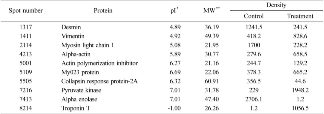
하였다 . Fig. 3 은 각각 spot 들에 대한 MALDI-TOF 분석 결과를 보여주고 있으며 , Table 1 은 MALDI-TOF 분석
결과 산삼배양액을 급여한 육계의 긴비골근과 얕은 흉 근에서 유의성 있는 변화를 보이는 총 10 종의 단백질의
동정 결과이며 각 단백질의 등전점 (pI) 과 분자량 (M.W)
을 나타내었다 . 동정된 단백질 중에서 desmin 은 약 5.2
배 , myosin light chain 1 은 약 7.5 배 , Actin polymerization inhibitor(heat shock 25 kDa protein) 은 약 1.8 배 , collapsin response mediator protein-2A 는 약 8.1 배 그리고 alpha
enolase 는 약 2.1 배씩 대조군에 비해 산삼배양액을 투여
한 육계의 긴비골근과 얕은 흉근에서 감소하는 경향을 보였고 , vimentin 은 약 2 배 , actin alpha 1 은 약 2.3 배 , my023 protein 은 약 1.7 배 , pyruvate kinase 는 약 8.5 배 그
Fig. 3. MALDI-TOF spectrum of protein spot (Troponin T). Aliquots (10 u l ) of peptide sample solution were loaded onto the MALDI-TOF MS sample plate together with 0.5 u l of the matrix solution (15 − 20 g/L 4HCCA in 70% acetonitrile).
The MALDI-MS measurements were obtained using a delayed-extraction reflectron time-of-flight mass spectrometer (Model M@LDI-R; Micromass, Manchster, UK).
Table 1. Protein changes of fibularis longus and superficial pectoral muscle after treated TCM-KWG group and control group in chicken
Spot number Protein pI
*MW
**Density
Control Treatment 1317 1411
4213 2114 5001 5109 5505 7216 7413 8214
Desmin Vimentin
Myosin light chain 1 Alpha-actin
Actin polymerization inhibitor My023 protein
Collapsin response protein-2A Pyruvate kinase
Alpha enolase Troponin T
4.89 4.92 5.08 5.89 6.27 6.69 6.32 7.01 -1.00 7.01
36.19 49.39 21.95 30.77 21.16 22.06 60.91 31.78 47.40 26.26
1241.5 418.2 279.6 1700 244.7 378.3 356.5 2706.1 229 1.2
241.5 828.6 228.2 658.5 129.2 665.2 1948.2 44.6 1056.5 1.2
*
pI = isoelectric point
**