Introduction
Opening wedge high tibial osteotomy (OWHTO) with medial plate fixation is a proven adequate and safe method for treatment of medial compartment osteoarthritis of the knee and varus deformity, particularly in young and/or active individuals1,2).
Advantages of OWHTO, compared to closed wedge osteotomy, include preservation of bone stock, predictable and adjustable correction, relatively easy exposure with avoidance of proximal tibio-fibular joint disruption, fibular nerve palsy, and compart- ment syndrome3).
However, OWHTO creates a gap in the metaphysis of the tibia.
To enhance bone healing and increase initial mechanical stabil- ity, a high degree OWHTO may require the application of bone graft/substitute to fill the osteotomy gap. Moreover, OWHTO has been associated with risk of nonunion, collapse and loss of correction4). Autologous bone graft is the “gold standard” to fill the bone defect, but iliac crest graft harvest has risks. The associ- ated complications include pain, thigh hypoesthesia, infection, pelvic bone fracture and discomfort wearing clothes5,6). Allograft to avoid donor side morbidity was successfully applied in HTO7). However, the use of allograft increases additional risks such as disease transmission, immunologic reactions and slow remodeling.
Is Bone Grafting Necessary in Opening Wedge High Tibial Osteotomy? A Meta-Analysis of Radiological Outcomes
Jae Hwi Han, MD
1, Hyun Jung Kim, PhD
2, Jae Gwang Song, MD
1, Jae Hyuk Yang, MD
3, Nikhl N Bhandare, MD
4, Aldrich Raymund Fernandez, MD
5, Hyung Jun Park, MD
1, and Kyung Wook Nha, MD
11Department of Orthopaedic Surgery, Inje University Ilsan Paik Hospital, Goyang; 2Department of Preventive Medicine, Institute for Evidence-based Medicine, Korea University College of Medicine, Seoul; 3Department of Orthopaedic Surgery, Veterans Health Service Medical Center, Seoul, Korea; 4Department of Orthopaedic Surgery, Bhandare Hospital, Panaji, India; 5Department of Orthopaedic Surgery, Army General Hospital, Bonifacio, Philippines
Purpose: Bone grafting in opening wedge high tibial osteotomy (OWHTO) is still controversial. The purpose of this study is to compare the radiological outcomes of OWHTO with bone graft (autogenous, allogenous, and synthetic bone graft) and those without bone graft.
Materials and Methods: PubMed, MEDLINE, EMBASE and Cochrane Register of Studies databases were searched using specific inclusion and exclusion criteria for radiological studies involving OWHTO with bone graft and without bone graft groups. All reported delayed union, nonunion and correction loss were analyzed. Data were searched from the time period of January 2000 through July 2014. In addition, a modified Coleman methodology score (CMS) system was used to assess the methodological quality of the included studies.
Results: Twenty-five studies with a mean CMS value of 77 (range, 61 to 85 score) were included. In total, 1,841 patients underwent OWHTO using 4 different procedures for bone graft: autobone graft (n=352), allobone graft (n=547), synthetic bone graft (n=541) and no bone graft (n=401). There was a similar tendency for delayed union, nonunion and correction loss rate among the osteotomy space filling methods.
Conclusions: The meta-analysis showed there was a similar tendency for radiological union and correction maintenance among patients undergoing OWHTO regardless of the type of bone in all of the studies. However, the currently available evidence is not sufficient to strongly support the superiority of OWHTO with bone graft to OWHTO without bone graft.
Keywords: Knee, Osteoarthritis, Osteotomy, Bone graft, Meta-analysis pISSN 2234-0726 · eISSN 2234-2451
Knee Surgery & Related Research
Received September 21, 2014; Revised (1st) May 10, 2015;
(2nd) September 14, 2015; Accepted September 17, 2015 Correspondence to: Kyung-Wook Nha, MD
Department of Orthopaedic Surgery, Inje University Ilsan Paik Hospital, 170 Juhwa-ro, Ilsanseo-gu, Goyang 10380, Korea
Tel: +82-31-910-7312, Fax: +82-31-910-7319 E-mail: [email protected]
207
This is an Open Access article distributed under the terms of the Creative Commons Attribution Non-Commercial License (http://creativecommons.org/licenses/by-nc/4.0/) which permits unrestricted non-commercial use, distribution, and reproduction in any medium, provided the original work is properly cited.
Copyright © 2015 KOREAN KNEE SOCIETY www.jksrr.org
Due to the limited autologous bone availability and the problem of donor-site morbidity, many efforts have been made to find adequate supporting material for augmentation after osteotomy.
Hydroxyapatite, β-tricalcium phosphate or the combination of both are the most commonly used synthetic augments in OWH- TO. In the meantime, some recent studies presented good short- term results of OWHTO without additional bone substitutes for filling the osteotomy gap8,9). Still, the most suitable material for filling the opening space in OWHTO has not been identified.
Over the past decade, there has been only one randomized con- trolled study on the comparison of autologous bone grafting and
no filling method, which reported similar outcomes between the two methods10). Although randomized controlled trials (RCTs) are considered to offer the ideal and highest level of evidence for patient care, numerous “good” surgical practices have evolved into the “standard of care” without being randomized against placebo or ineffective treatment options11), which probably ex- plains why only one RCT has been published. Considering that observational studies constitute the best available evidence12), we attempted to conduct a meta-analysis of observational studies for the purpose of providing surgeons with informed data for better decision-making in OWHTO.
Table 1. Inclusion and Exclusion Criteria
Inclusion criteria Exclusion criteria
Studies on patients who received OWHTO Studies on patients who received closing wedge HTO Studies reporting minimum 1-year follow-up data on radiological
outcomes and complications of OWHTO Studies reporting less than 1-year follow-up data on radiological outcomes and complications of OWHTO
Level I, II, III, or IV evidence Level V evidence (case report, technical note, letter to editor), biomechanical reports, and review articles
Articles written in English Articles written in languages other than English
Human subjects Non-human subjects
Study published or in press online between January 1, 2000 and May
1, 2014 Study published or in press online on or before January 1, 2000
Medial plate fixation for OWHTO Other devices (external fixator, staple) for OWHTO OWHTO: opening wedge high tibial osteotomy.
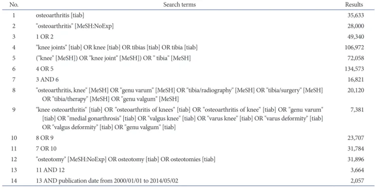
Table 2. Search Protocol
No. Search terms Results
1 osteoarthritis [tiab] 35,633
2 "osteoarthritis" [MeSH:NoExp] 28,000
3 1 OR 2 49,340
4 "knee joints" [tiab] OR knee [tiab] OR tibias [tiab] OR tibia [tiab] 106,972
5 ("knee" [MeSH]) OR "knee joint" [MeSH]) OR " tibia" [MeSH] 72,058
6 4 OR 5 134,573
7 3 AND 6 16,821
8 "osteoarthritis, knee" [MeSH] OR "genu varum" [MeSH] OR "tibia/radiography" [MeSH] OR "tibia/surgery" [MeSH]
OR "tibia/therapy" [MeSH] OR "genu valgum" [MeSH] 20,120
9 "knee osteoarthritis" [tiab] OR "osteoarthritis of knees" [tiab] OR "osteoarthritis of knee" [tiab] OR "genu varum"
[tiab] OR "medial gonarthrosis" [tiab] OR "valgus knee" [tiab] OR "varus knee" [tiab] OR "varus deformity" [tiab]
OR "valgus deformity" [tiab] OR "genu valgum" [tiab]
7,381
10 8 OR 9 23,707
11 7 OR 10 31,784
12 "osteotomy" [MeSH:NoExp] OR osteotomy [tiab] OR osteotomies [tiab] 31,896
13 11 AND 12 3,664
14 13 AND publication date from 2000/01/01 to 2014/05/02 2,057
The purpose of this study was to compare the radiological out- comes of OWHTO with bone graft (autogenous graft, allogenous graft, and synthetic bone) and those without bone graft. The hy- potheses were that the use of bone graft would produce superior radiological outcomes.
Materials and Methods
1. Eligibility Criteria
Published studies were included in this meta-analysis if they met the eligibility criteria described in Table 1. Patients included in this study were limited to osteoarthritis patients. There was no limitation in the age of patients. Fixation method was limited to plate fixation using either a locking or a non-locking plate.
2. Data Sources and Search Strategy
A literature search of online databases (MEDLINE, EMBASE and Cochrane Register of Studies) was performed. The search strategy used in the case of MEDLINE is presented in Table 2, which was modified for each of the other databases (Table 2).
Next, the references from the included studies were screened, and experts in the field were contacted for help in identifying additional studies. Two independent review authors (Han and Song) selected citations each based on the titles and abstracts.
The eligibility of the full papers of those citations initially thought to fulfill the inclusion criteria was then assessed. In cases where a consensus could not be reached, a third review author (Nha) was consulted.
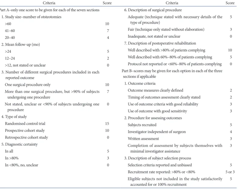
Table 3. Criteria of the Coleman Methodology Score for Studies
Criteria Score Criteria Score
Part A–only one score to be given for each of the seven sections 1. Study size–number of otsteotomies
>60 10
41–60 7
20–40 4
2. Mean follow-up (mo)
>24 5
12–24 2
>12, not stated or unclear 0
3. Number of different surgical procedures included in each reported outcome
One surgical procedure only 10
More than one surgical procedure, but >90% of subjects
undergoing one procedure 7
Not stated, unclear or <90% of subjects undergoing one
procedure 0
4. Type of study
Randomised control trial 15
Prospective cohort study 10
Retrospective cohort study 0
5. Diagnostic certainty
In all 5
In >80% 3
In <80%, no, unclear 0
6. Description of surgical procedure
Adequate (technique stated with necessary details of the
type of procedure) 5
Fair (technique only stated without elaboration) 3
Inadequate, not stated or unclear 0
7. Description of postoperative rehabilitation
Well described with >80% of patients complying 10 Well described with 60%–80% of patients complying 5 Protocol not reported or <60%–80% of patients complying 0 Part B–scores may be given for each option in each of the three
sections if applicable 1. Outcome criteria
Outcome measures clearly defined 2
Timing of outcomes assessment clearly stated 2 Use of outcome criteria with good reliability 3 Use of outcome with good sensitivity 3 2. Procedure for assessing outcomes
Subjects recruited 5
Investigator independent of surgeon 4
Written assessment 3
Completion of assessment by subjects themselves with
minimal investigator assistance 3
3. Description of subject selection process
Selection criteria reported and unbiased 5 Recruitment rate reported: >80% or <80% 5 or 3
Eligible subjects not included in the study satisfactorily
accounted for or 100% recruitment 5
3. Data Abstraction
A standardized form was used to extract data from the included papers on study characteristics, patient characteristics, surgical intervention, duration of follow-up, osteotomy size, postopera- tive rehabilitation protocol, radiological bone union period, nonunion rate, delayed union rate, correction loss rate and graft- related problems. The extracted data were then cross-checked for accuracy. Disagreements were settled by the Nha.
4. Statistical Analysis
Statistical analysis was performed using STATA/MP ver. 13.0 (Stata Corp., College Station, TX, USA). Effect sizes for dichoto- mous data (incidence of nonunion, delayed union, and loss of correction) were calculated using random-effect models and expressed as event rates. The mean values of the continuous data (the mean bone union period; the mean difference in bone union period) were compared using a t-test. For each effect size, a 95%
confidence interval was given. Heterogeneity was calculated according to the method of Higgins et al.13) and expressed as I2
(range, 0% [complete consistency] to 100% [complete inconsis- tency]). Analysis of comparative studies and level IV case series analysis were performed separately. Case series analysis was per- formed to investigate whether the case series support the results of the one RCT and four comparative studies.
5. Quality Assessment
The methodological quality of the included studies was assessed by the two review authors (Han and Song) using the 10 criti- cal appraisal criteria of the Coleman methodology score (CMS) (Table 3). The final scores ranged from 0 to 100, with a perfect score (100) indicating a study design that completely avoids the influences of chances, various biases, and confounding factors.
6. Included Studies
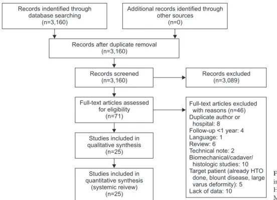
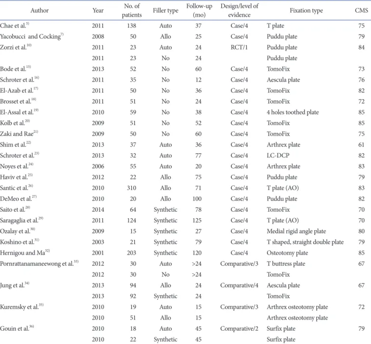
Based on the full-text review, the final meta-analysis included 25 studies on OWHTO. A flowchart illustrating the study selec- tion process is depicted in Fig. 114). The included studies are listed in Table 4. Twenty case series, one RCT and four comparative studies were included in the meta-analysis.
Results
1. Quality Assessment
The modified CMS value for individual study is presented in Table 3. The mean modified CMS value for all included studies was 77 (range, 61 to 85 score). The mean CMS value for each cri- terion is shown in Table 5.
2. Radiological Outcomes
The overall results are shown in Table 6.
Records excluded (n=3,089) Records indentified through
database searching (n=3,160)
Additional records identified through other sources
(n=0)
Records after duplicate removal (n=3,160)
Records screened (n=3,160)
Full-text articles assessed for eligibility
(n=71)
Studies included in synthesis (n=25) qualitative
Studies included in synthesis (systemic reivew)
(n=25) quantitative
Full-text articles excluded with reasons (n=46) Duplicate author or
hospital: 8 Follow-up <1 year: 4 Language: 1 Review: 6 Technical note: 2 Biomechanical/cadaver/
histologic studies: 10 Target patient (already HTO
done, blount disease, large varus deformity): 5 Lack of data: 10
Fig. 1. Flowchart of identification of open- ing wedge high tibial osteotomy studies.
HTO: high tibial osteotomy. Reprint from Moher et al.14)
1) Radiological bone union period
Only three studies with no-filling method, one study with al- logenous graft and one study with synthetic graft presented mean radiological bone union periods and statistical deviations, rendering any statistical analysis meaningless. The radiological outcomes and radiological bone union periods are described in Table 7 and plotted in Fig. 2. Note that allogenous graft groups required a longer period for union.
2) Nonunion and delayed union.
Table 6 lists all papers that were included in this meta-analysis.
Kolb et al.20) defined delayed union as prolonged osteotomy healing of more than four months, and defined nonunion as no evidence of healing six months after osteotomy. Shim et al.22) defined delayed union as the lack of bridging callus and the pres- ence of radiolucent areas within the opening wedge defect more than three months after surgery, and nonunion as no evidence of Table 4. Studies Included in the Meta-Analysis
Author Year No. of
patients Filler type Follow-up (mo) Design/level of
evidence Fixation type CMS
Chae et al.5) 2011 138 Auto 37 Case/4 T plate 75
Yacobucci and Cocking7) 2008 50 Allo 25 Case/4 Puddu plate 79
Zorzi et al.10) 2011 23 Auto 24 RCT/1 Puddu plate 84
2011 23 No 24 Puddu plate
Bode et al.15) 2013 52 No 60 Case/4 TomoFix 73
Schroter et al.16) 2011 35 No 12 Case/4 Aescula plate 76
El-Azab et al.17) 2011 50 No 36 Case/4 TomoFix 82
Brosset et al.18) 2011 51 No 24 Case/4 TomoFix 72
El-Assal et al.19) 2010 59 No 38 Case/4 4 holes toothed plate 85
Kolb et al.20) 2009 51 No 52 Case/4 TomoFix 85
Zaki and Rae21) 2009 50 No 60 Case/4 TomoFix 75
Shim et al.22) 2013 37 Auto 36 Case/4 Arthrex plate 61
Schroter et al.23) 2013 32 Auto 77 Case/4 LC-DCP 82
Noyes et al.24) 2006 55 Auto 20 Case/4 Arthrex plate 83
Haviv et al.25) 2012 22 Allo 75 Case/4 Puddu plate 79
Santic et al.26) 2010 310 Allo 71 Case/4 T plate (AO) 83
DeMeo et al.27) 2010 20 Allo 100 Case/4 Puddu plate 82
Saito et al.28) 2014 64 Synthetic 78 Case/4 TomoFix 70
Saragaglia et al.29) 2011 124 Synthetic 125 Case/4 T plate (AO) 70
Ozalay et al.30) 2009 15 Synthetic 27 Case/4 Medial rigid angle plate 80
Koshino et al.31) 2003 21 Synthetic 79 Case/4 T shaped, straight double plate 79
Hernigou and Ma32) 2001 203 Synthetic 120 Case/4 Osteotomy plate 85
Pornrattanamaneewong et al.33) 2012 30 Auto >24 Comparative/3 T buttress plate 67
2012 30 No >24 TomoFix
Jung et al.34) 2013 94 Allo 24 Comparative/4 Aescula plate 67
2013 92 Synthetic 24 TomoFix
Kuremsky et al.35) 2010 19 Auto 15 Comparative/3 Arthrex osteotomy plate 72
2010 51 Allo 15 Arthrex osteotomy plate
Gouin et al.36) 2010 18 Auto 45 Comparative/2 Surfix plate 79
2010 22 Synthetic 45 Surfix plate
CMS: Coleman methodology score, Auto: autogenous bone graft, Case: case series, Allo: allogenous bone graft, No: no filling, Synthetic: synthetic material filling, Comparative: comparative study, RCT: randomized controlled study.
healing within six months. Hernigou and Ma32) defined delayed union as the presence of pain at the osteotomy site after 45 days with evidence of loss of correction. Yacobucci and Cocking7), defining nonunion as the absence of radiological union requiring iliac crest bone grafting, reported two cases of nonunion in cor- rections of 15 degrees and an unrecognized fracture of the lateral hinge in one of those cases.
Nonunion and delayed union rates assessed according to the filler type are presented in Table 8. There was a similar tendency for nonunion and delayed union rates among studies classified according to the filler type. The nonunion and delayed union rates were also analyzed according to the type of fixation. Table 9 shows nonunion and delayed union rates assessed according to the fixation type. There was also a similar tendency for nonunion and delayed union rates among studies classified according to the fixation type. No series were reported on OWHTO using an
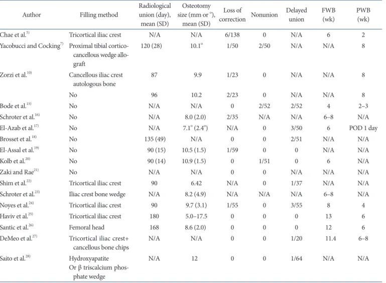
Table 6. Overall Results of Studies
Author Filling method Radiological
union (day), mean (SD)
Osteotomy size (mm or o),
mean (SD)
Loss of
correction Nonunion Delayed
union FWB
(wk) PWB
(wk)
Chae et al.5) Tricortical iliac crest N/A N/A 6/138 0 N/A 6 2
Yacobucci and Cocking7) Proximal tibial cortico- cancellous wedge allo- graft
120 (28) 10.1o 1/50 2/50 N/A N/A 8
Zorzi et al.10) Cancellous iliac crest
autologous bone 87 9.9 1/23 0 N/A N/A 8
No 96 10.2 2/23 0 N/A N/A 8
Bode et al.15) No N/A N/A 0 2/52 2/52 4 2–3
Schroter et al.16) No N/A 8.0 (2.0) 2/35 N/A N/A 6–8 N/A
El-Azab et al.17) No N/A 7.1o (2.4o) N/A 0 3/50 6 POD 1 day
Brosset et al.18) No 135 (49) N/A 0 0 2/51 N/A N/A
El-Assal et al.19) No 90 (15) 10.5 (1.5) 1/59 0 0 N/A N/A
Kolb et al.20) No 90 (14) 10.9 (1.5) 0 1/51 0 6 N/A
Zaki and Rae21) No N/A N/A 0 0 N/A N/A N/A
Shim et al.22) Tricortical iliac crest 90 6.42 N/A 0 1/37 N/A N/A
Schroter et al.23) Iliac crest bone wedge N/A 8.2 (4.9) N/A N/A N/A 6–8 N/A
Noyes et al.24) Tricortical iliac crest 90 9.7 (3.1) 1/55 0 3/55 8 4
Haviv et al.25) Tricortical iliac crest 180 5.0–17.5 0 0 0 13 6
Santic et al.26) Femoral head 168 8.6 (2.0) 0 0 0 12 6
DeMeo et al.27) Tricortical iliac crest+
cancellous bone chips N/A N/A 0 0 1/20 11.4 6–8
Saito et al.28) Hydroxyapatite Or β triscalcium phos-
phate wedge
N/A 12 0 0 1/64 N/A N/A
Table 5. Overall Coleman Methodology Score for Each Criterion Criteria (maximum score) Mean Standard
deviation Range Part A
Study size (10) 6.84 2.76 4–10
Mean follow-up (5) 4.4 1.22 2–5
No. of procedures (10) 10 0 10
Type of study (15) 7.8 4.58 0–15
Diagnostic certainty (5) 5 0 5
Surgery description (5) 4.4 1.22 3–5
Rehabilitation description (10) 7.6 4.35 0–10 Part B
Outcome criteria (10) 7.96 1.67 4–10
Procedure for outcomes (15) 8 2.41 3–12
Selection process (15) 15 0 15
Total score (100) 77 6.58 61–85
autologous bone graft with a locking plate.
3) Loss of correction
Yacobucci and Cocking7) reported one case of correction loss with lateral cortex fracture, wherein the correction angle was 15 degrees. Saragaglia et al.29) also reported one case of correction loss associated with screw breakage. Tables 9 and 10 list the inci- dence and rate of loss of correction. There was a similar tendency for the correction loss rate when compared according to the filler type and fixation method.
3. Comparative Studies
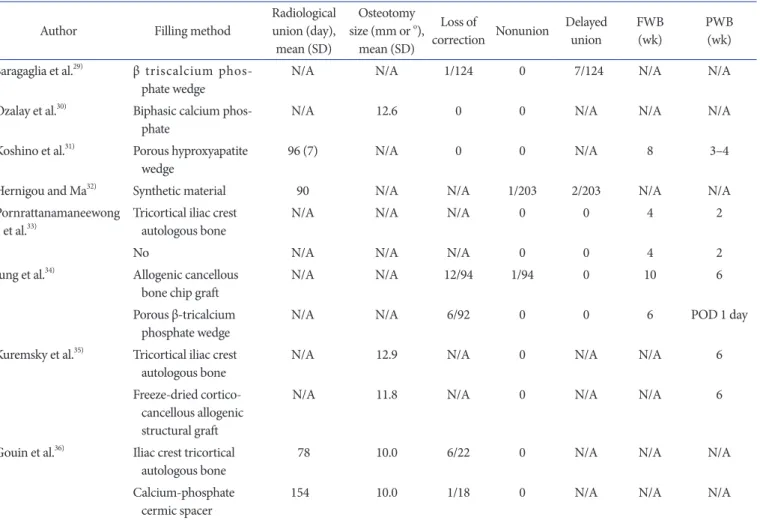
Only one randomized prospective study was identified. Zorzi et al.10) reported there was no significant difference in bone union period based on the review of a two-year comparison of 23 cases of OWHTO using autografts and another 23 cases of OWHTO without filling. As for comparative studies, there were four. Pornarattanamaneewong et al.33) reported that there were Table 6. Continued
Author Filling method Radiological
union (day), mean (SD)
Osteotomy size (mm or o),
mean (SD)
Loss of
correction Nonunion Delayed
union FWB
(wk) PWB
(wk) Saragaglia et al.29) β triscalcium phos-
phate wedge N/A N/A 1/124 0 7/124 N/A N/A
Ozalay et al.30) Biphasic calcium phos-
phate N/A 12.6 0 0 N/A N/A N/A
Koshino et al.31) Porous hyproxyapatite
wedge 96 (7) N/A 0 0 N/A 8 3–4
Hernigou and Ma32) Synthetic material 90 N/A N/A 1/203 2/203 N/A N/A
Pornrattanamaneewong
et al.33) Tricortical iliac crest
autologous bone N/A N/A N/A 0 0 4 2
No N/A N/A N/A 0 0 4 2
Jung et al.34) Allogenic cancellous
bone chip graft N/A N/A 12/94 1/94 0 10 6
Porous β-tricalcium
phosphate wedge N/A N/A 6/92 0 0 6 POD 1 day
Kuremsky et al.35) Tricortical iliac crest
autologous bone N/A 12.9 N/A 0 N/A N/A 6
Freeze-dried cortico- cancellous allogenic structural graft
N/A 11.8 N/A 0 N/A N/A 6
Gouin et al.36) Iliac crest tricortical
autologous bone 78 10.0 6/22 0 N/A N/A N/A
Calcium-phosphate
cermic spacer 154 10.0 1/18 0 N/A N/A N/A
SD: standard deviation, FWB: full weight-bearing, PWB: partial weight-bearing, N/A: not available, POD: postoperative day.
Table 7. Bone Union Period
Filler type Author Bone union period (day), mean (SD)
No Brosset et al.18) 135 (49)
El-Assal et al.19) 90 (15)
Kolb et al.20) 90 (14)
Auto Shim et al.22) 90
Noyes et al.24) 90
Allo Yacobucci and Cocking7) 120 (28)
Haviv et al.25) 180
Santic et al.26) 168
Synthetic Koshino et al.31) 96 (7)
Hernigou and Ma32) 90
SD: standard deviation, No: no filling, Auto: autogenous bone graft, Allo:
allogenous bone graft, Synthetic: synthetic material filling.
no significant differences of bone union period between their groups. Kuremsky et al.35) found that there was a 6-fold higher failure rate for the allograft group compared with the autograft group. Gouin et al.36), comparing an autologous bone graft group with a synthetic-material graft (BMCaPh) group, determined that correction loss was more frequent in the latter group (5%
vs. 27%). Specifically, the presence of lateral cortical hinge tears significantly increased the risk of correction loss in the BMCaPh group. These results indicated that the synthetic material was less tolerant to high mechanical stress, and that radiological union occurred significantly later in the BMCaPh group than in the autologous bone graft group. Jung et al.34) defined loss of correc- tion as less than 0 degrees of mechanical tibiofemoral angle at
Day
50 100 150 200
0 Hernigou and Ma32)
Koshino et al.31) Yacobucci and Cocking7) Santic et al.26) Haviv et al.25) Shim et al.22) Noyes et al.24) Kolb et al.20) El-Assal et al.19) Brosset et al.18)
Synthetic Allo Auto No
Fig. 2. Bone union period. No: no filling, Auto: autogenous bone graft, Allo: allogenous bone graft, Synthetic: synthetic material filling.
Table 8. Rates of Nonunion/Delayed Union by Filler Type Filler type Author Non-
union Event rate I2
(%) Delayed union Event
rate I2 (%)
No filling 3/313 0.018 0.0 7/263 0.03 0.0
Bode et al.15) 2/52 0.038 2/52 0.04 El-Azab et al.17) 0/50 0.010 3/50 0.06 Brosset et al.18) 0/51 0.008 2/51 0.04 El-Assal et al.19) 0/59 0.020 0/59 0.01 Kolb et al.20) 1/51 0.010 0/51 0.01 Zaki and Rae21) 0/50 0.023 N/A N/A
Auto 0/230 0.005 0.0 4/92 0.04 0.0
Chae et al.5) 0/138 0.004 N/A N/A Shim et al.22) 0/37 0.013 1/37 0.03 Noyes et al.24) 0/55 0.009 3/55 0.05
Allo 2/402 0.004 0.0 1/352 0.00 0.0
Yacobucci and
Cocking7) 2/50 0.040 N/A N/A
Haviv et al.25) 0/22 0.022 0/22 0.02 Santic et al.26) 0/310 0.002 0/310 0.00 DeMeo et al.27) 0/20 0.024 1/20 0.05
Synthetic 1/427 0.013 0.0 10/427 0.02 0.0
Saito et al.28) 0/64 0.004 1/64 0.02 Saragaglia et al.29) 0/124 0.031 7/124 0.06 Ozalay et al.30) 0/15 0.005 0/15 0.03 Koshino et al.31) 0/21 0.008 0/21 0.02 Hernigou and
Ma32) 1/203 0.009 2/203 0.01
N/A: not available, Auto: autogenous bone graft, Allo: allogenous bone graft, Synthetic: synthetic material filling.
Table 9. Nonunion/ Delayed Union/ Correction Loss for MOWHTO with Plate Fixation
Fixation type Nonunion Event rate I2 (%) Delayed union Event rate I2 (%) Correction loss Event rate I2 (%)
Non-locking plate 2/576 0.02 0 15/388 0.04 0 13/524 0.02 0
No filling 0/109 0.02 0 3/109 0.03 11 4/94 0.04 25.8
Auto 0/230 0.01 0 4/92 0.04 0 7/193 0.04 0
Allo 2/92 0.03 0 1/42 0.04 0 1/92 0.02 0
Synthetic 0/145 0.03 0a) 7/145 0.05 0 1/145 0.01 0
Locking plate 3/578 0.00 0 5/528 0.01 0 0/526 0.00 0
No filling 3/204 0.02 0 4/154 0.03 0 0/152 0.01 0
Auto N/A N/A N/A N/A N/A N/A N/A N/A N/A
Allo 0/310 0.00 0a) 0/310 0.00 0a) 0/310 0.00 0a)
Synthetic 0/64 0.00 0 1/64 0.02 0a) 0/64 0.01 0a)
MOWHTO: medial opening wedge high tibial osteotomy, N/A: not available, Auto: autogenous bone graft, Allo: allogenous bone graft, Synthetic:
synthetic material filling, I2: heterogeneity.
a)Only one case series.
the latest follow-up in the absence of an initial under-correction.
The rate of correction loss or varus recurrence was higher in the group receiving the Asecula plate (B. Braun Korea, Seoul, South Korea) with the β-tricalcium phosphate wedge. The outcomes of all of the comparative studies are documented in Table 11. There was no significant difference in bone union period between the autogenous graft groups and no filling groups (Fig. 3).
4. Starting Point of Weight-Bearing
All twenty studies included in this meta-analysis reported on the starting point of weight-bearing (Table 6). There was a tendency of delayed onset of weight-bearing in patients with allograft. Jung et al.34) permitted full weight-bearing walking one month after surgery to their allograft patients. Pornrattana- maneewong et al.33) allowed full weight-bearing walking at the same time point for both groups (tricortical iliac crest autologous
bone graft group and no filling group), and Zorzi et al.10) allowed partial weight-bearing walking at the same time point for both groups (cancellous iliac crest autologous bone graft group and no filling group); it must be noted, however, that the time point was not the endpoint of full weight-bearing walking but rather was the starting point of their rehabilitation protocol.
5. Bone Graft-Related Problem
Pornarattanamaneewong et al.33) and Kuremsky et al.35) de- scribed the graft harvest site as the source of pain that prolonged patient recovery time. Chae et al.5) also reported the graft harvest site as the source of pain for patients in the early postoperative period especially during trunk flexion. In addition, three of their patients underwent linear fracture of the iliac bone, which was treated conservatively. Schroter et al.23) reported that six patients suffered from hyperaesthesia or dysaesthesia at the iliac crest.
Gouin et al.36), Koshino et al.31) and Ozalay et al.30) all reported Table 10. Rates of Correction Loss by Filler Type
Filler type Author Loss of
correction Event rate I2 (%) Treatment
No filling 4/246 0.02 0.0
Schroter et al.16) 3/35 0.09 Two–medial dislocation of tibial plateau
Fixation with TomoFix and iliac creset autologous bone wedge No correlation with osteotomy size
Brosset et al.18) 0/51 0.01
El-Assal et al.19) 1/59 0.02 Partial loss of correction (4 degrees) No intervention
Kolb et al.20) 0/51 0.01
Zaki et al.21) 0/50 0.01
Auto 7/193 0.04 0.0
Chae et al.5) 6/138 0.04 No intervention
Noyes et al.24) 1/55 0.02 Revision
Allo 1/402 0.00 0.0
Yacobucci and Cocking7) 1/50 0.02 Correlation with nonunion and lateral hinge fracture Revision with Puddu plate and iliac crest autologous graft
Haviv et al.25) 0/22 0.02
Santic et al.26) 0/310 0.00
DeMeo et al.27) 0/20 0.02
Synthetic 1/224 0.01 0.0
Saito et al.28) 0/64 0.01
Saragaglia et al.29) 1/124 0.01 New osteotomy at the femoral level to correct the deformity 2 years later
Ozalay et al.30) 0/15 0.03
Koshino et al.31) 0/21 0.02
Auto: autogenous bone graft, Allo: allogenous bone graft, Synthetic: synthetic material filling.
poor incorporation of substitute material.
Discussion
In this meta-analysis, the RCT was found to have demonstrated similar bone union period, union rate and correction loss rate between the patients with autogenous bone graft and those with- out filling. One comparative study presented similar bone union period and union and delayed union rates between the autog- enous graft group and the no filling group. Another comparative study showed similar union and delayed union rates between al- logenous bone graft group and the synthetic material group. One comparative study reported the autogenous bone graft group showed shorter bone union period and lower correction rate compared to the synthetic material group. In analyzing the case
series, there was a similar tendency for nonunion, delayed union or correction loss rates among the groups classified according to the filler type. The bone union period showed a tendency to increase in the allogenous bone graft groups. The results of case series showed a similar tendency for union and delayed union rates among groups classified according to the graft type but a different was noted with regard to the correction loss rate.
The surgical techniques for opening wedge HTO underwent many modifications with regard to augmentation with bone grafts or bone substitutes. In general, autogenous bone graft is considered as the most successful bone filling material, owing to its osteoconductive, osteoinductive and osteogenic properties.
Nevertheless, autograft harvesting involves increased operative time as well as donor-site morbidity (hyper- or dysaesthesia, pain source and iliac crest fracture)24). Aryee et al.37) suggested that Table 11. Comparative Studies
Study Bone graft Bone union period
(day) p-value Opening
size (mm) Nonunion Delayed
union Loss of
correction Fixation type
Zorzi et al.10) Auto 86.7 0.128 9.9 0 N/A 1/23 Nonlocking (Puddu) plate
No 95.7 10.2 0 N/A 2/23 Nonlocking (Puddu) plate
Pornrattanamaneewong
et al.33) Auto 66.5 0.125 N/A 0 0 N/A Nonlocking (T buttress) plate
No 72.1 N/A 0 0 N/A Locking (TomoFix) plate
Jung et al.34) Allo N/A N/A 1/94 0 N/A Nonlocking (Aescula) plate
Synthetic N/A N/A 0 0 N/A Locking (TomoFix) plate
Kuremsky et al.35) Auto N/A N/A 12.9 0 N/A N/A Nonlocking (Arthrex opening wedge
osteotomy plate prototype set)
Allo N/A N/A 11.8 0 N/A N/A Nonlocking (Arthrex opening wedge
osteotomy plate prototype set)
Gouin et al.36) Auto 78 0.001 10 0 N/A 1/18 Locking (Surfix) plate
Synthetic 154 10 0 N/A 6/22 Locking (Surfix) plate
Auto: autogenous bone graft, N/A: not available, No: no filling, Allo: allogenous bone graft, Synthetic: synthetic material filling.
Zorzi et al.10)
Pornrattanamaneewong et al.33)
Overall (I-squared=0%, p=0.372)
Study Effect size (95% CI) Weight (%)a)
-12.7 0 12.7
89.07
10.93
100.00 9.00 (6.53, 11.47)
6.60 (1.45, 12.65)
8.68 (6.30, 10.96)
Day
Fig. 3. Bone union period of autogenous bone graft group and no filling group.
Forest plot of mean difference and 95%
con fidence interval (CI) for constant scores among patients assigned to Auto group or no filling group for opening wedge high tibial osteotomy. Mean dif- ference: 8.68 (day)=heterogeneity (I2)=0, p=0.372. a)Weights are from random ef- frects analysis.
iliac bone graft should be used only in high-risk patients (obese, smoker or opening gap larger than 10 mm). The use of bone al- lograft provides only an osteoconductive benefit, though, thanks to its availability without the need for additional surgical access, potential complications associated with the harvesting procedure of autogenous bone graft can be avoided26). However, allografts entail the risk of virus transfer38). Synthetic materials provide the osteoconductive benefit along with initial support; however sev- eral disadvantages such as soft-tissue irritation and infection have been reported39). van Hemert et al.40) reported synthetic bone augmentation does not aid much in primary stability and is not intended for load-bearing. Furthermore, it must be used in a me- chanically stable environment; otherwise, it cannot remodel into bone and shows slow incorporation into bone40). Seven studies in this meta-analysis noted the disadvantages of autogenous grafts and synthetic materials. There were no studies reporting any dis- advantages of allografts.
The recent trend of leaving the osteotomy with no graft is sup- ported by some authors3,41). Zorzi et al.10) included in this meta- analysis reported that there was no significant difference in the time to bone healing between patients that had an autologous bone graft and those that did not. They suggested that bone grafts could be reserved for special situations such as larger corrections and patients with conditions that impair bone healing. In the study, the no filling group also obtained satisfactory results and showed no differences compared with the other groups. Pornrat- tanamaneewong et al.33) included in this meta-analysis reported that a significantly higher percentage of osteotomies with medial defects was found in the no bone graft group and all defects re- mained until the time of the 2-year follow-up. Recent studies at- tributed such medial defects in OWHTO to low interfragmentary movement and tissue strain underneath the locking plate. They suggested that the use of dynamic locking screws in combination with early full load-bearing would increase the interfragmentary movement and tissue strain and potentially induce good bone healing underneath the plate42).
El-Assal et al.19) performed OWHTO without bone graft for the opening size of 14 mm in 9 cases, and the outcome was good.
Kolb et al.20) also performed OWHTO (up to 14 mm) and ob- tained favorable results. These two studies support the contention that OWHTO gaps of up to 14 mm in size can be healed without graft. Staubli et al.2) reported that the cases with opening sizes less than 15 mm did not have filling defects and showed good results overall. When an osteotomy size is smaller than 14 mm and rigid fixation and locking system are acquired, “no filling” of the osteotomy space can be a good option for OWHTO. OWHTO without bone graft is certainly very attractive particularly because
it is effective for reducing the length of surgery as well as patient morbidity.
The bone-healing process is complex and requires ideal biologi- cal and mechanical environments and factors. Kuremsky et al.35) included in this meta-analysis reported that lateral cortex fracture and opening size play a major role in bone union and construct failure. With higher varus angles, stability is largely dependent on the intact lateral hinge, which, if lost, would lead to the loss of resistance to axial compression forces and, thereby, loss of cor- rection. Stability plays a major role in this regard: the mechanical factors are functions of osteotomy stability, which depends on the osteotomy size, intact lateral cortex and rigid internal fixation43,44). Miller et al.45) detected a 58% reduction in axial stiffness and a 68% reduction in torsional stiffness in the case of lateral tibial cortex disruption. In case of lateral cortex fracture, there is insta- bility resulting from the insufficient primary stability provided by non-angular stable wedge plates46). Spahn et al.47) suggested that high body mass index (BMI) after weight-bearing could be the cause of correction loss. van Houten et al.48) noted that the use of tobacco was a major risk factor for development of nonunion in patients undergoing OWHTO. Rigid fixation, opening size and the presence/absence of lateral cortex fracture all affect bone union and maintenance of correction. In cases where there is a possibility of either correction loss or nonunion caused by lateral cortex fracture or large opening size, smoking or high BMI with non-rigid fixation, additional autologous bone graft is a better choice, with all due caution, to prevent harvest-site morbidity.
Limitations
There are several limitations to the present analysis. First, most of the reviewed studies were level IV case series; only one RCT and four comparative studies were found on the topic of OWHTO. Thus, the level IV study results could not be directly compared, which may affect the reliability of the analysis results.
Second, although the number of reviewed studies was large, the study designs were different: there were one RCT, four non-ran- domized trial studies and twenty uncontrolled trial studies. If the results are arranged according to the study design, the number of studies per one result analysis becomes less than 6 at the most.
On the assessment of publication bias, the power of the test of a meta-analysis based on the review of less than 10 studies is too low to derive definitive conclusions. In such a case, assessment for publication bias is not appropriate according to Cochrane handbook49). Accordingly, publication bias could not be assessed for our analysis.
Third, there were no precise common definitions of radiological
union, non-union, delayed union and loss of correction. Rather, these outcomes were assessed by the authors’ own subjective methods.
Fourth, there were not sufficient studies on OWHTO without bone graft. Finally, we did not include age, varus deformity angle and bone marrow density severity into the inclusion criteria.
These might have affected patient selection bias.
Conclusions
This meta-analysis evaluated the effects of four established methods of augmentation for osteotomy defect in OWHTO. Ac- cording to the published studies, all of the methods (autogenous bone grafting, allogenous bone grafting, synthetic material filling and no filling) appear to be appropriate operative treatment op- tions for osteotomy defect in OWHTO.
Conflict of Interest
No potential conflict of interest relevant to this article was re- ported.
References
1. Niemeyer P, Koestler W, Kaehny C, Kreuz PC, Brooks CJ, Strohm PC, Helwig P, Suedkamp NP. Two-year results of open-wedge high tibial osteotomy with fixation by medial plate fixator for medial compartment arthritis with varus malalignment of the knee. Arthroscopy. 2008;24:796-804.
2. Staubli AE, De Simoni C, Babst R, Lobenhoffer P. TomoFix:
a new LCP-concept for open wedge osteotomy of the medial proximal tibia: early results in 92 cases. Injury. 2003;34 Suppl 2:B55-62.
3. Lobenhoffer P, Agneskirchner JD. Improvements in surgical technique of valgus high tibial osteotomy. Knee Surg Sports Traumatol Arthrosc. 2003;11:132-8.
4. Akizuki S, Shibakawa A, Takizawa T, Yamazaki I, Horiuchi H.
The long-term outcome of high tibial osteotomy: a ten- to 20-year follow-up. J Bone Joint Surg Br. 2008;90:592-6.
5. Chae DJ, Shetty GM, Wang KH, Montalban AS Jr, Kim JI, Nha KW. Early complications of medial opening wedge high tibial osteotomy using autologous tricortical iliac bone graft and T-plate fixation. Knee. 2011;18:278-84.
6. Ahlmann E, Patzakis M, Roidis N, Shepherd L, Holtom P.
Comparison of anterior and posterior iliac crest bone grafts in terms of harvest-site morbidity and functional outcomes.
J Bone Joint Surg Am. 2002;84:716-20.
7. Yacobucci GN, Cocking MR. Union of medial opening- wedge high tibial osteotomy using a corticocancellous proxi- mal tibial wedge allograft. Am J Sports Med. 2008;36:713-9.
8. Lobenhoffer P, Agneskirchner J, Zoch W. Open valgus align- ment osteotomy of the proximal tibia with fixation by me- dial plate fixator. Orthopade. 2004;33:153-60.
9. Niemeyer P, Schmal H, Hauschild O, von Heyden J, Sud- kamp NP, Kostler W. Open-wedge osteotomy using an internal plate fixator in patients with medial-compartment gonarthritis and varus malalignment: 3-year results with re- gard to preoperative arthroscopic and radiographic findings.
Arthroscopy. 2010;26:1607-16.
10. Zorzi AR, da Silva HG, Muszkat C, Marques LC, Cliquet A Jr, de Miranda JB. Opening-wedge high tibial osteotomy with and without bone graft. Artif Organs. 2011;35:301-7.
11. Maier RV. What the surgeon of tomorrow needs to know about evidence-based surgery. Arch Surg. 2006;141:317-23.
12. Kral JG, Dixon JB, Horber FF, Rossner S, Stiles S, Torgerson JS, Sugerman HJ. Flaws in methods of evidence-based medi- cine may adversely affect public health directives. Surgery.
2005;137:279-84.
13. Higgins JP, Thompson SG, Deeks JJ, Altman DG. Measuring inconsistency in meta-analyses. BMJ. 2003;327:557-60.
14. Moher D, Liberati A, Tetzlaff J, Altman DG; PRISMA Group. Preferred reporting items for systematic reviews and meta-analyses: the PRISMA statement. PLoS Med.
2009;6:e1000097.
15. Bode G, von Heyden J, Pestka J, Schmal H, Salzmann G, Sudkamp N, Niemeyer P. Prospective 5-year survival rate data following open-wedge valgus high tibial osteotomy.
Knee Surg Sports Traumatol Arthrosc. 2015;23:1949-55.
16. Schroter S, Gonser CE, Konstantinidis L, Helwig P, Albrecht D. High complication rate after biplanar open wedge high tibial osteotomy stabilized with a new spacer plate (Posi- tion HTO plate) without bone substitute. Arthroscopy.
2011;27:644-52.
17. El-Azab HM, Morgenstern M, Ahrens P, Schuster T, Imhoff AB, Lorenz SG. Limb alignment after open-wedge high tibial osteotomy and its effect on the clinical outcome. Or- thopedics. 2011;34:e622-8.
18. Brosset T, Pasquier G, Migaud H, Gougeon F. Opening wedge high tibial osteotomy performed without filling the defect but with locking plate fixation (TomoFixTM) and early weight-bearing: prospective evaluation of bone union, pre- cision and maintenance of correction in 51 cases. Orthop
Traumatol Surg Res. 2011;97:705-11.
19. El-Assal MA, Khalifa YE, Abdel-Hamid MM, Said HG, Bakr HM. Opening-wedge high tibial osteotomy without bone graft. Knee Surg Sports Traumatol Arthrosc. 2010;18:961-6.
20. Kolb W, Guhlmann H, Windisch C, Kolb K, Koller H, Grutzner P. Opening-wedge high tibial osteotomy with a locked low-profile plate. J Bone Joint Surg Am.
2009;91:2581-8.
21. Zaki SH, Rae PJ. High tibial valgus osteotomy using the Tomofix plate: medium-term results in young patients. Acta Orthop Belg. 2009;75:360-7.
22. Shim JS, Lee SH, Jung HJ, Lee HI. High tibial open wedge osteotomy below the tibial tubercle: clinical and radiograph- ic results. Knee Surg Sports Traumatol Arthrosc. 2013;21:57- 63.
23. Schroter S, Mueller J, van Heerwaarden R, Lobenhoffer P, Stockle U, Albrecht D. Return to work and clinical outcome after open wedge HTO. Knee Surg Sports Traumatol Ar- throsc. 2013;21:213-9.
24. Noyes FR, Mayfield W, Barber-Westin SD, Albright JC, Heckmann TP. Opening wedge high tibial osteotomy: an operative technique and rehabilitation program to decrease complications and promote early union and function. Am J Sports Med. 2006;34:1262-73.
25. Haviv B, Bronak S, Thein R, Kidron A, Thein R. Mid-term outcome of opening-wedge high tibial osteotomy for varus arthritic knees. Orthopedics. 2012;35:e192-6.
26. Santic V, Tudor A, Sestan B, Legovic D, Sirola L, Rakovac I.
Bone allograft provides bone healing in the medial opening high tibial osteotomy. Int Orthop. 2010;34:225-9.
27. DeMeo PJ, Johnson EM, Chiang PP, Flamm AM, Miller MC.
Midterm follow-up of opening-wedge high tibial osteotomy.
Am J Sports Med. 2010;38:2077-84.
28. Saito T, Kumagai K, Akamatsu Y, Kobayashi H, Kusayama Y. Five- to ten-year outcome following medial opening- wedge high tibial osteotomy with rigid plate fixation in combination with an artificial bone substitute. Bone Joint J.
2014;96:339-44.
29. Saragaglia D, Blaysat M, Inman D, Mercier N. Outcome of opening wedge high tibial osteotomy augmented with a Biosorb® wedge and fixed with a plate and screws in 124 patients with a mean of ten years follow-up. Int Orthop.
2011;35:1151-6.
30. Ozalay M, Sahin O, Akpinar S, Ozkoc G, Cinar M, Cesur N. Remodeling potentials of biphasic calcium phosphate granules in open wedge high tibial osteotomy. Arch Orthop
Trauma Surg. 2009;129:747-52.
31. Koshino T, Murase T, Saito T. Medial opening-wedge high tibial osteotomy with use of porous hydroxyapatite to treat medial compartment osteoarthritis of the knee. J Bone Joint Surg Am. 2003;85:78-85.
32. Hernigou P, Ma W. Open wedge tibial osteotomy with acryl- ic bone cement as bone substitute. Knee. 2001;8:103-10.
33. Pornrattanamaneewong C, Numkanisorn S, Charean- cholvanich K, Harnroongroj T. A retrospective analysis of medial opening wedge high tibial osteotomy for varus osteo- arthritic knee. Indian J Orthop. 2012;46:455-61.
34. Jung WH, Chun CW, Lee JH, Ha JH, Kim JH, Jeong JH. Comparative study of medial opening-wedge high tibial osteotomy using 2 different implants. Arthroscopy.
2013;29:1063-71.
35. Kuremsky MA, Schaller TM, Hall CC, Roehr BA, Masonis JL. Comparison of autograft vs allograft in opening-wedge high tibial osteotomy. J Arthroplasty. 2010;25:951-7.
36. Gouin F, Yaouanc F, Waast D, Melchior B, Delecrin J, Passuti N. Open wedge high tibial osteotomies: Calcium-phosphate ceramic spacer versus autologous bonegraft. Orthop Trau- matol Surg Res. 2010;96:637-45.
37. Aryee S, Imhoff AB, Rose T, Tischer T. Do we need synthetic osteotomy augmentation materials for opening-wedge high tibial osteotomy. Biomaterials. 2008;29:3497-502.
38. Gaasbeek RD, Toonen HG, van Heerwaarden RJ, Buma P.
Mechanism of bone incorporation of beta-TCP bone substi- tute in open wedge tibial osteotomy in patients. Biomateri- als. 2005;26:6713-9.
39. Pornrattanamaneewong C, Harnroongroj T, Charean- cholvanich K. Loss of correction after medial opening wedge high tibial osteotomy: a comparison of locking plates with- out bone grafts and non-locking compression plates with bone grafts. J Med Assoc Thai. 2012;95 Suppl 9:S21-8.
40. van Hemert WL, Willems K, Anderson PG, van Heer- waarden RJ, Wymenga AB. Tricalcium phosphate granules or rigid wedge preforms in open wedge high tibial osteoto- my: a radiological study with a new evaluation system. Knee.
2004;11:451-6.
41. Brinkman JM, Lobenhoffer P, Agneskirchner JD, Staubli AE, Wymenga AB, van Heerwaarden RJ. Osteotomies around the knee: patient selection, stability of fixation and bone healing in high tibial osteotomies. J Bone Joint Surg Br.
2008;90:1548-57.
42. Roderer G, Gebhard F, Duerselen L, Ignatius A, Claes L.
Delayed bone healing following high tibial osteotomy re-