The clinical presentation of metastatic carcinoma in- volving the jugular foramen is rare. It shows usually hypo- vascular lytic bony destruction of the jugular foramen (1).
Hypervascular metastases such as those from renal and thyroid carcinoma may produce destructive vascu- lar lesions similar to paragangliomas (2).
The authors report a case of hypervascular metastasis from thyroid carcinoma in the jugular foramen simulat- ing glomus jugulare tumor.
Case Report
A 63-year-old woman presented with progressive headache, dizziness and hoarseness; Prior to hospitaliza- tion, she had suffered from the former two complaints for several months, and hoarseness for several years. A subsequent problem was left auricular pain.
Neurological examination indicated decreased gag re-
flex, left facial nerve palsy, and leftward deviation of the tongue. There was neither hearing impairment nor tin- nitus, nor any visual symptoms.
Initial brain CT revealed a lobulated inhomogeneous enhanced mass in the left jugular foramen and temporal bone. On temporal bone CT (Fig. 1), a large mass with irregular bony destruction in the left jugular foramen and temporal bone was seen, extending into the adja- cent posterior fossa and hypotympanum. The sigmoid s- inus plate was destroyed, and the ipsilateral carotico- jugular spine, jugular tubercle, and hypoglossal canal were eroded.
On T1-weighted MR imaging of the brain (Fig. 2A), in- homogeneous low signal intensity was seen, together with punctate vascular signal voids. The adjacent poste- rior fossa was severely indented by the mass. T2- weighted MR imaging (Fig. 2B) revealed inhomoge- neously mixed signal intensities, again with punctate vascular signal voids. On postcontrast T1-weighted MR images (Fig. 2C), the mass showed strong enhancement.
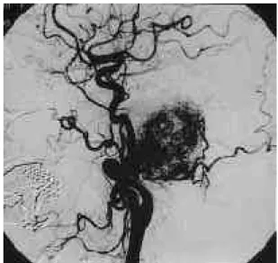
Angiography of the left common carotid artery (Fig. 3) demonstrated an extremely vascular mass with persis- tent vascular staining. The predominant source of vas- cular supply was the left occipital artery, while a small part of the mass was supplied from both the ascending
J Korean Radiol Soc 1999;41:1 0 97- 1 1 0 0
Metastatic Thy roid Carcinoma of Jugular Fo ra m e n Simulating Glomus Jugulare Tumor : A Case Re p o r t1
Eun Ja Lee, M.D., Dong Hun Yang, M.D., Chul Ku Jung, M.D.2, Si Won Kang, M.D.
We report a case of hy p e r vascular metastatic thyroid carcinoma of the jugular fora- men simulating a glomus jugulare tumor. Computed tomography(CT) revealed areas of irregular lytic bony destruction of the left jugular foramen, as well as characteristic i n vasion routes of a glomus jugulare tumor. Magnetic resonance(MR) imaging and an- g i o g r a p hy demonstrated a hy p e r vascular mass similar to a glomus tumor.
Index words :Skull, base
Skull, secondary neoplasms Skull, CT
Skull, MR A n g i o g r a p hy
1Department of Radiology, Taejon St. Mary’s Hospital, The Catholic University of Korea
2Department of Neurosurgery, Taejon St. Mary’s Hospiral, The Catholic University of Korea
Received June 16, 1999 ; Accepted August 30, 1999
Address reprint requests to : Eun Ja Lee, M.D., Department of Radiology, Taejon St, Mary’s Hospital, The Catholic University of Korea,
#520-2 Taehung-dong Taejon, 301-012, Korea.
Tel. 82-42-220-9625 Fax. 82-42-252-6807
─ 1 0 9 7 ─
pharyngeal and posterior auricular arteries. The venous phase showed occlusion of the left sigmoid sinus and the internal juglar vein. Preoperatively the left occipital artery was successfully embolized using coils.
Four days later, the mass was surgically removed and appeared to be a glomus jugulare. Postoperatively the pathologic diagnosis was metastatic papillary adenocar-
c i n o m a .
Work-up for the primary origin of the mass was per- formed; a mass in the right anterior part of the neck was palpable. Color Doppler ultrasonography of the thyroid gland demonstrated a heterogeneous solid mass in the
Eun Ja Lee, et al : Metastatic Thyroid Carcinoma of Jugular Foramen Simulating Glomus Jugulare Tumor
─ 1 0 9 8 ─ Fig. 1. Temporal bone CT scan
Axial CT scan shows a large expansile lesion in the left jugular foramen and temporal bone with irregular bony margin.
Sigmoid sinus is destroyed. Ipsilateral caroticojugular spine(s- mall arrows) and jugular tubercle(long arrow) are eroded.
Fig. 3. Left common carotid angiogram
Lateral arterial phase shows an extremely vascular mass. The vascular supply is predominantly from the occpital artery, and small part of the mass is supplied from the ascending pha- ryngeal and posterior auricular arteries. Venous phase reveals occlusion of left sigmoid sinus and internal jugular vein(not shown). Preoperative embolization of left occipital artery by the coils was performed successfully(not shown).
A B
Fig. 2. Brain MR imaging
Axial T1-weighted image(A) reveals a large lobulated mass in the left jugular foramen and temporal bone, extending into the poste- rior fossa. Ipsilateral cerebellar hemisphere is indented. The mass shows inhomogeneous low signal intensity with internal vascu- lar signal voids. Ipsilateral hypoglossal canal(arrow) is invaded by direct extension of the mass. On T2-weighted coronal image(B) , this mass shows inhomogeneous signal intensity with multiple vascular signal voids. Postcontrast coronal T1-weighted simage(C) shows strong enhancement.
C
lower pole of the right thyroid gland with internal blood flow. By means of sono-guided needle aspiration, papil- lary adenocarcinoma was confirmed.
Twenty days later, the patient underwent follow up MR imaging and was found to be free of residual tumor.
D i s c u s s i o n
As a complex bony canal, the jugular foramen trans- mits vessels and nerves from the posterior cranial fossa through the skull base into the carotid space. The jugu- lar foramen is subdivided into a pars nervosa anteriorly and a pars vascularis posteriorly by a fibrous or bony septum; it contains the glossopharyngeal nerve and infe- rior petrous sinus anteriorly(pars nervosa) and the va- gus nerve, spinal accessory nerve, jugular vein, and pos- terior meningeal artery posteriorly(pars vascularis). MR imaging is the modality of choice for soft tissue assess- ment but lesions affecting small cortical bony structures may require CT for further evaluation (1, 3).
Most malignant tumoral lesions of the jugular fora- men are seen on CT as areas of infiltrative bone destruc- tion, whereas schwannoma and meningioma cause s- mooth enlargement of the jugular foramen (4).
On contrast enhanced CT, a glomus jugulare tumor manifests as areas of irregular, lytic bony destruction of the jugular foramen, with significant enhancement. It tends to be extended in predictable patterns along path- ways of least resistance, frequently below the skull base, into the mastoid air cells, and into the hypotympa- num (1. 4). Erosion of the caroticojugular spine, jugular tubercle and hypoglossal canal, and progressive circular enlargement of the jugular fossa are common findings, with circular indentation of the adjacent posterior fossa dura and encroachment on the internal carotid artery. It frequently invades the jugular vein, sigmoid sinus or in- ferior petrosal sinus with subsequent formation of anas- tomotic venous channels (2, 5). On MR imaging, the tu- mor shows low signal intensity on T1-weighted images and high signal intensity on T2-weighted images.
Postcontrast T1-weighted MR images show strong en- hancement. A speckled appearance with multiple flow voids is typical in tumors larger than 2cm in diameter.
Angiography demonstrates an extremely vascular le- sion. The vascular supply is predominantly from the ex- ternal carotid artery, particularly the ascending pharyn- geal artery (4, 5). Preoperative embolization of the feed- ing artery dramatically reduces intraoperative blood loss and improves the chances of complete tumor re-
moval (5).
If they happen to destroy the jugular foramen, carci- noma, sarcoma, myeloma, metastasis, and other malig- nancies found in this orifice may be indistinguishable from a glomus jugulare tumor, as seen on CT (6, 7).
Metastatic carcinoma involving the jugular foramen is rare (1, 8), the common primary tumors being lung and breast carcinomas. Metastases to the jugular foramen are more aggressive, and clinical onset of their symp- toms is often more rapid than is that of a glomus jugu- lare tumor (4, 8). Metastases do not usually follow the characteristic routes of invasion of a glomus tumor and MR imaging shows that the flow voids are usually ab- sent. Angiography reveals that most are much less vas- cular than a glomus tumor. Hypervascular tumors such as metastatic thyroid carcinoma, metastatic renal cell carcinoma, metastatic pheochromocytoma, plasmacy- toma, and hemangiopericytoma in the jugular foramen may, however, be indistinguishable from a glomus jugulare tumor, as shown by cross-sectional imaging and on angiography (3, 4, 6, 8-10).
In our case, the patient’s clinical symptoms pro- gressed slowly and radiographic findings of the mass mimicked the usual roentgenographic presentation of a glomus jugulare. On CT, the mass manifested as areas of irregular and lytic bony destruction of the left jugular foramen and temporal bone, with significant enhance- ment. The mass followed the characteristic routes of in- vasion of a glomus jugulare tumor, and as seen on MR images was similar to this tumor. Conventional angiog- raphy revealed an extremely vascular lesion, supplied from an external carotid artery such as the occipital, as- cending pharyngeal, or posterior auricular.
Although the existence of an extensive vascular mass in the jugular fossa, together with lytic bony destruc- tion, is more consistent with a diagnosis of glomus jugu- lare tumor, hypervascular metastasis from thyroid car- cinoma must be differentiated.
R e f e r e n c e s
1. Chong VFH, Fan YF. Pictorial review: radiology of the jugular f o r a m e n . Clin Radiol 1 9 9 8 ; 5 3 : 4 0 5 - 4 1 6
2 . Chakeres DW, LaMasters DL. Paragangliomas of the temporal bone: high-resolution CT studies. R a d i o l o g y 1 9 8 4 ; 1 5 0 : 7 4 9 - 7 5 3 3 . Megerian CA, McKenna MJ, Nasol Jr JB. Non-paraganglioma
jugular foramen lesions masquerading as glomus jugulare tumors.
Am J Otol1 9 9 5 ; 1 6 : 9 4 - 9 8
4 . Caldemeyer KS, Mathews VP, Azzarelli B, Smith RR. The jugular foramen: a review of anatomy, masses, and imaging characteris- tics. R a d i o G r a p h i c s 1 9 9 7 ; 1 7 : 1 1 2 3 - 1 1 3 9
J Korean Radiol Soc 1999;41:1 0 97- 1 1 0 0
─ 1 0 9 9 ─
5 . Weber AL, Mckenna MJ. Radiologic evaluation of the jugular fora- men: anatomy, vascular variants, anomalies, and tumors.
Neuroimag Clin N Am1 9 9 4 ; 4 ; 5 7 9 - 5 9 8
6 . Lo WW, Solti-Bohman LG. Tumors of the temporal bone and the cerebellopontine angle. In: Som PM, Curtin HD,eds. Head and neck i m a g i n g. 3rd ed. St Louis, Mo: Mosby-Year Book 1996;1449-1534 7 . Reid CB, Fagan PA, Turner J. Low-grade myxoid chondrosarcoma
of the temporal bone: differential diagnosis and report of two cas- es. Am J Otol 1994;15: 419-422
8 . Hellier WP, Crockard HA, Cheesman AD. Metastatic carcinoma
of the temporal bone presenting as glomus jugulare and glomus tympanicum tumors: a description of two cases. J Laryngol Otol 1 9 9 7 ; 1 1 1 : 9 6 3 - 9 6 6
9 . Miyachi S, Negoro M, Saito K, Nehashi K, Sugita K. Myeloma manifesting as a large jugular tumor: case report. N e u r o s u r g e r y 1 9 9 0 ; 2 7 : 9 7 1 - 9 7 7
1 0 . Boileau MA, Grotta J, Borit A, et al. Metastatic renal cell carcino- ma simulating glomus jugulare tumor. J Surg Oncol 1 9 8 7 ; 3 5 : 2 0 1 - 2 0 3
Eun Ja Lee, et al : Metastatic Thyroid Carcinoma of Jugular Foramen Simulating Glomus Jugulare Tumor
─ 1 1 0 0 ─
경정맥구종양과 유사한 경정맥공의 전이성 갑상선암 : 1예보고1
1가톨릭대학교 의과대학 대전성모병원 방사선과
2가톨릭대학교 의과대학 대전성모병원 신경외과
이은자・양동헌・정철구2・강시원
저자들은 경정맥구종양과 유사한 경정맥공내에 과다혈관을 갖는 전이성 갑상선암의 한 증례를 보고하고자한 다. 전산화단층촬영에서 종양은 경정맥공내에 불규칙한 골파괴를 보였고 경정맥구종양의 특징적인 침범양상을 나타내었다. 자기공명영상과 혈관조영술에서는 경정맥구종양과 유사한 혈관과다의 종괴를 보였다.
대한방사선의학회지 1 9 99;41: 1 0 97- 1 1 0 0