Copyright © 2018 The Korean Society for Bone and Mineral Research
This is an Open Access article distributed under the terms of the Creative Commons Attribution Non-Commercial Li- cense (http://creativecommons.org/licenses/by-nc/4.0/) which permits unrestricted non-commercial use, distribu- tion, and reproduction in any medium, provided the original work is properly cited.
High Plasma Sphingosine 1-phosphate Levels
Predict Osteoporotic Fractures in Postmenopausal Women: The Center of Excellence for Osteoporosis Research Study
Mohammed-Salleh M. Ardawi1,2, Abdulrahim A. Rouzi1,3, Nawal S. Al-Senani1,3, Mohammed H. Qari1,4, Ayman Z. Elsamanoudy2, Shaker A. Mousa1,5
1Center of Excellence for Osteoporosis Research, Faculty of Medicine, King Abdulaziz University Hospital, King Abdulaziz University, Jeddah;
2Department of Clinical Biochemistry, Faculty of Medicine, King Abdulaziz University Hospital, King Abdulaziz University, Jeddah;
3Department of Obstetrics and Gynecology, Faculty of Medicine, King Abdulaziz University Hospital, King Abdulaziz University, Jeddah;
4Department of Hematology, Faculty of Medicine, King Abdulaziz University Hospital, King Abdulaziz University, Jeddah, Saudi Arabia;
5The Pharmaceutical Research Institute, Albany College of Pharmacy and Health Sciences, Rensselaer, NY, USA
Background: Higher sphingosine 1-phosphate (S1P) plasma levels are associated with decreased bone mineral density (BMD), and increased risk of prevalent vertebral fracture.
So, we hypothesized that postmenopausal women with increased baseline plasma S1P levels have a greater risk for future incident fracture (osteoporosis-related fractures [ORFs]).
Methods: This study was conducted in a prospective longitudinal cohort of 707 women recruited in 2004 and followed up annually for a mean period of 5.2±1.3 years. They were postmenopausal (aged ≥50 years). The primary outcome measure was the time to the first confirmed ORF event using radiographs and/or a surgical report. Results: The plasma S1P levels (μmol/L) were significantly higher in the women with incident fracture (7.23±0.79) than in those without ORFs (5.02±0.51; P<0.001). High S1P levels were strongly associated with increased fracture risk. After adjustment for age and other con- founders, the hazard ratio (HR) was 6.12 (95% confidence interval [CI], 4.92-7.66) for each 1-standard deviation increase in plasma S1P levels. The women in the highest quartile of S1P levels had a significant increase in fracture risk (HR, 9.89; 95% CI, 2.83-34.44). Results were similar when we compared plasma S1P levels at the 1-year visit. Conclusions: The associations between plasma S1P levels and fracture risk were independent of BMD and other confounders. These findings demonstrate that high plasma S1P level at baseline and at years 1 to 5 is a strong and independent risk factor for future [ORFs] among post- menopausal women and could be a useful biomarker for fracture risk assessment in this population.
Key Words: Osteoporosis, Osteoporotic fractures, Postmenopausal, Sphingosine 1 phos- phate
INTRODUCTION
Incident fracture (osteoporosis-related fractures [ORFs]) have major economic and social impacts, with serious morbidities and increased mortality following hip and/or vertebral fractures.[1,2] Many postmenopausal women at risk of fracture Corresponding author
Ayman Z. Elsamanoudy
Department of Clinical Biochemistry, Faculty of Medicine, King Abdulaziz University Hospital, P.O. Box 20724, Jeddah 21465, Saudi Arabia
Tel: +966-59-506-2375 Fax: +966-6952063
E-mail: [email protected] Received: February 23, 2018
Revised: April 14, 2018 Accepted: April 18, 2018
No potential conflict of interest relevant to this article was reported.
Original Article
pISSN 2287-6375 eISSN 2287-7029
will be missed based on bone mineral density (BMD) as- sessment alone,[1,2] a finding that is related to the multi- ple determinants of bone fragility, including age, fracture history, and deterioration of bone quality.[2] Identifying new biomarker(s) independent of BMD to improve fracture prediction is needed. Bone turnover markers (BTMs), particu- larly those of bone resorption, can predict fracture risk among postmenopausal women.[3] Such prediction may improve if BTMs are assessed along with BMD.[4,5] One possible new biomarker is plasma sphingosine 1-phos- phate (S1P).
S1P is a bioactive lysophospholipid, autocrine-paracrine signaling molecule that regulates multiple cellular process- es, including migration, proliferation, differentiation, and apoptosis, in many cell types.[6-8] S1P plays an important role in bone metabolism as a coupling factor between bone resorption and formation. It is also produced by osteoclasts to stimulate proliferation, migration, and survival of osteo- blasts, possibly leading to bone formation stimulation.[6-8]
Conversely, S1P augments osteoclastogenesis via induction of receptor activator of nuclear factor-κB ligand (RANKL) in osteoclasts and T-cells.[9] A concentration gradient of S1P is formed between circulation and bone tissue whereby osteoclast precursor cell movement is facilitated by che- mo-repulsion mediated by S1P receptor 2 (S1PR2) signal- ing.[10] Such observations suggest that S1P affects osteo- clasts, leading to increased bone resorption.[8] Furthermore, S1PR2-deficient mice exhibit moderate osteoporosis due to a decrease in osteoclastic bone resorption.[11] Recently in a case-control study, Kim et al.[12] demonstrated mark- edly higher S1P levels in 69 postmenopausal women with vertebral fractures than those in 69 non-fractured age and body mass index (BMI) matched controls, with S1P levels inversely correlated with BMD at various sites and positive- ly correlated with bone resorption markers (BRMs). These data suggest that circulating S1P level could serve as a new biomarker of fracture risk. However, Heilmann et al.[13] us- ing systemic FTY720 (an S1P analog) treatment did not improve fracture healing in experimental mice although several in vitro studies demonstrated osteoanabolic effects [14,15] and improved bone defect healing.[16]
We hypothesized that postmenopausal women with high- er circulating S1P levels have a greater ORF risk. We exam- ined the association between plasma S1P levels and ORF risk in a prospective cohort of postmenopausal women
aged ≥50 years in the Center of Excellence for Osteoporo- sis Research (CEOR) study.
METHODS
1. Study designThe CEOR study is an ongoing prospective, population- based cohort of 912 women aged ≥50 years residing in the Jeddah area of Western Saudi Arabia who were recruit- ed in 2004. The study investigated the determinants of bone loss and fractures among healthy postmenopausal women. The rationale and design of the study as well as sampling and data collection have been described previ- ously.[5,17] The baseline evaluation included 3,015 wom- en. At enrollment, women were eligible for the study if they were aged ≥50 years, had been postmenopausal for
>1 year (with serum FSH levels >15 mIU/L), were inde- pendently mobile and on an unrestricted diet. Exclusion criteria included a cancer history (n=11), regardless of type and stage) and current diagnosis of or prior therapy for os- teoporosis (n=15). Women were also ineligible at enroll- ment if they had diseases or conditions that could interfere with bone metabolism or if they used anti-osteoporotic treatments (n=36), bisphosphonates, estrogen, calcitonin, fluoride salts, calcitriol, strontium or selective estrogen-re- ceptor modulators). Women were also excluded if they had fever (oral temperature ≥38.0°C) or abnormal findings on complete blood cell counts including platelets and/or leu- kocytes (n=305). Women with abnormal hepatic, renal or thyroid function tests were also excluded (n=1,736). All women provided written informed consent for study par- ticipation. The study was approved by the Human Research Ethics Committee of CEOR, King Abdulaziz University. Dur- ing the follow-up, of the 912 women initially recruited (ful- filling inclusion criteria), 148 were lost to follow-up, 20 died and 37 withdrew. The present study investigated 707 heal- thy postmenopausal women (mean age, 61 years; range, 53-93 years) from the CEOR cohort at the sixth annual fol- low-up for whom blood and urine samples were obtained at the baseline and follow-up visits. Women were followed with the intention to treat from the baseline visit to frac- ture, completion of the follow-up period, withdrawal from the study, or loss to follow-up or death, if no fracture had occurred. The participants received vitamin D and calcium supplementation to maintain the serum 25-hydroxy-vita-
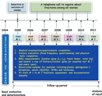
min D (25[OH]D) concentration >50 nmol/L and eura para- thyroidism. Among the women who completed the study, plasma S1P and BTM levels, along with other analytes, were evaluated during the baseline visit, the 1-year follow-up visit, and annually thereafter (Fig. 1). Additional baseline assessments (using a questionnaire) included information on medical history, menopausal age, years since meno- pause, socioeconomics, education level, family history of fragility/fracture(s), and occurrence of a fall(s) during the past year. Information on smoking, physical activity (PA), vitamins and medication use were also recorded. Height and weight, as well as hip and waist circumferences, were measured while the women were wearing lightweight clo- thing with no shoes. BMI and the waist-to-hip ratio were also calculated. Cumulative PA scores from 0 to 27 were constructed based on the 7-day PA record described previ- ously.[1] Women were classified into either sedentary (PA- score ≤14) or moderate/high activity (PA-score >14) groups.
Hand-grip strength was measured using a JAMAR-Plus hand dynamometer (Sammons Preston, Bolingbrook, IL, USA) on both hands. Dietary intake of calories, protein, cal- cium and vitamin D were assessed using a food-frequency questionnaire.[17]
2. Assessment of fractures
Research physicians at CEOR monitored the enrolled women for incident fractures, which were recorded using
CEOR’s information system network. Two research physi- cians classified fracture events independently according to the International Statistical Classification of Diseases and Related Health Problems, 10th revision (ICD-10).[18] A ra- diologist and/or an orthopedic surgeon reviewed all coded events for final fracture classification. Only low-trauma fractures (i.e., falls from standing height or less) were con- sidered and classified as incident fragility fractures. Inci- dent fractures were reported during annual follow-ups, and all peripheral fractures were confirmed by radiographs or a surgical report, as described previously.[17]
We excluded all non-ORFs (i.e., accident-related fractures such as a traffic road accident or a fall from a height higher than a chair or the first rung of a ladder or severe trauma;
and all finger, toe, skull, and facial fractures). The specific fractures and fracture follow-up times were recorded at different sites during a follow-up period when a woman sustained an incident first fracture. The follow-up period was calculated as the time from the baseline visit to the first fracture, death, or the end of the study, whichever oc- curred first.[17] For women lost during the follow-up, the period was computed as the time from enrollment to the date of last contact. For incident vertebral fractures, lateral X-ray films of the thoracic and lumbar spine were obtained at baseline and annual visits for all women, and at the end of the study. All prevalent and incident vertebral fractures were identified by a radiologist as described by Genant et al.[19]. A prevalent fracture was defined as a vertebral body with a semi-quantitative grade ≥1. Any new vertebral frac- ture was identified by an increase of at least one grade in a vertebral body that was defined as normal at baseline. We excluded vertebral fractures due to major trauma, and/or vertebral deformities with non-osteoporosis causes (e.g., osteoarthritis and/or bone diseases). All vertebral and non- vertebral fractures were included in the final analyses.
3. Assessment of BMD
The BMD (lumbar spine [L1-4] and the mean of the prox- imal right and left femur [total and sub-regions]) was mea- sured by dual energy X-ray absorptiometry using the Lunar Prodigy Model (Lunar Corp., Madison, WI, USA). Quality- control procedures were performed according to the man- ufacturer’s recommendations.[17]
Fig. 1. Study design and measurements.
4. Measurement of plasma S1P levels and analytes
During the baseline and follow-up visits, venous blood samples were collected between 9:00 and 11:00 a.m. after an overnight fast under standardized conditions. Second- void morning urine samples were collected on the same day of blood sampling. The blood was centrifuged, and plasma (sodium citrate- or EDTA-treated), serum, and urine samples were stored in liquid nitrogen at -190°C at the Bio- bank Unit of CEOR until they were analyzed. Plasma (treat- ed with sodium citrate) S1P was measured using a com- petitive ELISA kit (Echelon Biosciences, Salt Lake City, UT, USA) according to the manufacturer’s instructions.[20] All plasma samples showing hemolysis or clotting were dis- carded. However, no sample was collected immediately following any ORF. For women with ORFs, samples (n=9 women) were collected at least 3 months following the in- cident, and adjustments were made for the collection time.
[17] The lower detection limit was 0.06 µmol/L with intra- assay and inter-assay coefficients of variations equaling 6.3% and 5.9%, respectively. Duplicate samples were as- sayed, and all results were reported as means. Plasma sam- ples were diluted by a factor of 1/10. Further validation studies were performed for this assay. Linearity was as- sessed by serially diluting samples with sample diluents (dilutions 1:15, 1:20, and 1:30) and comparing observed values with expected values (observed recoveries range, 101%-109%) (data not shown). Additionally, recovery of spiked standards was tested by adding different S1P con- centrations (three different concentrations) to eight differ- ent human plasma samples presented with various levels of endogenous S1P. Spiked recovery ranged from 90% to 106% (data not shown). No sample tested had S1P values below the lower limit of detection or above the upper limit of quantitation corresponding to 2.0 µmol/L. Other bio- chemical tests were performed at the Diagnostic laborato- ry of CEOR according to standard methods as described previously [21,22].
5. Fracture risk assessment tools evaluation BMD (g/cm2) was determined as described before.
Part of the withdrawn fasting blood sample was used for serum preparation to measure the biochemical BTMs. More- over, urine samples were obtained in morning. Serum bone alkaline phosphatase (s-bone ALP), serum procollagen type
1 N-terminal propeptide (s-P1NP) , urinary cross-linked C- terminal telopeptide of type 1 collagen (CTX) and serum N-terminal telopeptide of collagen type I (NTx) were mea- sured using competitive inhibition enzyme linked immu- noassay by the commercially available kits.
Serum follicle stimulating hormone (FSH), luteinizing hormone, E2 and intact-parathyroid hormone were also, measured by commercially available immunoassays using Elecsys autoanalyzer (Roche Diagnostics GmbH, D-68298.
Mannheim, Germany). The intra- and inter-assay coefficient of variations (CVs) were less than 4.0%. Serum 25(OH)D was measured by direct competitive chemiluminescence immunoassay using LIASON autoanalyzer (DiaSorin Inc., Stillwater, MN,. USA). The intra- and inter-assay CVs were 7.8% and 3.8% respectively.
6. Statistical analysis
Results are presented as means±standard deviation (SD), and categorical variables are expressed as frequen- cies. Data were analyzed using SPSS version 17.0 software (SPSS Inc., Chicago, IL, USA). Analysis of variance or the Mann-Whitney test was used, as appropriate, to examine differences among the groups for different variables and χ2 test for categorical variables to test for significant differ- ences among groups. Correlation analysis was performed using Pearson’s test. Partial correlation analysis was per- formed to adjust for various possible confounders. The re- lationship between incident fracture risk and various plas- ma S1P levels was evaluated using SD score changes and quartile-based analysis. The SD score was calculated using the formula (S1Pi–S1Pm)÷SD, where S1Pi is the individual S1P level, S1Pm is the mean cohort S1P level, and SD is the SD of cohort S1P levels. This calculation was used to deter- mine the change in incident fracture risk for each incre- mental increase of 1-SD in S1P levels. Similar calculations were made for BTMs. Plasma S1P and BTMs were catego- rized into Quartiles, and the lowest Quartiles were com- pared to the fourth (highest) or other Quartiles. Quartiles were calculated for the following variables: age, years since menopause, dietary calcium intake, serum 25(OH)D, PA score, hand-grip strength, and BMD measurement at all sites. Cox proportional hazards regression analysis was used to estimate fracture risk. Data were used to estimate the hazard ratios (HRs) by Quartile and per SD increase in plasma S1P and other BTMs, and results were reported as
HRs with estimation of 95% confidence intervals (CIs). In the Cox model, the follow-up time describes the time from the baseline visit to (a) the first fracture; or (b) completion of the follow-up period; or (c) death, if no fracture had oc- curred. The first fracture was considered the analysis end- point. HRs were computed by comparing women with and without a fracture. Cox regression analysis was performed for different times: 2.0, 3.0, 5.0, and 6.0 years after collect- ing samples of the baseline visit. All fracture risk estimates were adjusted for age, BMI, and other confounders as ap- propriate. The population attributable risk (PAR) propor- tion was calculated according to the formula {Px(RR–1)÷
[1+Px(RR–1)]}×100, where P is the percentage of the pop- ulation exposed, and RR is the relative risk.[23]
RESULTS
1. Baseline characteristics
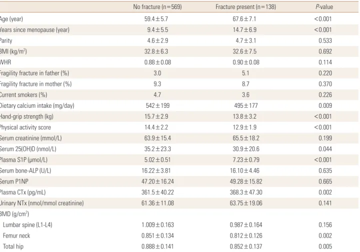
Baseline characteristics of the participants are presented in Table 1. During the mean follow-up period of 5.2±1.3 years, 138 women sustained ORFs: 44 vertebral fractures and 104 non-vertebral fractures (52 wrist, 12 hip, 21 rib, 8 humerus, 4 ankle, 5 metatarsal, and 2 pelvic) were docu- mented. Women who had no fracture (n=569) were used as age and BMI matched control group in all analyses. As expected, women with ORFs were older, had higher num- ber of years since menopause, lower values for BMD and lower hand-grip strength and PA-score values. Plasma S1P levels were significantly higher in women with ORFs than in women without fractures (7.23±0.79 vs. 5.02±0.51
Table 1. Baseline characteristics of postmenopausal women studied according to fracture
No fracture (n=569) Fracture present (n=138) P-value
Age (year) 59.4±5.7 67.6±7.1 <0.001
Years since menopause (year) 9.4±5.5 14.7±6.9 <0.001
Parity 4.6±2.9 4.7±3.1 0.533
BMI (kg/m2) 32.8±6.3 32.6±7.5 0.692
WHR 0.88±0.08 0.90±0.08 0.114
Fragility fracture in father (%) 3.0 5.1 0.220
Fragility fracture in mother (%) 9.3 8.7 0.370
Current smokers (%) 4.7 3.6 0.226
Dietary calcium intake (mg/day) 542±199 495±177 0.009
Hand-grip strength (kg) 15.7±2.9 13.8±3.2 <0.001
Physical activity score 14.4±2.2 12.9±1.9 <0.001
Serum creatinine (mmol/L) 63.9±15.4 65.5±18.2 0.199
Serum 25(OH)D (nmol/L) 35.2±23.3 30.9±20.6 0.044
Plasma S1P (µmol/L) 5.02±0.51 7.23±0.79 <0.001
Serum bone-ALP (U/L) 16.22±3.81 16.10±4.46 0.635
Serum P1NP 47.20±16.24 49.28±15.82 0.665
Plasma CTx (pg/mL) 361.5±40.22 368.3±47.30 0.002
Urinary NTx (nmol/mmol creatinine) 61.36±11.08 63.75±19.06 0.141
BMD (g/cm2)
Lumbar spine (L1-L4) 1.009±0.163 0.987±0.164 0.156
Femur neck 0.851±0.134 0.812±0.126 0.002
Total hip 0.888±0.141 0.852±0.137 0.005
The data is presented as mean±standard deviation.
To convert 25(OH)D values to ng/mL divide by 2.496; to convert creatinine values to mg/dL, divide by 88.4.
BMI, body mass index; WHR, waist-to-hip ratio; 25(OH)D, 25-hydroxy-vitamin D; S1P, sphingosine 1-phosphate; ALP, alkaline phosphatase; P1NP, procol- lagen type 1 N-terminal propeptide; CTX, C-terminal telopeptide of type 1 collagen; NTx, N-terminal telopeptide of collagen type I; BMD, bone mineral density.
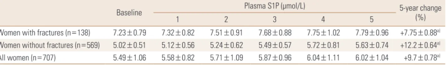
Table 2. Plasma sphingosine 1-phosphate levels in the study participants at baseline and 1, 2, 3, 4, and 5 years
Baseline Plasma S1P (µmol/L) 5-year change
1 2 3 4 5 (%)
Women with fractures (n=138) 7.23±0.79 7.32±0.82 7.51±0.91 7.68±0.88 7.75±1.02 7.79±0.96 +7.75±0.88a) Women without fractures (n=569) 5.02±0.51 5.12±0.56 5.24±0.62 5.49±0.57 5.72±0.81 5.63±0.74 +12.2±0.64a)
All women (n=707) 5.49±1.06 5.58±0.82 5.71±1.09 5.87±0.96 6.04±1.11 6.02±1.04 +9.7±0.78a)
The data is presented as mean±standard deviation. The change in plasma S1P is percent change of baseline.
a)All changes in plasma S1P levels (repeated-measures analysis of variance) are significant at P<0.001.
S1P, sphingosine 1-phosphate.
Table 3. Hazard ratios for osteoporosis-related fractures by sphingo- sine 1-phosphate cut-off values
Analysis Continuous (per SD
analysis)a) Quartile-specific analysisb) Plasma S1P (µmol/L)
Unadjusted 6.22 (5.02–7.82) 10.41 (2.92–33.37) Adjusted for age 6.07 (4.87–7.58) 9.92 (2.84–34.58) Multivariate 1c) 6.12 (4.92–7.66) 9.89 (2.83–34.44) The data is presented as HR (95% confidence interval) and all values were statistically significant.
a)S1P levels were analyzed as a continuous measure. The HR is for each increment of 1-SD above the mean value. b)S1P levels were analyzed as a dichotomous variable. The HR for women in the highest quartile of S1P levels, as compared with women in the three lower quartiles. c)The analysis included adjustments for age, body mass index, physical activ- ity score, dietary calcium intake, serum 25(OH)D, hand-grip strength, and bone mineral density of total hip.
SD, standard deviation; S1P, sphingosine 1-phosphate; HR, hazard ratio;
25(OH)D, 25-hydroxy-vitamin D.
µmol/L; P<0.001) with no significant difference in BTMs except for plasma CTX (p-CTX) (P=0.002). The mean plas- ma S1P values at baseline and at different time-point mea- surements are shown in Table 2. The plasma S1P levels re- mained stable during the follow-up period with only mi- nor but significant increases (1.94%/year; P<0.001) for all women studied. Approximately 76% to 93% of women re- mained in the same plasma S1P Quartile at baseline and at the 5-year interval (data not shown). Of those at the high- est Quartile at baseline, 89% remained at the highest Quar- tile also 5 years later (data not shown). Women who had fractures during the follow-up period had constantly high plasma S1P at baseline and at different time-point mea- surements (Table 2).
Stratifying the participants into three groups according to the number of incident fracture (i.e., no fracture, single or multiple fractures) demonstrated that plasma S1P levels (µmol/L) were significantly higher in women with single (7.03±0.71) or multiple fractures (7.92±0.64) than in those without fractures (5.02±0.51), (P-value for trending P<0.001).
After adjustment for BMD value of the lumbar spine (L1-4) or total hip, statistical significance persisted. Plasma S1P levels in vertebral fractures (7.12±0.87) was not signifi- cantly different from women with non-vertebral fractures (7.27±0.75).
2. Plasma S1P levels in relation to fracture risk and BMD
High plasma S1P levels were associated with increased incident fracture risk (Table 3). After adjusting for age and other potential confounders, the ORF risk for each incre- ment increase of 1-SD of the S1P level was greater than 6-fold among postmenopausal women. Women in the highest Quartile (risk group) had an increased incident fracture risk, so the risk was more than 10-fold as high as the risk in the lower three Quartiles combined (reference
group) (Table 3). Significant negative correlations were ob- served between the plasma S1P levels and BMD at differ- ent sites studied, and the adjustments for controlling fac- tors strengthened the statistical significance (Table 4). When we included BMD in multivariate regression analyses, risk estimates were not significantly affected (Table 3). Figure 2 shows the cumulative incidence of fractures in the cohort according to the specific Quartile of the plasma S1P level.
3. Possible confounding variables
Circulating S1P levels were positively correlated with age (r=0.339; P<0.001), age at menopause (r=0.103; P=0.006), BMI (r=0.212; P<0.001), and years since menopause (r=
0.189 P<0.001). Furthermore, in the women of the present cohort, plasma S1P showed a significant positive correla- tion with BRMs: p-CTX (r=0.166; P<0.001) and urinary NTx (u-NTx) (r=0.253; P<0.001) (data not shown). Conversely, bone formation markers (BFMs) s-P1NP (r=-0.014; P=0.708) and s-bone ALP (r=0.028; P=0.457) were not associated
with plasma S1P, respectively, before or after adjustment for covariates (Table 4).
4. How long can plasma S1P predict fractures?
Circulating S1P levels could predict ORFs 1 year from the baseline visit with HR (95% CI, 6.36 [5.08-7.94]) for a 1-SD increase and 10.23 (2.87-34.46) for levels lower than the highest Quartile (data not shown). HRs for incident frac- ture prediction was of similar magnitude or slightly lower but remained significant when the follow-up was extend- ed to 2, 3, and 5 years after enrollment (Table 5). There was no apparent increase in the risk of incident fractures in Quartile 2 as compared with quartile 1, but it became sig- nificant for Quartiles 3 and 4 (Fig. 3). There were positive significant correlations between plasma S1P levels mea- sured at baseline visit and at 1 year (r=0.812; P<0.001), 2
years (r=0.851; P<0.001), 3 years (r=0.802; P<0.001), and 5 years (r=0.846; P<0.001), after the basal visit.
5. BTMs level in relation to fracture risk and the prediction of fractures
Baseline BRMs were associated with fracture risk: the HRs per SD for p-CTX, and u-NTx were 1.29, and 1.41, respec- tively (data not shown). These values persisted after adjust- ment for age and other potential covariates. When fracture risk was analyzed for Quartiles, women in the highest Quar- Table 4. Correlation between plasma sphingosine 1-phosphate lev-
els and bone mineral density at various sites, bone turnover markers and other potential confounding factors among postmenopausal wom- en studied
Variable γ P-valuea) γ P-valueb)
Bone mineral density (g/cm2)
Lumbar spine (L1-4) -0.073c) 0.043c) -0.096c) 0.031c) Neck femur -0.085c) 0.023c) -0.101c) 0.001c) Trochanter -0.071c) 0.058c) -0.085c) 0.036c) Total hip -0.078c) 0.038c) -0.094c) 0.002c) Bone turnover markers
s-bone ALP (U/L) 0.028 0.457 0.031 0.446
s-P1NP (µg/L) -0.014 0.708 0.012 0.760
p-CTX (pg/mL) 0.166 <0.001 0.170 <0.001 u-NTx (nmol/mmol creatinine) 0.253 <0.0001 0.260 <0.0001 Others
s-25(OH)D (nmol/L) -0.027 0.468 -0.018 0.560
s-PTH (pmol/L) 0.028 0.456 0.030 0.462
s-FSH (IU/L) -0.033 0.385 -0.023 0.411
s-LH (IU/L) -0.061 0.103 -0.072 0.094
s-E2 (pg/mL) -0.091c) 0.016c) -0.098c) 0.011c) To convert 25-hydroxyvitamin-D values to ng/mL divide by 2.496.
a)P-values were determined by Pearson’s correlation analysis with re- spect to plasma S1P levels. b)P-values were determined by partial corre- lation analysis with respect to plasma S1P levels adjusted for age, body mass index, physical activity-score, current smoking, hand-grip strength and dietary calcium intake. c)The statistically significant.
s-bone ALP, serum bone alkaline phosphatase; s-P1NP, serum procolla- gen type 1 N-terminal propeptide; p-CTX, plasma C-terminal telopeptide of type 1 collagen; u-NTx, urinary N-terminal telopeptide of collagen type I; s-25(OH)D, serum 25-hydroxy-vitamin D; s-PTH, serum parathyroid hormone; s-FSH, serum follicle stimulating hormone; s-LH, serum lutein- izing hormone; s-E2, serum estradiol; S1P, sphingosine 1-phosphate.
Fig. 2. Cumulative incidence of fractures among the studied women with sphingosine 1-phosphate levels in the highest quartile compared with that in all other quartiles. CI, confidence interval.
40
30
20
10
0
Cumulative incidence of fracture (%)
1 2 3 4 5 6 7 Follow-up (year)
RR, 9.8 (95% CI, 2.8-34.4)
P<0.001 Highest quartile
Lower quartiles
Table 5. Hazard ratio with 95% confidence interval per standard de- viation or highest quartile of plasma sphingosine1-phosphate levels (µmol/L) for osteoporosis-related fractures from baseline (time point zero) to 2, 3, and 5 years after baseline
Fractures HR (95% CI)
Per SD Highest quartile 0-2 years (n=51) 5.96 (4.82-7.42) 9.94 (2.89-32.76) 0-3 years (n=87) 6.14 (4.96-7.98) 9.88 (2.76-32.44) 0-5 years (n=110) 6.32 (5.10-7.94) 10.61 (2.95-31.66) 0-mean follow-up years (n=148) 6.22 (5.02-7.82) 10.41 (2.92-33.37) All values were statistically significant. Women with fractures are com- pared with women without fractures. Follow-up starts from baseline visit, and the annual follow-up times are 2, 3, and 5 years and the complete period was 5.2±1.3 years. HR is given per SD increase or per highest quartile of plasma sphingosine 1-phosphate (µmol/L). The lowest quar- tiles are used as the reference group (HR, 1.0).
HR, hazard ratio; CI, confidence interval; SD, standard deviation.
tiles of BRMs exhibited higher fracture risk. These values persisted following adjustment for age and other potential covariates (data not shown). However, BFMs s-P1NP and s- bone ALP were not associated with fracture risk either when analyzed per SD increase or in quartiles of the BFMs and none was able to predict incident fractures (data not shown).
Plasma CTX, and u-NTx levels were able to predict ORFs 1 year from baseline visit with HRs (95% CIs, 1.28 [1.26-1.54];
1.36 [1.08-1.50]) for 1-SD increase and 1.46 (1.10-1.97), and 1.38 (1.10-1.80) for levels lower than the highest quartiles, respectively (data not shown). The HRs for incident fractures prediction was of similar magnitude or slightly higher but remained significant when the follow-up period was ex- tended to 2, 3, and 5 years (data not shown).
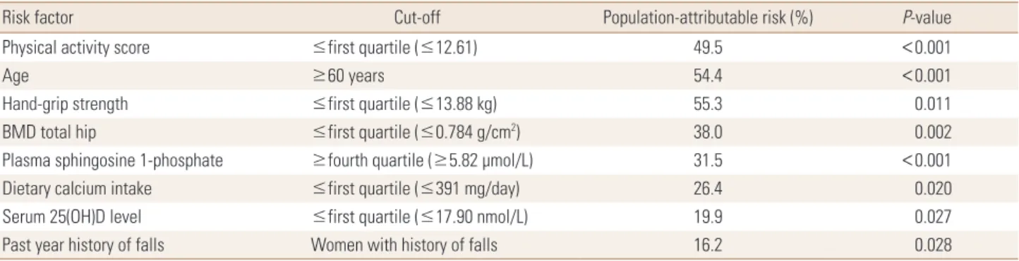
6. PAR
The incident fracture risk attributable to plasma S1P lev- els (in the highest Quartile) was estimated at 31.5%. The PAR for incident fracture independent risk factors among women studied is detailed in Table 6.
DISCUSSION
Our study is novel because it is the first longitudinal co- hort being followed for 5.2±1.3 years with annual sam- pling and measurement of S1P as well as assessment of in- cident fractures at each visit. Thus, the fracture risk was not based on one measurement but on several determinations of the predictor plasma S1P. This study also includes a large number of patients (n=707), and both vertebral and non- vertebral fractures were assessed. This study is therefore more informative than that reported by Kim et al.[12], which used a cross-sectional sampling approach of 69 women with vertebral fractures only as a case-control study com- pared to 69 postmenopausal women without vertebral fractures and based on only one measurement of S1P.
In this study, we report a significant association between increased plasma S1P and incident fracture risk in a popu- lation of postmenopausal women followed prospectively for 5.2±1.3 years. We demonstrated that S1P levels were significantly higher with increasing number of incident fractures even after adjustment for possible confounders.
A novel aspect of this study is an examination of the rela- tionship between the risk of fracture and plasma S1P levels in postmenopausal women. The association was indepen- dent of the known risk factors for fragility fractures, includ- ing BMD, prior fractures, recent falls, smoking, low PA-score, and nutritional deficiencies, suggesting that plasma S1P Table 6. Factors independently associated with all osteoporosis-related fractures in relation to population-attributable risk among 707 Saudi post- menopausal women
Risk factor Cut-off Population-attributable risk (%) P-value
Physical activity score ≤first quartile (≤12.61) 49.5 <0.001
Age ≥60 years 54.4 <0.001
Hand-grip strength ≤first quartile (≤13.88 kg) 55.3 0.011
BMD total hip ≤first quartile (≤0.784 g/cm2) 38.0 0.002
Plasma sphingosine 1-phosphate ≥fourth quartile (≥5.82 µmol/L) 31.5 <0.001
Dietary calcium intake ≤first quartile (≤391 mg/day) 26.4 0.020
Serum 25(OH)D level ≤first quartile (≤17.90 nmol/L) 19.9 0.027
Past year history of falls Women with history of falls 16.2 0.028
BMD, bone mineral density; 25(OH)D, 25-hydroxy-vitamin D.
Fig. 3. Multivariable-adjusted hazard ratios for the risk of osteoporo- sis-related fractures, according to the quartile of plasma sphingosine 1-phosphate (S1P) concentration. Plasma S1P (µmol/L) quartiles were:
Q1<4.85; Q2=4.86-5.24; Q3=5.25-5.81; and Q4≥5.82. The y axis is on a log scale. The reference group is quartile 1. The I bars denote 95% confidence intervals.
Plasma S1P quartile
0 1 5 10 15 20 25 30 35 40 Hazard ratio
1.58 3.81
9.89
could be a new potential biomarker of fracture risk inde- pendent of BMD in postmenopausal women.
Generally, plasma S1P levels in our cohort were similar to those reported for Korean postmenopausal women us- ing the same assay method.[24] Our study is also consis- tent with the most recent report describing higher plasma S1P levels associated with a greater risk of vertebral frac- tures in Korean postmenopausal women (a case-control study of 69 women with vertebral fractures versus 69 wom- en without fractures).[12] BRMs (p-CTX and u-NTx) were significantly associated with ORFs for the same follow-up period, with much lower HR values than those obtained for plasma S1P. No association was obtained for BFMs and fracture risk. Our observations of BTMs in relation to the prediction of ORFs are consistent with those of our recent study [5] and those of Ivaska and colleagues [25] and oth- ers [4].
In this study, the majority of women that were classified with high plasma S1P (remaining stable over the follow-up period) during one time interval were then classified with high plasma S1P 5.2±1.3 years later. This suggests that a single measurement of plasma S1P could be sufficient to identify women at high risk for ORFs. Thus, plasma S1P me- tabolism, clearance, and/or production were stable among the women studied. However, we did observe minor chang- es in the longitudinal plasma S1P levels. Such changes may be related to aging and not analytical variability because plasma S1P measurements were performed under stan- dardized conditions using a validated assay. Plasma levels in women are influenced by age. We observed a positive association between S1P levels and age, confirming previ- ous studies in Korean women.[24] The S1P production in- creases as women age, but decreased S1P production, clear- ance, and/or hormonal changes may also contribute to in- creases in S1P levels. Further work is needed in this regard.
Other biological variability, such as diet, PA, and/or other lifestyle factors, cannot be excluded.
The estimated PAR of increased S1P levels was significant, and S1P levels in the highest quartile produced 31.5% of incident fractures. The PARs were lower than those for S1P compared with the well-known fracture risk factors, includ- ing low PA-score, low hand-grip strength, and low total hip BMD, among others [5] (Table 6). High S1P levels had an effect that was equal to or greater in magnitude to estab- lished ORF risk factors for this cohort.[5,26]
The mechanisms connecting S1P levels and incident fracture risk remain unclear. S1P mediates bone destruc- tion by inducing the formation of osteoclasts.[9,27] A gra- dient exists for S1P in which it is more abundant in circula- tion than in the bone marrow.[28,29] When the difference in S1P levels between these two compartments increases, osteoclast precursors are present in a limited amount in the bone marrow cavity, thus promoting bone resorption.
[8,10,11] Accordingly, bone resorption is promoted, and BMD decreases as the plasma levels of S1P become higher in the circulation,[24] resulting in higher fracture risk.[12]
Additionally, increased plasma S1P levels may lead to en- hanced osteoblast-dependent osteoclastogenesis, which is usually associated with bone loss.[9] Such mechanisms are consistent with our present observations whereby high- er plasma S1P levels were only associated with BRMs (Ta- ble 4), and plasma S1P levels were negatively associated with BMD at various sites studied. It is possible that one and/or both mechanisms contribute to the effects of S1P on bone. However, because S1P levels within bone mar- row plasma are lower than those in circulation, the S1P systemic mechanism is suggested to be more clinically im- portant than the autocrine mechanism. It is interesting that in the present study plasma S1P levels were associat- ed with the risk and number of ORFs, even after additional adjustment for BMD. Such observations could be inter- preted by the potential effect of S1P on bone resorption.
Thus, elevated osteoclastic activity among postmenopaus- al women with relatively higher bone resorption results in the deterioration of bone quality and microarchitecture that will contribute to higher risk for fracture out of pro- portion to the decreased bone mass.[30] It is possible that compromised bone strength by higher levels of S1P may have been mainly due to deteriorated bone quality rather than low bone mass and might contribute to an increased risk of incident fractures. Taken together, these observa- tions provide evidence that S1P controls migratory traffick- ing of osteoclasts precursors, dynamically regulating bone mineral homeostasis, and possibly identifying a critical control point in osteoclastogenesis.[10]
The present study has several strengths. This is the first report demonstrating that high plasma S1P levels predict incident fractures in a large prospective population-based study. The study had a large sample size for which women were randomly selected from the local population using
strict inclusion criteria. We obtained detailed lifestyle char- acteristics and BTM and BMD measurements along with a large number of fracture outcomes over a follow-up period of 5.2±1.3 years, plus multiple measurements of plasma S1P in relation to BTMs during annual visits. We standard- ized the sampling time after all of the participants fasted overnight in order to minimize pre-analytic variations due to circadian rhythm and/or food intake. Moreover, both platelets and mast cells can secrete S1P when activated by thrombin or IgE-bound antigen, respectively.[6] Such pos- sible effects were minimized by our inclusion criteria that excluded women who may have had infectious or immune disorders during recruitment. However, neither platelets nor mast cells were shown to play a role in controlling S1P homeostasis in the blood.[31] Plasma S1P levels were mea- sured using a commercially well-validated assay method and no significant differences were evident between basal mean plasma S1P levels and S1P levels in those who died during the follow-up period (data not shown).
The principal limitation of this study relates to the accu- racy of self-reported data collection in relation to lifestyle information that may have been subject to report bias. Dur- ing recruitment, women with osteoporosis were excluded from the study because we wanted to examine and treat fractures and bone loss in a cohort of healthy postmeno- pausal women. The effect that this exclusion may have had on the study results is unknown. Finally, the number of in- cident fractures is relatively low for calculating the HRs in relation to a given type of fracture.
The association between higher plasma S1P levels and ORF risk should be confirmed in other larger population studies. Proof of a causal relationship between elevated S1P levels and bone disease could be established by inter- vention studies that aim to lower plasma S1P levels. Indeed, it was previously demonstrated that treatment with FTY720 relieved ovariectomy-induced osteoporosis in mice by de- creasing the number of mature osteoclasts that were at- tached to bone surfaces.[11] The mechanism of action of S1P is completely different from that of conventional ther- apies. Controlling the recruitment and migration of osteo- clast precursors was suggested to represent a promising new therapeutic target in the treatment of bone disease,[8]
including osteoporosis.
Finally, the current study could confirm the results of the previously published works of Kim et al.[32] and Bae et
al.[33]. Our study could conclude that high S1P level is sig- nificantly associated with increased risk of incident fracture in postmenopausal women. Moreover, S1P in combination with BMD and biochemical markers of bone turnover could be used as a reliable predictors of the risk of incident frac- ture over a period extending up to 5.0 years duration.
ACKNOWLEDGEMENT
We are grateful to the Ministry of Higher Education for financial support to the Center of Excellence for Osteopo- rosis Research at King Abdulaziz University, Jeddah, Saudi Arabia. We thank the subjects who participated in this study and staff and colleagues at CEOR, King Abdulaziz Universi- ty Hospital, and the Primary Care Health Centers, for their invaluable assistance during the execution of this study.
We thank our colleagues at the Departments of Diagnostic Radiology and Orthopedics at New Jeddah Clinic Hospital and Al-Khandara Clinic Hospital for their unlimited support throughout this study. Special thanks are due to Ms. Veron- ica Orbacedo for her excellent secretarial help.
Preliminary data of the present study were presented as oral presentations at the World Congress on Osteoporosis, Osteoarthritis and Musculoskeletal Diseases, 2-5 April, 2014, Seville, Spain. Published in Osteoporos Int 25:S233; and at the American Society for Bone and Mineral Research (AS- BMR) 2014–36th Annual Meeting of the ASBMR, 12-15 Sep- tember 2014, Texas, USA. Published in J Bone Miner Res 29.
REFERENCES
1. Garnero P, Delmas PD. Contribution of bone mineral den- sity and bone turnover markers to the estimation of risk of osteoporotic fracture in postmenopausal women. J Musculoskelet Neuronal Interact 2004;4:50-63.
2. Sattui SE, Saag KG. Fracture mortality: associations with epidemiology and osteoporosis treatment. Nat Rev Endo- crinol 2014;10:592-602.
3. Chopin F, Biver E, Funck-Brentano T, et al. Prognostic inter- est of bone turnover markers in the management of post- menopausal osteoporosis. Joint Bone Spine 2012;79:26- 31.
4. Garnero P, Cloos P, Sornay-Rendu E, et al. Type I collagen racemization and isomerization and the risk of fracture in postmenopausal women: the OFELY prospective study. J
Bone Miner Res 2002;17:826-33.
5. Ardawi MS, Rouzi AA, Al-Sibiani SA, et al. High serum scle- rostin predicts the occurrence of osteoporotic fractures in postmenopausal women: the Center of Excellence for Os- teoporosis Research Study. J Bone Miner Res 2012;27:2592- 602.
6. Proia RL, Hla T. Emerging biology of sphingosine-1-phos- phate: its role in pathogenesis and therapy. J Clin Invest 2015;125:1379-87.
7. Rosen H, Stevens RC, Hanson M, et al. Sphingosine-1-phos- phate and its receptors: structure, signaling, and influence.
Annu Rev Biochem 2013;82:637-62.
8. Ishii M, Kikuta J. Sphingosine-1-phosphate signaling con- trolling osteoclasts and bone homeostasis. Biochim Bio- phys Acta 2013;1831:223-7.
9. Ryu J, Kim HJ, Chang EJ, et al. Sphingosine 1-phosphate as a regulator of osteoclast differentiation and osteoclast- osteoblast coupling. EMBO J 2006;25:5840-51.
10. Ishii M, Kikuta J, Shimazu Y, et al. Chemorepulsion by blood S1P regulates osteoclast precursor mobilization and bone remodeling in vivo. J Exp Med 2010;207:2793-8.
11. Ishii M, Egen JG, Klauschen F, et al. Sphingosine-1-phos- phate mobilizes osteoclast precursors and regulates bone homeostasis. Nature 2009;458:524-8.
12. Kim BJ, Koh JM, Lee SY, et al. Plasma sphingosine 1-phos- phate levels and the risk of vertebral fracture in postmeno- pausal women. J Clin Endocrinol Metab 2012;97:3807-14.
13. Heilmann A, Schinke T, Bindl R, et al. Systemic treatment with the sphingosine-1-phosphate analog FTY720 does not improve fracture healing in mice. J Orthop Res 2013;
31:1845-50.
14. Grey A, Chen Q, Callon K, et al. The phospholipids sphin- gosine-1-phosphate and lysophosphatidic acid prevent apoptosis in osteoblastic cells via a signaling pathway in- volving G(i) proteins and phosphatidylinositol-3 kinase.
Endocrinology 2002;143:4755-63.
15. Lotinun S, Kiviranta R, Matsubara T, et al. Osteoclast-spe- cific cathepsin K deletion stimulates S1P-dependent bone formation. J Clin Invest 2013;123:666-81.
16. Sefcik LS, Aronin CE, Awojoodu AO, et al. Selective activa- tion of sphingosine 1-phosphate receptors 1 and 3 pro- motes local microvascular network growth. Tissue Eng Part A 2011;17:617-29.
17. Rouzi AA, Al-Sibiani SA, Al-Senani NS, et al. Independent predictors of all osteoporosis-related fractures among
healthy Saudi postmenopausal women: the CEOR Study.
Bone 2012;50:713-22.
18. World Health Organization. International statistical classi- fication of diseases and related health problems 10th revi- sion (ICD-10). Geneva, CH: World Health Organization; 1992.
19. Genant HK, Wu CY, van Kuijk C, et al. Vertebral fracture as- sessment using a semiquantitative technique. J Bone Min- er Res 1993;8:1137-48.
20. Lai WQ, Irwan AW, Goh HH, et al. Anti-inflammatory ef- fects of sphingosine kinase modulation in inflammatory arthritis. J Immunol 2008;181:8010-7.
21. Ardawi MS, Maimani AA, Bahksh TA, et al. Reference inter- vals of biochemical bone turnover markers for Saudi Ara- bian women: a cross-sectional study. Bone 2010;47:804- 14.
22. Ardawi MS, Al-Kadi HA, Rouzi AA, et al. Determinants of serum sclerostin in healthy pre- and postmenopausal wom- en. J Bone Miner Res 2011;26:2812-22.
23. Hennekens CH, Buring JE. Measure of disease frequency and association. In: Mayrent SL, editor. Epidemiology in medicine. Boston, MA: Little, Brown and Company; 1997.
p.87-95.
24. Lee SH, Lee SY, Lee YS, et al. Higher circulating sphingo- sine 1-phosphate levels are associated with lower bone mineral density and higher bone resorption marker in hu- mans. J Clin Endocrinol Metab 2012;97:E1421-8.
25. Ivaska KK, Gerdhem P, Väänänen HK, et al. Bone turnover markers and prediction of fracture: a prospective follow- up study of 1040 elderly women for a mean of 9 years. J Bone Miner Res 2010;25:393-403.
26. Grey A, Xu X, Hill B, et al. Osteoblastic cells express phos- pholipid receptors and phosphatases and proliferate in response to sphingosine-1-phosphate. Calcif Tissue Int 2004;74:542-50.
27. Roelofsen T, Akkers R, Beumer W, et al. Sphingosine-1-phos- phate acts as a developmental stage specific inhibitor of platelet-derived growth factor-induced chemotaxis of os- teoblasts. J Cell Biochem 2008;105:1128-38.
28. Peest U, Sensken SC, Andréani P, et al. S1P-lyase indepen- dent clearance of extracellular sphingosine 1-phosphate after dephosphorylation and cellular uptake. J Cell Bio- chem 2008;104:756-72.
29. Maeda Y, Seki N, Sato N, et al. Sphingosine 1-phosphate receptor type 1 regulates egress of mature T cells from mouse bone marrow. Int Immunol 2010;22:515-25.
30. Armas LA, Recker RR. Pathophysiology of osteoporosis:
new mechanistic insights. Endocrinol Metab Clin North Am 2012;41:475-86.
31. Rivera J, Proia RL, Olivera A. The alliance of sphingosine- 1-phosphate and its receptors in immunity. Nat Rev Im- munol 2008;8:753-63.
32. Kim BJ, Shin KO, Kim H, et al. The effect of sphingosine-
1-phosphate on bone metabolism in humans depends on its plasma/bone marrow gradient. J Endocrinol Invest 2016;
39:297-303.
33. Bae SJ, Lee SH, Ahn SH, et al. The circulating sphingosine- 1-phosphate level predicts incident fracture in postmeno- pausal women: a 3.5-year follow-up observation study.
Osteoporos Int 2016;27:2533-41.