Ⅰ. INTRODUCTION
The objective of root canal preparation is to clean and shape the root canal system, while maintaining the original configuration. A continu-
ously tapering, conical, funnel-shaped canal with the smallest diameter at the end-point and the largest at the orifice is perceived to be the most appropriate for filling with gutta-percha1).
Overpreparation of the coronal third of canal is one of the aberrations that may occur during root canal preparation and it may weaken the tooth2) and root perforation is a possible consequence of canal preparation that may result in treatment failure3). When using stainless steel hand files, Hedstrom files have been recommended to use in a up-and-down motion with the file rasping the
Shaping Ability of Four Rotary Nickel-Titanium Instruments to Prepare Root Canal at Danger Zone
Seok-Dong Choi, Myoung-Uk Jin, Ki-Ok Kim1, Sung-Kyo Kim*
Department of Conservative Dentistry, School of Dentistry, Kyungpook National University, Department of Dentistry, College of Medicine, Konkuk University1
The aim of this study was to evaluate the shaping abilities of four different rotary nickel-titanium instru- ments with anticurvature motion to prepare root canal at danger zone by measuring the change of dentin thickness in order to have techniques of safe preparation of canals with nickel-titanium files.
Mesiobuccal and mesiolingual canals of forty mesial roots of extracted human lower molars were instru- mented using the crown-down technique with ProFile, GTTM Rotary file, Quantec file and ProTaperTM. In each root, one canal was prepared with a straight up-and-down motion and the other canal was with an anticurvature motion. Canals were instrumented until apical foramens were up to size of 30 by one opera- tor. The muffle system was used to evaluate the root canal preparation. After superimposing the pre- and post-instrumentation canal, change in root dentin thickness was measured at the inner and outer sides of the canal at 1, 3, and 5 ㎜ levels from the furcation. Data were analyzed using two-way ANOVA.
Root dentin thickness at danger zone was significantly thinner than that at safe zone at all levels (p < 0.05).
There was no significant difference in the change of root dentin thickness between the straight up-and- down and the anticurvature motions at both danger and safe zones in all groups (p > 0.05).
ProTaper removed significantly more dentin than other files especially at furcal 3 ㎜ level of danger and safe zones (p < 0.05)
Therefore, it was concluded that anticurvature motion with nickel-titanium rotary instruments does not seem to be effective in danger zone of lower molars. [J Kor Acad Cons Dent 29(5):446-453, 2004]
Key words : Shaping ability, Ni-Ti rotary instrument, Root canal preparation, Danger zone, Anticurvature filing, ProTaper
ABSTRACT
* Corresponding author: Sung-Kyo Kim Department of Conservative Dentistry,
School of Dentistry, Kyungpook National University 188-1, 2-Ga, Samdeok-Dong, Jung-Gu, Daegu, Korea, 700-412 Tel : 82-53-420-5935 Fax : 82-53-426-8958 Email: [email protected]
canal wall during its outward movement. The fil- ing was to direct against the canal wall farthest from the furcation to avoid the weakening of the canal wall4). When using stainless steel rotary instruments, Gates-Glidden burs have been rec- ommended to direct apically and laterally away from the furcation too4). This use of Hedstrom files and the Gates-Glidden burs creates a flared preparation in the coronal half to two-thirds of the canal, resulting in a straighter access to the apical portion of the canal. However, overflare weakens the tooth and may cause a perforation.
Stripping perforation on the furcal side of the root canal can be prevented by limiting the circumfer- ential filing to the areas of greatest bulk4).
During the last decade, various kinds of root canal instruments made of nickel-titanium were developed. These recently developed nickel-titani- um files of increased taper and various designs make the crown-down preparation easy, which allows easier access to the apical canal and better distribution of irrigant with less apical extrusion of canal debris. To protect danger zone of lower molars, some precautions or special technique may be needed when root canal is enlarged with rotary nickel-titanium instruments. For this rea- son, canal preparation with anticurvature motion is worth to be investigated in order to elucidate its effect on protecting the danger zone when using different taper and designs of nickel-titani- um rotary instruments.
Therefore, the aim of this study was to evaluate the shaping abilities of four different rotary nick- el-titanium instruments with anticurvature motion to prepare root canal at danger zone in order to have techniques of safe preparation of canals with nickel-titanium files.
Ⅱ. MATERIALS AND METHODS
Forty mesial roots of extracted human lower first and second molars were used in this study.
Calculus and soft tissue debris were removed with scalers after storing the teeth in 5.25% sodium
tion, size 10 K-files were introduced to determine the patency of canals and working lengths were established 1 mm from the apical foramen.
Fabrication of a muffle system
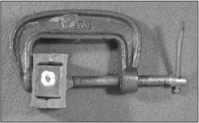
The muffle system introduced by Bramante5)and modified by McCann et al.6) was used to evaluate the root canal preparation. Teeth were mounted in the muffle with roots embedded in the acrylic resin. During root canal shaping procedure, two muffle halves were held together with a common C clamp (Figure 1).
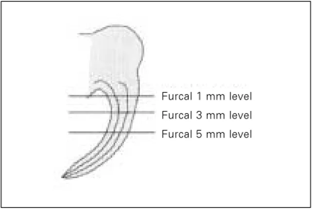
After sectioning the embedded roots horizontally at 1, 3, and 5 ㎜ levels from the furcation with a microtome (IsometTM, Buehler Co., Lake Bluff, IL, U.S.A., Figure 2), teeth were remounted in the muffle system.
Root canal preparation
Forty roots were divided into four groups according to the rotary nickel-titanium instru- ments used: ProFile�, GTTM Rotary (Dentsply- Maillefer, Ballaigues, Switzerland), Quantec (Analytic Endodontics, Glendora, CA, U.S.A.), and ProTaperTM (Dentsply-Maillefer). Both the mesiobuccal and mesiolingual canals were instru-
Figure 1. Both halves of the muffle with C clamp to maintain muffle-to-muffle and muffle-to-resin relationships. A tooth is embedded in acrylic resin in the muffle. C clamp also serves as a convenient handle during experimental procedure.
mented using the crown-down technique with one of the above rotary nickel-titanium instruments.
In each root, one canal was prepared with a straight up-and-down motion and the other canal was with anticurvature motion. Canals were instrumented until apical diameter had attained a size of 30. Each file was examined before and after use for any defects, and was wiped regularly to remove debris. Each instrument was used in only one canal before being replaced. Root canals were irrigated after each instrument use with 5 mL of water using a 27-gauge needle (Endo-Tips, Ultradent Products Inc., Utah, U.S.A.). RC-PrepTM
(Stone Pharmaceuticals, Philadelphia, U.S.A.) was used as a lubricant. Canals were prepared with a crown-down technique according to the recommended sequences of the manufacturers by one operator who was a postgraduate student of Endodontic Program with one year of clinical experience of Ni-Ti rotary files. The operator practiced each instrumentation technique twenty times before experiment.
In the ProFile group, Orifice Shaper #3 and #2 were used until resistance was encountered (12 to 15 ㎜) for coronal shaping followed by 0.06/#25 and .06/#20 files to resistance (within 1 to 2 ㎜ of the working length). For apical shaping, 0.04/#25 and 0.04/#30 files were used to the working length. In the GT Rotary group, 0.12/#20, 0.10/#20, 0.08/#20 and 0.06/#20 files were used sequentially until progression became difficult for coronal flaring. A 0.12/#20 file was used to a depth of 12 ㎜. 0.10/#20 to a depth of 14 ㎜, a 0.08/#20 to 16 ㎜ and a 0.06/#20 to the working length. 0.04/#25 and 0.04/#30 files were used for apical shaping. In the Quantec group, a Quantec LX #1 (0.06/#25, 17 ㎜) was used to a depth of 11 to 12 ㎜ for coronal flaring. For the deep canal shaping, 0.12/#25, 0.10/#25, 0.08/#25, and 0.06/#25 files were inserted in the canal as deep as possible. A 0.12/#25 file was used to 12 ㎜, a Figure 2. Levels of cross-section. Roots were sectioned
horizontally 1, 3, and 5 ㎜levels from the furcation.
A B
Figure 3. Computer captured images. A, before root canal instrumentation; B, after canal instrumentation.
Furcal 1 mm level Furcal 3 mm level Furcal 5 mm level
0.10/#25 to 14 ㎜, a 0.08/#25 to 16 ㎜ and final- ly 0.06/#25 and 0.05/#25 files were used to the working length. Apical shaping was done by an Accessory Quantec LX 0.02/#30 file. In the ProTaper group, a Shaping file No.1 (S1) was used first in the canal and moved apically to just short of the working length (16 ㎜). SX files were then used sequentially to resistance (13 to 14 ㎜) followed by S1 and S2 to working length for the shaping of the coronal two-thirds of the canal. Apical one-third was finished using F1, F2 and F3 sequentially to the working length with only one pecking motion for each instru- ment.
Assessment of shaping ability
Shaping ability of each instrument was assessed by the evaluation of the root dentin thickness change. Pre- and post-instrumentation canal images were observed under a stereomicroscope (SZ40, Olympus Optical Co., Ltd., Tokyo, Japan), and stored in a computer using a CCD camera (GP-KR222, Panasonic, Osaka, Japan) and a commercial digitizing image program (micro VIDEO Studio 200 program, Pinnacle system, Brauschweig, Germany, Figure 3). The pre- and post-instrumentation canal images were superim- posed and changes in root thickness were mea- sured at distal side (danger zone) and mesial side (safe zone) of the canal using a computer program (Auto�CAD 2000, Autodesk Corp., San Rafael.
CA, U.S.A.).
Statistical analysis
All numerical data in the text and tables are expressed as mean ± S. D. The data of the differ- ence in root dentin thickness were analyzed sta- tistically using two-way ANOVA.
Ⅲ. RESULTS
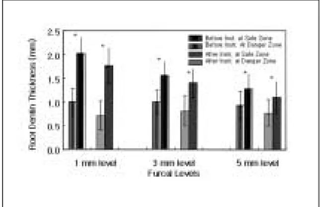
Root dentin thicknesses and its changes, in gen- eral, are shown in Figure 4. Before instrumenta- tion, root dentin thickness at danger zone was significantly thinner than that at safe zone at each furcal level of 1, 3, and 5 ㎜ (n = 80, p <
0.05). Regardless of the instruments and instru- mentation techniques, root dentin thickness at danger zone remained significantly thinner than that at safe zone after instrumentation at all fur- cal levels (n = 80, p < 0.05). However, there was a tendency to remove more dentin at safe zone than at danger zone at 1 ㎜ level compared to 3 and 5 ㎜ levels without statistical significance.
In all instrument groups, there was no signifi- cant difference in the change of root dentin thick- ness between the straight up-and-down and the anticurvature motions at both danger and safe zones (p > 0.05, Tables 1 and 2).
Among instrumentation groups, ProTaper group removed significantly more dentin than in another groups at the furcal 3 ㎜ level of the danger zone and at 1 and 3 ㎜ levels of safe zone (p < 0.05, Table 1).
No perforations were noted in the study.
Figure 4. Thickness of root dentin (㎜) at danger and safe zones before and after root canal instrumentation (mean ± S.D., n = 80 each).
*Root dentin thickness at danger zone was significantly thinner than that at safe zone (p
< 0.05).
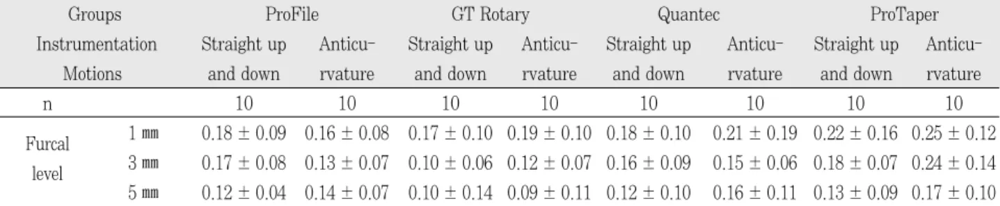
Table 1.Change of root dentin thickness (mm) by canal preparation at danger zone (mean ± S.D.)
Groups ProFile GT Rotary Quantec ProTaper
Instrumentation Straight up Anticu- Straight up Anticu- Straight up Anticu- Straight up Anticu- Motions and down rvature and down rvature and down rvature and down rvature
n 10 10 10 10 10 10 10 10
Furcal 1 ㎜ 0.18 ± 0.09 0.16 ± 0.08 0.17 ± 0.10 0.19 ± 0.10 0.18 ± 0.10 0.21 ± 0.19 0.22 ± 0.16 0.25 ± 0.12 level 3 ㎜ 0.17 ± 0.08 0.13 ± 0.07 0.10 ± 0.06 0.12 ± 0.07 0.16 ± 0.09 0.15 ± 0.06 0.18 ± 0.07 0.24 ± 0.14 5 ㎜ 0.12 ± 0.04 0.14 ± 0.07 0.10 ± 0.14 0.09 ± 0.11 0.12 ± 0.10 0.16 ± 0.11 0.13 ± 0.09 0.17 ± 0.10
No statistical significance was shown between straight up-and-down motion and anticurvature motion.
Table 2.Change of root dentin thickness (mm) by canal preparation at safe zone (mean ± S.D.)
Groups ProFile GT Rotary Quantec ProTaper
Instrumentation Straight up Anticu- Straight up Anticu- Straight up Anticu- Straight up Anticu- Motions and down rvature and down rvature and down rvature and down rvature
n 10 10 10 10 10 10 10 10
Furcal 1 ㎜ 0.14 ± 0.07 0.12 ± 0.06 0.22 ± 0.10 0.23 ± 0.18 0.23 ± 0.13 0.27 ± 0.13 0.30 ± 0.17 0.29 ± 0.17 level 3 ㎜ 0.10 ± 0.09 0.08 ± 0.06 0.07 ± 0.05 0.12 ± 0.08 0.15 ± 0.11 0.20 ± 0.11 0.18 ± 0.14 0.17 ± 0.13 5 ㎜ 0.13 ± 0.09 0.06 ± 0.07 0.11 ± 0.11 0.10 ± 0.07 0.12 ± 0.16 0.19 ± 0.12 0.13 ± 0.09 0.06 ± 0.07
No statistical significance was shown between straight up-and-down motion and anticurvature motion.
Ⅳ. DISCUSSION
Cross sections through the coronal third of fur- cated roots reveal that canals are not typically centered anatomically within their roots. Instead, they are often skewed toward the furcal-side con- cavities7). The mesial root of mandibular first molars was reported to have a distal surface con- cavity with a root thickness in this area of about 0.7 ㎜8), and is subject to perforation. In the pre- sent study, cross-section of the mesial roots of mandibular molars showed that root dentin thick- ness at distal side (danger zone) was significantly thinner than that at mesial side (safe zone) at all levels from the furcation too. This finding was in agreement with the previous reports7,8). Because root curvature is toward the distal side in gener- al, this distal side is especially subject to strip perforation.
ing to the areas of greatest bulk to prevent strip- ping perforation on the furcal side of the root canal4). Coronal flaring can reduce these undesir- able aberrations, and it has also been recom- mended that they should be used like files, with an anticurvature motion toward the safe zone of the tooth9).
The nickel-titanium files were found to have two to three times more elastic flexibility of the stain- less steel files in bending and torsion, as well as superior resistance to fracture10). Because of its high flexibility, the load on the cutting blade is greatly reduced in curved canals, which reduces stress on the instrument and the possibility of fracture. Several studies have reported that canals prepared with rotary nickel-titanium files of high taper were excellently tapered and that the use of these files reduced the incidence of canal aberration11-16)and have a decreased tenden- cy for canal transportation and therefore remain
better centered17).
It was suggested that the coronal two-thirds of the canal can easily be moved and relocated away from furcal danger and toward the greatest bulk of dentin when Gates Glidden burs are used in a step-back technique18). Gates Glidden burs were used to cut and remove dentin on just one or two of the outer walls of the canal and away from fur- cal danger.
Isom et al.18) compared root thickness in the mesial canals of lower molars before and after flaring with Gates Glidden burs by using a muffle system. Gates Glidden burs were used with either a straight up-and-down motion or with an antic- urvature motion. At a level 2 ㎜ apical to the fur- cation, the anticurvature method removed more dentin than the straight up and down18). However, there was no significant difference in the change of root dentin thickness between the straight up- and-down and the anticurvature motions in the present study. The reason of the difference of the result between the stainless steel instrument and nickel-titanium ones may be the difference of their flexibility. As nickel-titanium files induce less transportation of the canal, it may have less effectiveness in anticurvature filing. One of the rotary nickel-titanium instruments, LightSpeed caused significantly less transportation in 60 mesial canals in mandibular molars than did stainless steel or nickel-titanium manual files19). Therefore, the anticurvature motion of nickel- titanium rotary instruments seems to be less effective to cut the dentin of safe zone than stain- less steel ones do.
Shape of the prepared root canal may be influ- enced by the design and taper of instruments. In the present study, ProTaper group removed sig- nificantly more dentin than other groups, espe- cially at furcal 3 ㎜ level at danger zone. The taper of the ProTaper files is bigger than the oth- er files at the same level of the root canal, which may result in greatest reduction in the thickness of root canal dentin. Yun and Kim20)studied shap- ing abilities of four nickel-titanium rotary instru-
that ProTaper cut more dentin than any other instruments tested.
Muffle system was used in this study. The mod- el system allowed direct comparison of the four instrumentation techniques at three canal levels.
Although the technique described by Bramante et al.5) is an excellent method for the comparison of original and shaped canals, some problems21)were encountered. First, during sectioning, 0.4 mm over of root material was lost. The additional loss of root material was caused by the lateral move- ment of band saw. Secondly, not all sections were at right angles to the canal. In curved canals the loss of root material and some oblique-sectioned surfaces acted as ledges that hindered the pas- sage of the file through the canal to the working length. In severely curved canals, this even pre- vented further instrumentation, irrespective of the technique used. Further investigations should try to minimize the width of dentine lost during root sectioning. This is best achieved by reducing the lateral movements of the band saw or by using a different sectioning technique.
Therefore, it was concluded that there was no difference between up-and-down motion and anti- curvature one in removing root dentin with nick- el-titanium rotary instruments in the condition of the present study, which indicates that anticur- vature motion with nickel-titanium rotary instru- ments may not as effective as that with stainless steel ones. ProTaper removed more root dentin than GT Rotary, Quantec, and ProFile especially at furcal 3 ㎜ level.
Further research is needed to evaluate influence of working length, apical diameter and canal cur- vature on the shaping ability of rotary nickel-tita- nium instruments to prepare root canal at danger zone.
REFERENCES
1. Schilder H. Cleaning and shaping the root canal. Dent Clin Nor Am 18:269-296, 1974.
2. Buchanan LS. Paradigm shifts in cleaning and shap- ing. J Calf Dent Assoc 19:23-26, 28-33, 1991.
9:439, 1983.
4. Goerig AC, Michelich RJ, Schultz H. Instrumentation of root canals in molar using step-down technique. J Endod 8:550-554, 1982.
5. Bramante CM, Berber A, Borges RP. A methodology for evaluation of root canal instrumentation. J Endod 13:243-245, 1987.
6. McCann JT, Keller DL, LaBounty GL. A modification of the muffle model system to study root canal mor- phology. J Endod 16:114-116. 1990.
7. Ruddle C. Cleaning and shaping the root canal system.
p231-291, in Cohen S, Burns R. Pathways of the pulp.
8th ed, Mosby, St Louis, 2002.
8. Bower RC. Furcation morphology relative to periodon- tal treatment. J Periodontol 50:23-27, 1979.
9. Abou-Rass M, Frank AL, Glick DH. The anticurvature filing method to prepare the curved canal. J Am Dent Assoc 101:792-794, 1982.
10. Walia H, Brantley WA, Gerstein H. An initial investi- gation of the bending and torsional properties of Nitinol root canal files. J Endod 14:346-351, 1988.
11. Kavanagh D, Lumley PJ. An in vitro evaluation of canal preparation using ProFile .04 and .06 taper instruments.Endod Dent Traumatol 14:16-20, 1998.
12. Bryant ST, Dummer PM, Pitoni C, Bourba M, Moghal S. Shaping ability of .04 and .06 taper ProFile rotary nickel-titanium instruments in simulated root canals.
Int Endod J 32:155-164, 1999.
13. Park HS, Lee MK, Kim JJ, Lee JY. A study on the shape of a canal prepared with ProFiles in curved
canal. J Kor Acad Conserv Dent 24:633-638, 1999.
14. Lee JH, Cho YB. The effect of some canal preparation techniques on the shape of root canals. J Kor Acad Conserv Dent 24:337-345, 1999.
15. Kum KY, Spangberg L, Cha BY, Jung IY, Lee SJ, Lee CY. Shaping ability of three ProFile rotary instrumen- tation techniques in simulated resin root canals. J Endod 26:719-723, 2000.
16. Park H. A comparison of Greater Taper files, ProFiles, and stainless steel files to shape curved root canals.
Oral Surg Oral Med Oral Pathol Dent Radiol Endod 91:715-718, 2001.
17. Short JA, Morgan LA, Baumgartner JC. A comparison of canal centering ability of four instrumentation tech- niques. J Endod 23:503-507, 1997.
18. Isom TL, Marshall JG, Baumgartner JC. Evaluation of root thickness in curved canals after flaring. J Endod 21:368-371, 1995.
19. Grosson CR, Haller RH, Dove SB, Del Rio CE. A com- parison of root canal preparation using NiTi hand, NiTi engine-driven, and K-Flex endodontic instruments. J Endod 21:173-176, 1995.
20. Yun HH, Kim SK. A comparison of the shaping abili- ties of four nickel-titanium rotary instruments in sim- ulated root canals. Oral Surg Oral Med Oral Pathol Dent Radiol Endod 95:228-233, 2003.
21. Portenier FL, Barbakow F. Preparation of the apical part of the root canal by the Lightspeed and step-back techniques. Int Endod J 31:103-111, 1998.
네 가지 전동 Ni-Ti 파일의 danger zone에서의 근관성형력
최석동∙진명욱∙김기옥1∙김성교*
경북대학교 치과대학 치과보존학교실, 건국대학교 의과대학 치과학교실1
하악 대구치 근심치근의 danger zone에서 수종의 전동 니켈-티타늄 파일의 근관성형력을 근관성형 전후 치질 두께의 변화를 측정하여 평가하고자 하였다. 기구에 따라 총 40개의 하악 대구치를 10개씩 Profile�, GT Rotary file, Quantec 및 ProTaper 4개 군으로 나누고 각 치아당 2개의 근관을 straight up-and-down과 anticurvature 군으로 나누어 근단부 근관을 모두 30번 크기로 일정하게 확대하였다. 수정된 Bramante 법을 사용하였으며 술전 및 술후의 근관 상아질 두께를 치수저 하방 1, 3 및 5 ㎜ 지점에서 측정, 이원변량분석법으로 통계분석하였다.
모든 군의 danger zone과 safe zone에서의 straight up-and-down 동작과 anticurvature 동작 사이에는 치근상아 질 두께변화에 현저한 차이를 나타내지 않았다 (p > 0.05).
ProTaper는 danger zone과 safe zone 모두에서 다른 기구에 비해 많은 량의 근관상아질 삭제를 보였으며 특히 분지 부 3 ㎜ 수준에서 현저하였다 (p < 0.05).
국문초록