INTRODUCTION
Among gynecologic cancers, epithelial ovarian cancer (EOC)
is second only to cervical cancer in incidence and number of deaths caused [1]. Patients with ovarian cancer are staged accor
ding to the International Federation of Obstetrics and Gyne
cology (FIGO) system [2], which determines patient ma na ge
ment and provides prognostic information (fiveyear survival rates are 32.5% and 18.6% for stages IIIC and IV, respectively) [3]. Cytoreductive surgery is the treatment of cho ice for patients with ovarian cancer. “Optimal” cytoreductive surgery (residual disease <1 cm) is a very strong predictor of survival [4], and even af
Thoracic metastasis in advanced ovarian cancer:
comparison between computed tomography and video- assisted thoracic surgery
Oleg Mironov1, Evis Sala2, Svetlana Mironov1, Harpreet Pannu1, Dennis S. Chi3, Hedvig Hricak1
1Department of Radiology, Memorial Sloan-Kettering Cancer Center, New York, USA, 2Department of Radiology,
Addenbrooke’s Hospital, University of Cambridge, Cambridge, UK, 3Department of Surgery, Memorial Sloan-Kettering Cancer Center, New York, USA
Received Apr 25, 2011, Revised Jun 3, 2011, Accepted Jun 28, 2011 Correspondence to Hedvig Hricak
Department of Radiology, Memorial Sloan-Kettering Cancer Center, 1275 York Ave., New York, NY 10065, USA. Tel: 1-212-639-7284, Fax: 1-212-794- 4010, E-mail: [email protected]
Copyright © 2011. Asian Society of Gynecologic Oncology, Korean Society of Gynecologic Oncology
Objective: To determine which computed tomography (CT) imaging features predict pleural malignancy in patients with advanced epithelial ovarian carcinoma (EOC) using videoassisted thoracic surgery (VATS), pathology, and cytology findings as the reference standard.
Methods: This retrospective study included 44 patients with International Federation of Obstetrics and Gynecology (FIGO) stage III or IV primary or recurrent EOC who had chest CT ≤30 days before VATS. Two radiologists independently reviewed the CT studies and recorded the presence and size of pleural effusions and of ascites; pleural nodules, thickening, enhancement, subdiaphragmatic tumour deposits and supradiaphragmatic, mediastinal, hilar, and retroperitoneal adenopathy; and peritoneal seeding. VATS, pathology, and cytology findings constituted the reference standard.
Results: In 26/44 (59%) patients, pleural biopsies were malignant. Only the size of leftsided pleural effusion (reader 1: rho=
-0.39, p=0.01; reader 2: rho=-0.37, p=0.01) and presence of ascites (reader 1: rho=-0.33, p=0.03; reader 2: rho=-0.35, p=0.03) were significantly associated with solid pleural metastasis. Pleural fluid cytology was malignant in 26/35 (74%) patients. Only the presence (p=0.03 for both readers) and size (reader 1: rho=0.34, p=0.04; reader 2: rho=0.33, p=0.06) of rightsided pleural effusion were associated with malignant pleural effusion. Interobserver agreement was substantial (kappa=0.78) for effusion size and moderate (kappa=0.46) for presence of solid pleural disease. No other CT features were associated with malignancy at biopsy or cytology.
Conclusion: In patients with advanced EOC, ascites and leftsided pleural effusion size were associated with solid pleural meta
stasis, while the presence and size of rightsided effusion were associated with malignant pleural effusion. No other CT features evaluated were associated with pleural malignancy.
Keywords: Computed tomography, Malignant/diagnosis, Ovarian neoplasms, Pleural effusion, Pleural neoplasms, Thoracic surgery/
videoassisted
ter the threshold for optimal cytoreduction has been reached, it is important to remove as much of the residual tumor as possible [5].
Circulation of peritoneal fluid throughout the abdomen and pelvis commonly results in diaphragmatic tumor implants, and in turn, peritonealpleural communication through the diaphragm may allow transdiaphragmatic spread of tumor into the tho
rax [6]. Malignant thoracic involvement indicates FIGO stage IV disease, for which treatment options include neoadjuvant chemotherapy and, in patients who are candidates for optimal abdominal cytoreduction, thoracic debulking [7,8]. Neoadju
vant chemotherapy followed by interval debulking surgery is a suitable alternative [9,10] that is supported by the outcome
of a recent multicentre randomised controlled trial [11].
Several studies have demonstrated that the thorax frequently harbors undiagnosed pleural disease at the time of the initial diagnosis, and that this is likely to affect survival even in cases of optimal debulking [1214]. In addition, a recent study found that moderatetolarge pleural effusion on preoperative com
puted tomography (CT) was associated with a decrease in overall survival in patients with stage III or IV EOC after control
ling for age, preoperative CA125, surgical stage, ascites, and cytoreductive status [15]. Therefore, accurate identification of the presence and extent of thoracic disease, including both solid metastasis and malignant pleural effusion, is important for determining prognosis and selecting appropriate treat
Fig. 1. Postmenopausal female with stage IV high-grade papillary serous carcinoma. (A) Contrast-enhanced computed tomography (CT) scan demonstrates bilateral pleural effusions (white arrows), larger on the right side, at the time of initial diagnosis. Right-sided pleural tap revealed positive cytology. Subsequently, bilateral video-assisted thoracic surgery was performed and revealed the presence of microscopic pleural metastasis bilaterally. Pleural effusions were treated with talc pleurodesis. (B) Post-treatment follow-up CT images demonstrate residual loculated small bilateral effusions (black arrow) and talc-related hyperdensity within the right pleural space (white arrow). (C) Contrast- enhanced CT scan 4 months after initial diagnosis and neoadjuvant chemotherapy showing residual disease in the abdomen and pelvis (white arrow) that was subsequently treated with optimal debulking. (D) Contrast-enhanced CT scan 6 months after optimal debulking showing gross pleural metastases (white arrows).
ment in patients with advanced EOC [15] (Figs. 1 and 2).
Pleural effusions in ovarian cancer have a higher likelihood of malignancy when they are moderate to large in size and as
sociated with enlarged superior diaphragmatic lymph nodes [6]. However, studies on CT indicators of malignant pleural effusion have used cytology rather than surgical findings as the reference standard [6,16,17]. The accuracy of pleural fluid cytologic examination ranges between 40% and 87% [18
20]. Videoassisted thoracic surgery (VATS) has been shown to enable accurate pathological diagnosis and intrathoracic resection of pleural metastasis in patients with ovarian cancer [21]. In a small retrospective study from our institution, 4/10 patients (40%) with negative cytological findings had macro
scopic pleural lesions on VATS [13], and in another such study, preoperative CT identified solid pleural disease in only one third of patients (2/6) who had macroscopic disease on VATS [14]. Therefore, the aims of our study were to compare chest CT to VATS, pathology, and cytology findings and determine possible CT imaging features that may be predictive of pleural malignancy in patients with advanced EOC.
MATERIALS AND METHODS 1. Patients
Our retrospective, crosssectional imaging study was com
pliant with Health Insurance Portability and Accountability Act. The institutional review board approved the study and is
sued a waiver of informed consent. Patients were selected by
means of a computerized review of institutional gynecologic surgery and radiology databases. Consecutive patients with primary or recurrent FIGO stage III and IV ovarian cancer who underwent CT of the chest up to 30 days before VATS from January 1, 1997 through December 31, 2009 were included in the study. If a patient had more than one CT study within the 30 days before VATS, the most recent one was used. A total of 44 patients (3 with FIGO stage III and 41 with FIGO stage IV) were included in the analysis, of whom 41 had primary and 3 had recurrent disease. Indications for VATS were malignant pleural effusion on cytology (12/44), suspected malignant nodules or lymph nodes on CT (16/44) or moderate to large (defined as occupying 1/3 or more of the lung field on chest Xray) and/or recurrent pleural effusions (16/44).
2. CT examinations
Most of the CT examinations (32/44) were performed with intravenous contrast medium, and the slice thickness ranged from 5 to 8 mm. For CT examinations performed outside our institution, the type of CT scanner and the amount of con
trast material used were unknown. At our institution, CT was performed with various scanners (GE Medical Systems, Mil
waukee, WI, USA). Our standard CT protocols were tailored to the individual scanners. Before October 1, 2000, studies were obtained with a conventional nonhelical scanner; afterwards they were obtained with a helical scanner with one to 64 de
tector rows.
For all CT examinations performed at our institution, a dy
namic power injection of 150 mL of nonionic intravenous Fig. 2. Photographs obtained during video-assisted thoracic surgery show (A) white tumor plaques (white arrow) and collapsed lung (black arrow), (B) tumor plaques on the pleura (arrow), and (C) biopsy of pleural tumor.
contrast material was administered at a rate of 2.5 mL/second.
Time delay to scanning varied with the type of scanner used but was determined on the basis of the typical time to portal venous phase imaging. All CT studies that were done outside our institution were digitized and sent to our enterprisewide picture archiving and communication system.
3. Image analysis
Images were retrospectively and independently analyzed by two radiologists (SM and ES) who were aware that the patients had ovarian cancer but were blinded to the patients’
clinical data, prospective CT reports, and pathology findings.
Both readers were fellowshiptrained in body imaging with 8 years of experience in oncologic imaging.
For each patient, the radiologists recorded the presence and size of pleural effusion. Pleural effusion size was categorized based on visual estimation as small (occupying less than 1/3 of the visualized hemithorax), moderate (1/3 to 2/3 of the hemithorax), or large (more than 2/3 of the hemithorax). Pre
vious work has suggested visual approximation to be about 85% accurate compared to 3D CT reconstruction [22]. The radiologists recorded the presence of pleural nodules, pleural thickening (greater than 3 mm), pleural enhancement and subdiaphragmatic deposits. They also noted the presence of supradiaphragmatic (greater than 5 mm), mediastinal, hilar, and retroperitoneal lymphadenopathy. In addition, they esti
mated the size of ascites and recorded the presence of perito
neal seeding if present.
4. Reference standard
Results from VATS served as the reference standard. All pro
cedures were performed at our institution by gynecologic oncology and thoracic surgical teams whose members’ expe
rience ranged from 5 to 25 years. VATS was performed based on CT findings. Rightsided VATS was performed in 27 of the 44 patients, leftsided VATS in 8 and bilateral in 9. The proce
dure included supradiaphragmatic lymph node dissection if indicated (in 12 patients), drainage of effusion (in 35 patients), and pleural biopsy of any suspicious areas or of random areas if no suspicious areas were identified. A research study assis
tant (OM) reviewed the intraoperative notes and pathology reports retrospectively to determine whether any effusion was drained preoperatively or intraoperatively and to record cyto
pathological and histopathological results (for 9 patients, cy
tology results were not available either because there was no effusion or because macroscopic pleural disease was present and thus cytologic evaluation was not completed, as it would not have been clinically relevant).
5. Statistical analyses
Differences in age were compared using the Wilcoxon rank
sum test. Differences in imaging features between benign and malignant findings at cytopathology or histopathology were evaluated using Fisher’s exact test and Spearman's rank cor
relation. Kappa statistics along with 95% confidence intervals were calculated to assess interreader agreement and were interpreted as follows: к<0.00, no agreement; 0.00≤к≤0.20, slight agreement; 0.21≤к≤0.40, fair agreement; 0.41≤к≤0.60, moderate agreement; 0.61≤к≤0.80, substantial agreement;
and 0.81≤к≤1.0, almost perfect agreement. All pvalues less than 0.05 were considered statistically significant. All statisti
cal analyses were performed by using commercially available software (SPSS ver. 16.0, SPSS Inc, Chicago, IL, USA; SAS ver. 9.0, SAS Institute, Cary, NC, USA).
RESULTS
1. Patient characteristics
The patients’ median age was 56 years (range, 40 to 81 years). The median time from CT to surgery was 7 days (range, 2 to 30 days). Age did not differ significantly between patients with and without solid pleural metastasis (p=0.66) or be
tween patients with and without malignant pleural effusions (p=0.16).
2. Predicting pleural malignancy
In 26/44 patients (59%), pleural biopsies were positive for malignancy. For both readers, the size of leftsided pleural ef
fusion (rho=-0.39, p=0.01 and rho=-0.37, p=0.01 for readers 1 and 2, respectively) and the presence of ascites (rho=-0.33, p=0.03 and rho=-0.35, p=0.03 for readers 1 and 2, respec
tively) were the only imaging features associated with malig
nant pleural involvement on CT, there was no difference in the presence of pleural nodules, pleural enhancement, pleural thickening, subdiaphragmatic deposits, supradiaphragmatic lymphadenopathy, mediastinal lymphadenopathy, retroperi
toneal lymphadenopathy, hilar lymphadenopathy, supracla
vicular lymphadenopathy or the presence of peritoneal seed
ing between patients with and without pleural malignancy at biopsy (Tables 1 and 2).
In 26 (74%) of the 35 patients who had pleural fluid cytology available, the results indicated malignant pleural effusion. The only features associated with malignant pleural effusion were the presence (p=0.03 for both readers) and size (rho=0.34, p=0.04 and rho=0.33, p=0.06 for readers 1 and 2, respectively) of any rightsided pleural effusion (Tables 1 and 2).
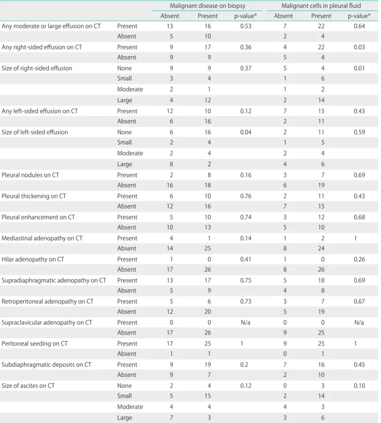
Table 1. Comparison of CT features in patients with and without pleural malignancy as assessed by reader 1
Malignant disease on biopsy Malignant cells in pleural fluid
Absent Present p-value* Absent Present p-value*
Any moderate or large effusion on CT Present 13 16 0.53 7 22 0.64
Absent 5 10 2 4
Any right-sided effusion on CT Present 9 17 0.36 4 22 0.03
Absent 9 9 5 4
Size of right-sided effusion None 9 9 0.37 5 4 0.01
Small 3 4 1 6
Moderate 2 1 1 2
Large 4 12 2 14
Any left-sided effusion on CT Present 12 10 0.12 7 15 0.43
Absent 6 16 2 11
Size of left-sided effusion None 6 16 0.04 2 11 0.59
Small 2 4 1 5
Moderate 2 4 2 4
Large 8 2 4 6
Pleural nodules on CT Present 2 8 0.16 3 7 0.69
Absent 16 18 6 19
Pleural thickening on CT Present 6 10 0.76 2 11 0.43
Absent 12 16 7 15
Pleural enhancement on CT Present 5 10 0.74 3 12 0.68
Absent 10 13 5 10
Mediastinal adenopathy on CT Present 4 1 0.14 1 2 1
Absent 14 25 8 24
Hilar adenopathy on CT Present 1 0 0.41 1 0 0.26
Absent 17 26 8 26
Supradiaphragmatic adenopathy on CT Present 13 17 0.75 5 18 0.69
Absent 5 9 4 8
Retroperitoneal adenopathy on CT Present 5 6 0.73 3 7 0.67
Absent 12 20 5 19
Supraclavicular adenopathy on CT Present 0 0 N/a 0 0 N/a
Absent 17 26 9 25
Peritoneal seeding on CT Present 17 25 1 9 25 1
Absent 1 1 0 1
Subdiaphragmatic deposits on CT Present 9 19 0.2 7 16 0.45
Absent 9 7 2 10
Size of ascites on CT None 2 4 0.12 0 3 0.10
Small 5 15 2 14
Moderate 4 4 4 3
Large 7 3 3 6
*The p-values are derived from Fisher's exact test and indicate the significance of the association between each computed tomography (CT) finding and the presence of malignancy on biopsy or cytology.
3. Interobserver agreement
Interobserver agreement was substantial (κ=0.78) for pleural effusion size and moderate for presence of solid pleural dis
ease (κ=0.46). Agreement on individual pleural findings varied from fair to substantial, with κ values of 0.21 for agreement on pleural nodules, 0.48 for agreement on pleural thickening, and Table 2. Comparison of CT features in patients with and without pleural malignancy as assessed by reader 2
Malignant disease on biopsy Malignant cells in pleural fluid
Absent Present p-value* Absent Present p-value*
Any moderate or large effusion on CT Present 13 16 0.53 7 22 0.64
Absent 5 10 2 4
Any right-sided effusion on CT Present 9 17 0.36 4 22 0.03
Absent 9 9 5 4
Size of right-sided effusion None 9 9 0.69 5 4 0.16
Small 3 4 1 6
Moderate 2 3 1 4
Large 4 10 2 12
Any left-sided effusion on CT Present 12 10 0.12 7 15 0.43
Absent 6 16 2 11
Size of left-sided effusion None 6 16 0.08 2 11 0.59
Small 2 4 1 5
Moderate 5 5 4 6
Large 5 1 2 4
Pleural nodules on CT Present 7 14 0.37 3 15 0.26
Absent 11 12 6 11
Pleural thickening on CT Present 6 13 0.36 2 15 0.12
Absent 12 13 7 11
Pleural enhancement on CT Present 10 15 1 6 18 0.33
Absent 6 8 3 3
Mediastinal Adenopathy on CT Present 9 8 0.21 2 10 0.44
Absent 8 18 7 15
Hilar adenopathy on CT Present 2 1 0.54 1 1 0.50
Absent 11 22 7 19
Supradiaphragmatic adenopathy on CT Present 13 22 0.45 8 18 0.39
Absent 5 4 1 8
Retroperitoneal adenopathy on CT Present 4 5 1 1 5 1
Absent 13 19 7 19
Supraclavicular adenopathy on CT Present 4 1 0.07 1 2 1
Absent 13 25 8 23
Peritoneal seeding on CT Present 16 22 1 8 25 0.45
Absent 2 4 1 1
Subdiaphragmatic deposits on CT Present 15 19 0.49 6 13 0.63
Absent 3 7 1 7
Size of ascites on CT None 4 8 0.001 1 4 0.646
Small 1 13 2 11
Moderate 8 2 4 6
Large 3 2 1 3
*The p-values are derived from Fisher's exact test and indicate the significance of the association between each computed tomography (CT) finding and the presence of malignancy on biopsy or cytology.
0.61 for agreement on pleural enhancement. Agreement on the amount of ascites was fair (κ=0.37).
DISCUSSION
The management of patients with malignant pleural effu
sions and/or intrathoracic metastasis from primary or recurrent advanced EOC is not standardized. The Gynecologic Cancer Intergroup Ovarian Cancer Consensus Conference statement, 2004 [23] and the evidence from randomized controlled tri
als [11,24] indicate that disease outside the peritoneum pre
cludes optimal debulking and therefore these patients should undergo neoadjuvant chemotherapy followed by cytoreduc
tive surgery. Still other studies have reported that patients with malignant pleural effusions as the only extraperitoneal manifestation of the disease have a more favorable prognosis than patients with other sites of stage IV disease such as liver or distant lymph nodes [2527]. A study from our institution in patients who had optimal cytoreduction showed a worse prognosis for those with than for those without malignant pleural effusions [12]. Therefore, it is important for patients with pleural effusions to be appropriately triaged for intratho
racic cytoreduction or neoadjuvant chemotherapy depending on the feasibility of optimal cytoreduction [13,14,2830].
In a very recent study of 203 patients with FIGO stage III and IV EOC who underwent CT before primary cytoreductive surgery at our institution, the presence of moderatetolarge pleural effusion on preoperative CT was independently associ
ated with poorer overall survival (reader 1: hazard ratio [HR], 2.26, 95% confidence interval [CI], 1.31 to 3.91, p<0.01; reader 2: HR, 2.25, 95% CI, 1.26 to 4.01, p=0.02) after controlling for age, preoperative CA125, surgical stage, ascites and cytore
ductive status. Since many patients who had pleural effusions did not have the pleural fluid removed and evaluated for ma
lignant cells, it was unclear if the poor prognosis associated with moderatetolarge pleural effusions was due to the size of the effusions, the likelihood that they were malignant, the possibility that they were associated with bulky intrathoracic disease, or a combination of the above. We have previously reported that as many as twothirds of patients with pleural effusions have gross intrathoracic disease on VATS [14]. There
fore, at our institution VATS is now routinely performed in pa
tients with moderatetolarge pleural effusions.
In our present study the size of pleural effusion and the presence of ascites on CT were associated with malignant pleural effusion. However, CT features generally considered suggestive of solid pleural malignancy such as solid pleural nodules, pleural thickening and enhancement were not asso
ciated with pleural malignancy at biopsy or cytology obtained during VATS. Our results agree with those of a prior study of 15 patients with advanced ovarian cancer, in which video
assisted thoracoscopy was done to evaluate unilateral or bi
lateral pleural effusions or (in one patient) to assess the effects of neoadjuvant chemotherapy on pleural metastases [31].
The study found that preoperative CT had sensitivity of just 14% and specificity of only 25% for determining pleural status when videoassisted thoracoscopy was used as the reference standard [31].
Several studies have investigated the use of positron emis
sion tomography (PET) or PET/CT as an alternative imaging test for diagnosing pleural metastases in patients with me
sothelioma and nonsmallcell lung cancer; using pathology from VATS or cytology obtained from thoracocentesis as the reference standard. They found very high accuracy ranging from 92% to 97.5% [3224]. In the primary staging of ovarian cancer, Kitajima et al. [35] found that integrated FDGPET/con
trastenhanced CT was more accurate than CT alone. The in
cremental value of PET/CT in perlesion accuracy was greater in extrapelvic sites, particularly metastatic lymph nodes in the abdomen; however, almost all patients (39/40) in the study had stage III disease [35]. Further studies are needed to assess the potential of PET/CT for evaluating thoracic metastases from ovarian cancer.
Our study had a number of limitations. First, selection bias may have affected our results, since patients had VATS be
cause of suspicion of pleural malignancy based either on CT findings or pleural cytology. Optimally, the same study could be repeated with patients who routinely undergo VATS re
gardless of CT findings. Second, we chose an arbitrary period of ≤ 30 days between imaging and surgery. However, we re
viewed only the most recent imaging study for each patient so the interval between CT examination and surgery would be as short as possible and the analysis of the relationship between CT and surgical findings would be valid. Third, we combined patients with stage III, stage IV, recurrent disease and different histological subtypes. Subgroup analysis was not feasible given the limited number of patients who under
went VATS. Finally, although the size and laterality of pleural effusion are unlikely to be affected by scanning technique or equipment, we cannot account for the variations in CT tech
nique and equipment that occurred during the large time
span of this study. Detection of pleural metastasis could have been difficult on digitized images from other institutions, and this could account for the fact that interreader agreement was lower for the presence of solid pleural disease (κ=0.46) than for the size of pleural effusion (κ=0.78). Since a number of the CT examinations were performed at outside institutions
it was not possible to apply a computerassisted method of effusion volume estimation, which may be more accurate than the visual method that was used in our study.
Our study also had some important advantages compared to other published studies. First, to our knowledge, our sample size was larger than that of any other published study comparing VATS and CT findings of pleural disease in patients with advanced primary or recurrent EOC. Second, we relied on independent CT readings by two radiologists with substantial experience in oncologic imaging rather than consensus evalu
ation of CT images.
In conclusion, we found that in patients with advanced or re current epithelial ovarian cancer, CT imaging findings gen
erally considered suggestive of malignant pleural involvement such as nodularity, thickening and enhancement of the pleura do not predict the presence of pleural malignancy on VATS.
Therefore, VATS should be considered to evaluate thoracic in
volvement in ovarian cancer and facilitate appropriate mana
ge ment.
CONFLICT OF INTEREST
No potential conflicts of interest relevant to this article were reported.
REFERENCES
1. Sankaranarayanan R, Ferlay J. Worldwide burden of gy nae
cological cancer: the size of the problem. Best Pract Res Clin Obstet Gynaecol 2006;20:20725.
2. FIGO Committee on Gynecologic Oncology, Denny L, Ha
cker NF, Gori J, Johns HW III, Ngan HY, et al. Staging cla ssi
fications and clinical practice guidelines for gyne co logic cancers. Oxford: Elsevier; 2000.
3. Ozols RF. Update on Gynecologic Oncology Group (GOG) trials in ovarian cancer. Cancer Invest 2004;22 Suppl 2:1120.
4. Chi DS, McCaughty K, Diaz JP, Huh J, Schwabenbauer S, Hummer AJ, et al. Guidelines and selection criteria for se
condary cytoreductive surgery in patients with recurrent, platinumsensitive epithelial ovarian carcinoma. Cancer 2006;106:19339.
5. Morgan RJ Jr, Alvarez RD, Armstrong DK, Boston B, Burger RA, Chen LM, et al. NCCN Clinical Practice Guidelines in Oncology: epithelial ovarian cancer. J Natl Compr Canc Netw 2011;9:82113.
6. Kim KW, Choi HJ, Kang S, Park SY, Jung DC, Cho JY, et al.
The utility of multidetector computed tomography in
the diagnosis of malignant pleural effusion in the patients with ovarian cancer. Eur J Radiol 2010;75:2305.
7. Diaz JP, AbuRustum NR, Sonoda Y, Downey RJ, Park BJ, Flores RM, et al. Videoassisted thoracic surgery (VATS) evaluation of pleural effusions in patients with newly dia
g nosed advanced ovarian carcinoma can influence the primary management choice for these patients. Gynecol Oncol 2010;116:4838.
8. Munstedt K, Franke FE. Role of primary surgery in advan ced ovarian cancer. World J Surg Oncol 2004;2:32.
9. Huober J, Meyer A, Wagner U, Wallwiener D. The role of neo adjuvant chemotherapy and interval laparotomy in advanced ovarian cancer. J Cancer Res Clin Oncol 2002;
128:15360.
10. Schwartz PE. Cytoreductive surgery for the management of stage IV ovarian cancer. Gynecol Oncol 1997;64:13.
11. Vergote I, Trope CG, Amant F, Kristensen GB, Ehlen T, Johnson N, et al. Neoadjuvant chemotherapy or primary surgery in stage IIIC or IV ovarian cancer. N Engl J Med 2010;363:94353.
12. Eitan R, Levine DA, AbuRustum N, Sonoda Y, Huh JN, Fran
klin CC, et al. The clinical significance of malignant pleu ral effu sions in patients with optimally debulked ovarian car ci
no ma. Cancer 2005;103:1397401.
13. Juretzka MM, AbuRustum NR, Sonoda Y, Downey RJ, Flores RM, Park BJ, et al. The impact of videoassisted tho racic sur
gery (VATS) in patients with suspected advanced ova rian malignancies and pleural effusions. Gynecol Oncol 2007;
104:6704.
14. Chi DS, AbuRustum NR, Sonoda Y, Chen SW, Flores RM, Downey R, et al. The benefit of videoassisted thora co sco pic surgery before planned abdominal exploration in patients with suspected advanced ovarian cancer and moderate to large pleural effusions. Gynecol Oncol 2004;94:30711.
15. Mironov O, Ishill NM, Mironov S, Vargas HA, Zheng J, Moskowitz CS, et al. Pleural effusion detected at CT prior to primary cytoreduction for stage III or IV ovarian carcinoma: effect on survival. Radiology 2011;258:77684.
16. Leung AN, Muller NL, Miller RR. CT in differential diagnosis of diffuse pleural disease. AJR Am J Roentgenol 1990;154:
48792.
17. Traill ZC, Davies RJ, Gleeson FV. Thoracic computed tomo
graphy in patients with suspected malignant pleural effu
sions. Clin Radiol 2001;56:1936.
18. Grunze H. The comparative diagnostic accuracy, effi cien cy and specificity of cytologic technics used in the dia g no sis of malignant neoplasm in serous effusions of the pleural and pericardial cavities. Acta Cytol 1964;8:15063.
19. Sahn SA. Pleural diseases related to metastatic malignan
cies. Eur Respir J 1997;10:190713.
20. Sallach SM, Sallach JA, Vasquez E, Schultz L, Kvale P. Volume of pleural fluid required for diagnosis of pleural malignancy.
Chest 2002;122:19137.
21. Lim MC, Lee HS, Jung DC, Choi JY, Seo SS, Park SY. Patholo
gi cal diagnosis and cytoreduction of cardiophrenic lymph node and pleural metastasis in ovarian cancer patients using videoassisted thoracic surgery. Ann Surg Oncol 2009;16:
19906.
22. Mergo PJ, Helmberger T, Didovic J, Cernigliaro J, Ros PR, Staab EV. New formula for quantification of pleural effusions from com puted tomography. J Thorac Imaging 1999;14:1225.
23. du Bois A, Quinn M, Thigpen T, Vermorken J, AvallLundqvist E, Bookman M, et al. 2004 consensus statements on the management of ovarian cancer: final document of the 3rd International Gynecologic Cancer Intergroup Ovarian Can
cer Consensus Conference (GCIG OCCC 2004). Ann Oncol 2005;16 Suppl 8:viii712.
24. van der Burg ME, van Lent M, Buyse M, Kobierska A, Colombo N, Favalli G, et al. The effect of debulking surgery after induction chemotherapy on the prognosis in advanced epithelial ovarian cancer: Gynecological Cancer Cooperative Group of the European Organization for Research and Treatment of Cancer. N Engl J Med 1995;332:62934.
25. Bonnefoi H, A'Hern RP, Fisher C, Macfarlane V, Barton D, Blake P, et al. Natural history of stage IV epithelial ovarian cancer. J Clin Oncol 1999;17:76775.
26. Penson RT, Skates SJ, Fuller AJ Jr, Seiden MV. Clinical course of stage IV epithelial ovarian cancer. J Clin Oncol 1999;17:
33612.
27. Aletti GD, Podratz KC, Cliby WA, Gostout BS. Stage IV ovarian cancer: disease sitespecific rationale for postoperative treatment. Gynecol Oncol 2009;112:227.
28. Eisenkop SM. Thoracoscopy for the management of advan
ced epithelial ovarian cancer: a preliminary report. Gynecol Oncol 2002;84:31520.
29. Bristow RE, Tomacruz RS, Armstrong DK, Trimble EL, Montz FJ. Survival effect of maximal cytoreductive surgery for ad vanced ovarian carcinoma during the platinum era: a metaanalysis. J Clin Oncol 2002;20:124859.
30. Munkarah AR, Hallum AV 3rd, Morris M, Burke TW, Levenback C, Atkinson EN, et al. Prognostic significance of residual disease in patients with stage IV epithelial ovarian cancer.
Gynecol Oncol 1997;64:137.
31. CohenMouly S, Badia A, Bats AS, Barthes F, Bensaid C, Riquet M, et al. Role of videoassisted thoracoscopy in patients with ovarian cancer and pleural effusion. Int J Gynecol Cancer 2009;19:16625.
32. Erasmus JJ, McAdams HP, Rossi SE, Goodman PC, Coleman RE, Patz EF. FDG PET of pleural effusions in patients with nonsmall cell lung cancer. AJR Am J Roentgenol 2000;
175:2459.
33. Orki A, Akin O, Tasci AE, Ciftci H, Urek S, Falay O, et al. The role of positron emission tomography/computed to mo graphy in the diagnosis of pleural diseases. Thorac Cardiovasc Surg 2009;57:21721.
34. Yildirim H, Metintas M, Entok E, Ak G, Ak I, Dundar E, et al.
Clinical value of fluorodeoxyglucosepositron emission tomography/computed tomography in differentiation of malignant mesothelioma from asbestosrelated benign pleural disease: an observational pilot study. J Thorac Oncol 2009;4:14804.
35. Kitajima K, Murakami K, Yamasaki E, Kaji Y, Fukasawa I, Inaba N, et al. Diagnostic accuracy of integrated FDGPET/contrast
enhanced CT in staging ovarian cancer: comparison with en hanced CT. Eur J Nucl Med Mol Imaging 2008;35:1912
20.