1. Introduction
During the last decade, there has been a great interest in investigating Mg and its alloys for application as tempo- rary implant materials in cardiovascular and orthopaedic devices1-6). This is due to the unique property of Mg that degrades spontaneously in physiological solutions and due to the proximity of the mechanical properties to that of the natural bone1). The dissolved ions are tolerable for human body system and even beneficial for some metabol- ic reactions7). However, its application was limited due to the relatively poor corrosion resistance of Mg when exposed to human body fluid. Consequently, the implant will liberate strong hydrogen gas and cause loss of me- chanical integrity before the replaced tissue or bone com- pletely heals.
Great efforts have been done to improve the corrosion
resistance by alloying, surface treatment, and coating1-5). Conventional Mg alloys (AZ and AM) commonly used Al as the main alloying element that could enhance the corrosion resistance and mechanical properties of Mg al- loys2,3,5,6). Implant devices such as screws and pins used to fix bone fracture, are expected to handle tension and compression stresses during service. Therefore, Mg is preferably used in the alloys form. AZ31 and AZ91 are mostly studied magnesium alloys but there is only few reports in exploring AM (containing Al and Mn) Mg series alloys for biodegradable materials. Small amounts of Mn are tolerable and essential in the human body3). In fact, addition of small amount of Mn is a commercial way of mitigating the detrimental effect of impurities thus im- proving corrosion resistance3,8).
Ca is another interesting alloying element that had been explored for Mg based alloys. It was reported3,4,5,9) that Ca enhanced the corrosion resistance and mechanical properties of Mg alloys. Ca can be tolerated by human
Role of Ca in Modifying Corrosion Resistance and Bioactivity of Plasma Anodized AM60 Magnesium Alloys
Anawati Anawati1, Hidetaka Asoh1,2, and Sachiko Ono1,2,†
1Research Institute for Science and Technology
2Department of Applied Chemistry, Kogakuin University, 2665-1 Nakano, Hachioji, Tokyo 192-0015, Japan (Received January 28, 2016; Revised June 26, 2016; Accepted June 26, 2016)
The effect of alloying element Ca (0, 1, and 2 wt%) on corrosion resistance and bioactivity of the as-received and anodized surface of rolled plate AM60 alloys was investigated. A plasma electrolytic oxidation (PEO) was carried out to form anodic oxide film in 0.5 mol dm-3 Na3PO4 solution. The corrosion behavior was studied by polarization measurements while the in vitro bioactivity was tested by soaking the specimens in Simulated Body Fluid (1.5xSBF). Optical micrograph and elemental analysis of the substrate surfaces indicated that the number of intermetallic particles increased with Ca content in the alloys owing to the formation of a new phase Al2Ca. The corrosion resistance of AM60 specimens improved only slightly by alloying with 2 wt% Ca which was attributed to the reticular distribution of Al2Ca phase existed in the alloy that might became barrier for corrosion propagation across grain boundaries. Corrosion resistance of the three alloys was significantly improved by coating the substrates with anodic oxide film formed by PEO. The film mainly composed of magnesium phosphate with thickness in the range 30 - 40 µm. The heat resistant phase of Al2Ca was believed to retard the plasma discharge during anodization and, hence, decreased the film thickness of Ca-containing alloys. The highest apatite forming ability in 1.5xSBF was observed for AM60-1Ca specimens (both substrate and anodized) that exhibited more degradation than the other two alloys as indicated by surface observation. The increase of surface roughness and the degree of supersaturation of 1.5xSBF due to dissolution of Mg ions from the substrate surface or the release of film compounds from the anodized surface are important factors to enhance deposition of Ca-P compound on the specimen surfaces.
Keywords : magnesium, biodegradable, bioactivity, anodization, corrosion.
†Corresponding author: [email protected]
body3). It is a major component in human bone and essen- tial in chemical signalling with cells10). However, most of the reported works were focused on the effect of Ca on microstructure, mechanical properties and corrosion behavior3,4,5,9) of the substrate. There has been lack of re- ports on the effect of alloying element Ca on anodic oxide film characteristics formed on Mg alloys. In this work, the effect of alloying element Ca on the corrosion resist- ance and apatite forming ability (bioactivity) of the anodic oxide film formed by plasma electrolytic oxidation (PEO) on AM60 magnesium alloy was investigated.
2. Experimental Procedure
The specimens used were rolled plates of commercial AM60 magnesium alloys with Ca content 0, 1, and 2 wt%.
The chemical composition of the AM60 alloy is listed in Table 1. The specimen was cut into pieces to give a working area of 5 cm2 with thickness of 1 mm. For micro- structure observation, the specimens were ground to #1200 grit paper. The surface microstructure was studied by opti- cal microscope (Olympus BX51M type).
Prior to anodization, pretreatment was applied in a mixed acid solution of 8 vol% HNO3-1 vol% H3PO4 for 20 s and then washed with deionized (DI) water before dipping in 5 wt% NaOH solution at 80 °C for 1 min.
Anodizing was carried out by plasma electrolytic oxida- tion technique in 0.5 moldm-3 Na3PO4 solution at a con- stant current of 200 Am-2 at 25 °C for 20 min. The oxide film thickness was measured by using a coating thickness meter of dual type (SME-1) from Sanko.
The corrosion behavior of the specimens was inves- tigated by polarization test in physiological solution 0.9 wt% NaCl solution at 37 °C based on ASTM standard and our earlier work11). The polarization test was per- formed by using a potentiostat instrument from IviumStat started from potential -1.65 VAg/AgCl and terminated when the current output reach 30 mA at a sweep rate 1 mV s-1. The polarization measurement was conducted immedi- ately upon immersion of the specimen in the test solution.
Pt wire was used as a counter electrode and silver chloride was used as a reference electrode. The corrosion potential and current densities were determined by Tafel plot.
The apatite forming ability of the specimens was eval- uated by an in-vitro test in 1.5SBF with 1.5 times of con- centration of SBF10 for 7 days. The SBF10 was prepared as previously reported12). The solution pH was adjusted to 7.4 at 36.4 °C. Sodium azide (NaN3) was added to the resulting solution to inhibit bacterial growth. The specimens were exposed to the solution with a surface- to-volume ratio of 20 ml cm-2. The solution was refreshed after 3 days. After the test, the specimen was rinsed thor- oughly with DI water and then dried in air stream.
The elemental analysis was performed by using an en- ergy dispersive X-ray spectroscopy (EDS, JEOL EX- 54175JMU) attached to SEM (JEOL JSM-6380LA).
3. Results and Discussion
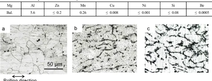
Fig. 1 shows the optical microscope images showing the microstructure of the three alloys. The base alloy ex- hibited grain sizes in the range of 10 - 50 µm (Fig. 1a).
Table 1. Chemical composition of AM60 alloy in wt%
Mg Al Zn Mn Cu Ni Si Be
Bal. 5.6 ≤ 0.2 0.26 ≤ 0.008 ≤ 0.001 ≤ 0.08 ≤ 0.0005
Fig. 1. The optical micrographs of a) AM60, b) AM60-1Ca, and c) AM60-2Ca alloys. The scale bar and rolling direction in a) apply to all images. The coarse lines appeared in image b) and c) were the grinding lines.
Addition of 1 and 2 wt% Ca in the alloy did not affect the grain sizes significantly but it modified the composi- tion and the number of intermetallic particles. Similar as previously reported8,9), the microstructure of AM60 was composed of α-Mg matrix and β-Mg17Al12 as analyzed by EDS. A new phase of Al2Ca was formed along grain boundaries when Ca was present in the alloys. The number of Al2Ca phases increased with Ca content in the alloys.
Some of these phases appeared continuous along grain boundaries while others existed as spheroidized particles (Fig. 1b). The grains of AM60-1Ca were coarser compare to those of the other two alloys. In the alloy containing 2 wt% Ca, the Al2Ca phases became more continuous and formed a network (Fig. 1c). Formation of Al2Ca phases was attributed to the low solubility of Ca in Mg. The equilibrium phase diagram indicated that the maximum solid solubility of Ca in Mg is 0.82 at %13). Above the solubility limit, the excess of Ca was assumed to dissolve in the intermetallics by replacing Mg in β-Mg17Al12 to form Al2Ca phase5,9). The Al2Ca network formed in AM60-2Ca was beneficial to stop the corrosion prop- agation of localized attack into adjacent grains8). The po- larization measurements indicated that the current den- sities of AM60 alloys decreased gradually by increasing Ca content in the alloys as tabulated in Table 1. The effect of Ca on corrosion potential was only observed on AM60-2Ca which exhibited higher corrosion potentials of -1.55 VAg/AgCl than that of the other two alloys (-1.57 VAg/AgCl).
Anodization of the three alloys for 20 min resulted in the formation of porous oxide film with average thickness of 42 µm, 36 µm, and 35 µm for AM60, AM60-1Ca, and AM60-2Ca specimens, respectively. The film thick-
ness decreased with increasing Ca content in the alloys owing to the increase number of Al2Ca phase in the alloys.
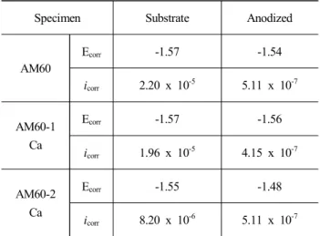
The Al2Ca phase is a heat resistant phase and a stable line compound with a relatively high melting point14). The presence of Al2Ca phase delayed the appearance of a big sparks/plasma which responsible for the rapid thickening of the oxide film. However, since the type of film was porous, thicker oxide did not necessarily provide better corrosion protection. The film density and composition are more important properties that affect the corrosion be- havior of anodized specimen. The oxide film composed of magnesium phosphate regardless of Ca content in the alloy, as analyzed by EDS. The anodic film formed on the three alloys greatly enhanced the corrosion resistance during polarization test in NaCl solution as indicated by lower corrosion current density down to two order of mag- nitude relative to the substrates (Fig. 2). The corrosion potential and corrosion current densities values are listed in Table 2. The corrosion potential of anodized AM60-1Ca specimen was nearly similar as of the substrate while slight increase of corrosion potential was obtained for ano- dized AM60 compare to the substrate. Combination of the noblest corrosion potential and the lowest corrosion current density was obtained for anodized specimen of AM60-2Ca alloy as well as the substrate itself as compare to the other two alloys. The relatively small differences in corrosion potential when comparing the three alloys in both substrate and anodized condition is due to the fact that the potential is more a representative of the corroding solid solution matrix than of the intermetallics8).
The apatite forming ability of the substrates and the anodized specimens were analyzed after SBF soaking for
Fig. 2. Potentiodynamic polarization of substrate and anodized AM60, AM60-1Ca, and AM60-2Ca in 0.9% NaCl solution.
Table 2. Corrosion potentials (VAg/AgCl) and current densities (A cm-2) data obtained from polarization measurements in 0.9 wt%
NaCl solution of the three alloys in both substrate and anodized form
Specimen Substrate Anodized
AM60
Ecorr -1.57 -1.54
icorr 2.20 x 10⁻⁵ 5.11 x 10⁻⁷
AM60-1 Ca
Ecorr -1.57 -1.56
icorr 1.96 x 10⁻⁵ 4.15 x 10⁻⁷
AM60-2 Ca
Ecorr -1.55 -1.48
icorr 8.20 x 10⁻⁶ 5.11 x 10⁻⁷
7 days. The specimen’s appearances on both sides of sur- faces of substrates and anodized specimens after SBF soaking are shown in Fig. 3. All of the substrates surfaces (left sides) had been attacked by a relatively uniform corrosion. Only few pits, indicated by arrows in Fig. 3, were observed near the edges of the Ca-containing specimens. The corroded surfaces of AM60 substrate showed variation of whitish and dark-grey areas while the Ca-containing alloys were uniformly dark-grey. The discoloration suggested variation in the deposit layer composition. The dark-grey areas apparently propagated preferentially along the roll- ing lines vertically from the lower part of the specimen’s edge towards the upper part of the specimen visible as dark-grey furrows in Fig. 3 (AM60 substrate). EDS analy- sis on the surface layers of the three specimens over an area of 650 µm x 485 µm detected similar elements com- position of Mg, O, Cl, Ca and P. The O concentration was almost similar about 50 mass% for the three alloys
while Cl concentration decreased with increasing Ca con- tent in the alloys, 1.18, 0.53, and 0.36 mass% for the AM60, AM60-1Ca, and AM60-2Ca alloys, respectively.
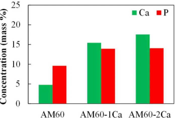
The Ca and P concentrations were plotted and shown in Fig. 4. The EDS elemental analysis indicated that the whit- ish areas corresponded to the corroded areas covered mainly by corrosion product of Mg(OH)2 and MgCl2. The Mg(OH)2 layer gave a typical white dull appearance8). The dark-grey areas were also corroded areas but covered by additional Ca-P compounds.
Fig. 4 shows the quantitative EDS analysis results of the substrate surfaces after SBF soaking for 7 days. The graph plotted the concentrations of Ca and P which are the main elements of apatite. It is obvious that higher Ca and P concentrations were obtained on the Ca-containing alloys indicating higher deposition of Ca-P compound on the surfaces, than that on the base alloy. The Ca concen- tration of the substrate surfaces notably increased from 4.7 to 15.4 mass% by addition of 1 wt% Ca in the alloys.
The Ca concentration further increased slightly to 17.6 mass% on 2 wt% Ca-containing alloy surface while the P concentration was relatively similar at 14.0 mass%.
Thus, it is concluded that the deposit layer formed on Ca-containing alloys was highly contained Ca-P com- pounds rather than Mg(OH)2 or MgCl2 phases. The Mg(OH)2 layer which formed initially was probably de- composed into Ca-P compounds. The absence of whitish areas in Ca-containing alloys indicated uniform deposition of Ca-P compounds on the surface while only local depo- sition was observed on the AM60 substrate.
The corrosion resistance of anodized specimens in 1.5xSBF is better than of the substrates. All of the ano- dized specimens show no visible corroded areas or pit (Fig. 3). The surface appearance of anodized specimens after SBF immersion was similar to those before immersion.
Fig. 3. The specimen’s appearance of both surfaces of substrates and anodized specimens after soaking in 1.5SBF for 7 days. The scale bar in a) applies to all images.
Fig. 4. Concentration (mass %) of Ca and P of the substrates after SBF soaking for 7 days.
Fig. 5. Concentration (mass%) of Ca and P of the anodized specimens after SBF soaking for 7 days.
The results of immersion test on anodized specimens were in agreement with the polarization data that indicated an im- provement in corrosion resistance by anodization. However, the apatite forming ability of the anodic film was relatively low. Fig. 5 shows the obtained Ca and P concentrations of the anodized specimens after SBF immersion. The high P concentration stems from the film which composed of magnesium phosphate. The highest Ca concentration (7.4 mass%) was obtained on the film formed on AM60-1Ca alloy followed by AM60-2Ca alloy. SEM investigation on the surface after SBF immersion indicated a superficial dissolution of the anodic film surfaces had occurred on the film formed on AM60-1Ca alloy while the film formed on the other two alloys remained intact. The release of oxide film component such as phosphate to the solution may increase the degree of supersaturation of 1.5xSBF relative to apatite1,2,4,6) and therefore triggered higher dep- osition of Ca-P compound on the film surface of AM60- 1Ca alloy. Another factor is the increase of surface rough- ness which became preferential site for Ca-P compound deposition.
4. Conclusions
Addition of alloying element Ca modified the micro- structure, corrosion behaviour in NaCl solution, and bio- activity in 1.5SBF of AM60 alloys in both substrate and anodized specimens. Al2Ca phase was formed along grain boundaries and the number phase increased with Ca con- tent in the alloys. The presence of heat resistant Al2Ca phase delayed the initiation of big sparks during anodizing and hence decreased the film thickness. The network of Al2Ca phase existed in AM60-2Ca enhanced the corrosion resistance of the specimens both substrate and anodized during polarization in NaCl solution. The AM60-1Ca specimens of both substrate and anodized exhibited higher apatite forming ability than the other two alloys. The rea- son was due to higher dissolution of substrate or anodized surfaces experienced by this alloy which further con-
tributed to the increase in the degree of supersaturation of the solution relative to the apatite and the increase in surface roughness that triggered deposition of Ca-P com- pound on the surface.
Acknowledgments
We acknowledge the Light Metal Educational Foundation, Inc., and the Ministry of Education, Culture, Sports, Science and Technology of Japan (MEXT)- Supported Program for the Strategic Research Foundation at Private Universities, 2013-2017.
References
1. L. C. Li, J. C. Gao, Y. Wang, Surf. Coat. Tech., 185, 92 (2004).
2. F. Witte, V. Kaese, H. Haferkamp, E. Switzer, A. Meyer- Lindberg, C. J. Wirth, H. Windhagen, Biomaterials, 26, 3557 (2005).
3. G. Song, Corros. Sci., 49, 1696 (2007).
4. Z. Li, X. Gu, S. Lou, Y. Zheng, Biomaterials, 29, 1329 (2008).
5. M. B. Kannan, R. K. S. Raman, Biomaterials, 29, 2306 (2008).
6. S. Virtanen, Mater. Sci. Eng. B, 176, 1600 (2011).
7. J. Vormann, Mol. Aspects Med., 24, 27 (2003).
8. O. Lunder, Corrosion Rev., 15, 439 (1997).
9. B. Kondori, R. Mahmudi, Mater. Sci. Eng. A, 527, 2014 (2010).
10. J. Z. Ilich, J. E. Kerstetter, J. Am. Coll. Nutr., 19, 715 (2000).
11. Anawati, H. Tanigawa, H. Asoh, S. Ono, Corros. Sci., 70, 212 (2013).
12. L. Müller, F. A. Müller, Acta Biomater. 2, 181 (2006).
13. A. A. Nayeb-Hashemi, J. B. Clark, Bulletin of Alloy Phase Diagrams, 8, 58 (1987).
14. H. I. Kaplan, J. Hryn and B. Clow, Magnesium Technology 2000, p. 279 (TMS, 2000).