1)
Introduction
Production of the high-energy compounds that fuel the biochemical, biophysical and mechanical functions of the body is coupled with ongoing generation of potentially cytotoxic reactive oxygen species (ROS).
ROS can attack, denature or modify structural and functional molecules and thereby cause cytotoxicity, tissue injury and dysfunction. Oxidative stress, aris- ing as a result of an imbalance between ROS produc- tion and antioxidant defenses has been implicated in the pathogenesis of tissue injury and dysfunction in a wide range of human diseases including atherosclero- sis, infection, inflammation, neoplasm, degenerative disorders, metabolic diseases and radiation injury1, 2). There is now accumulating evidence that ROS may participate in renal vascular dysfunction in cardio- vascular-renal diseases. Renal dysfunction is a cen-
Correspondence: Ja-Ryong Koo, M.D., Department of Internal Medicine, Chunchon Sacred Heart Hospital, Hallym University, Chunchon, Kangwon-Do, Korea Tel : 033)252-9970 Fax : 033)255-4291
E-mail : [email protected]
tral cause of hypertension and a common conse- quence of diabetes mellitus. These pathophysiological conditions set up a vicious cycle of repeated renal injury and are the two leading causes of end-stage renal failure.
Oxidative stress is associated with both diabetes and hypertension in humans and in experimental animal models. This review will briefly discuss the production, metabolism and targets of ROS, the role of ROS in renal dysfunction during diabetes mellitus and, finally, highlight recent studies that suggest a role for oxygen radicals in the renal vasculature during hypertension.
ROS production and antioxidant defense
1. Superoxide production
A free radical can be defined as any molecular species capable of independent existence that contains an unpaired electron in an atomic orbital2). Many rad- icals are highly reactive and can either donate an electron to or extract an electron from other mole- cules, therefore behaving as oxidants or reductants.
Ox idative Stress in Diabetes and H ypertension
Ja-Ryong Koo, M. D.
Division of Nephrology, Department of Internal Medicine, Hallym University, Chunchon, Korea
Oxidative stress implies an increased production of reactive oxygen species (ROS) or a decreased capacity to metabolize them. Recent studies suggested that oxidative stress is implicated in the patho- genesis of tissue injury and dysfunction in diabetes and hypertension, which are two major causes of ESRD. In these diseases, oxygen radicals are increased and contribute to diabetic nephropathy and hypertension by enhancing renal vascular tone, sensitivity to vasoconstrictors, inflammatory cell infil- tration and tubuloglomerular feedback. ROS induced nitric oxide inactivation and induction of stress activated signaling pathways are main underlying mechanisms for ROS induced tissue injury and dysfunction. Treatment with pharmacological antioxidant agents such as TEMPOL (superoxide dis- qmutase mimetic) reverses many of these injury and dysfunction underlying the role of oxidative stress in the pathogenesis of diabetic nephropathy and hypertension.
Key Words : Reactive oxygen species, Pathogenesis, Diabetes mellitus, Hypertension
The most important free radicals in many disease states are oxygen derivatives, particularly superoxide (O2-.) and the hydroxyl radical (OH.). Superoxide is produced endogenously during mitochondrial respira- tion and by NADPH oxidase, xanthine oxidase, cyclo- oxygenase and lipoxygenase, nitric oxide synthase (NOS) and cytochrome P4503). Several molecules, in- cluding adrenaline, flavine nucleotides, thiol com- pounds, and glucose, can oxidise in the presence of oxygen to produce superoxide, and these reactions are greatly accelerated by the presence of transition metals such as iron or copper2). The electron trans- port chain in the inner mitochondrial membrane per- forms the reduction of oxygen to water. During this process free radical intermediates are generated, which are generally tightly bound to the components of the transport chain.
However, there is a constant leak of a few elec- trons into the mitochondrial matrix and this results in the formation of superoxide. The activity of several other enzymes, such as cytochrome p450 oxidase in the liver and enzymes involved in the synthesis of adrenal hormones, also results in the leakage of a few electrons into the surrounding cytoplasm and hence superoxide formation. There might also be con- tinuous production of superoxide by vascular endothe- lium to neutralize nitric oxide, production of super- oxide by other cells to regulate cell growth and differentiation, and the production of superoxide by phagocytic cells during the respiratory burst.
2. ROS metabolism and antioxidant system
Superoxide spontaneously gains an electron to form hydrogen peroxide; however, three isoforms of super- oxide dismutase (SOD) also catalyze this reaction.
Mn-SOD is located in mitochondria and two isoforms of Cu,Zn-SOD are located either extracellularly or intracellularly. Once produced, hydrogen peroxide can be scavenged to water by catalase or by glutathione (GSH) peroxidase in the presence of GSH.
Decomposition of hydrogen peroxide in the pres- ence of Fe2+ produces a hydroxyl radical, also known as the Fenton reaction2). Superoxide and hydrogen peroxide also can react together directly to produce the hydroxyl radical under the presence of transition metal ion, also known as Haber-Weiss reaction2).
(Fenton reaction) : Fe2++H2O2→ Fe3++OH.+OH- (Haber-Weiss reaction) : Fe3++O2-
→Fe2++O2 and Fe2++ H2O2→Fe3++OH.+OH-
Net result : O2-
+H2O2→OH-+OH.+O2
The hydroxyl radical (OH.), or a closely related species, is probably the final mediator of most free radical induced tissue damage. All of the ROS de- scribed above exert most of their pathological effects by giving rise to hydroxyl radical formation. The rea- son for this is that the hydroxyl radical reacts, with extremely high rate constants, with almost every type of molecule found in living cells including sugars, amino acids, lipids, and nucleotides.
3. Targets of ROS
The three main cellular targets of ROS are lipids, proteins, and DNA. Extensive lipid peroxidation in bi- ological membranes causes alterations in fluidity, per- meability and membrane potential, and eventual rup- ture of the cell. Oxidation of proteins changes their primary structure, including the overall charge, fold- ing and hydrophobicity, which can lead to increased aggregation and degradation. Oxygen-radical-induced damage of DNA includes changes in both DNA structure and chemistry with the result being strand breakage.
In addition to their ability to directly inflict damage upon cellular macromolecules, ROS play a significant role in activating stress-sensitive signaling pathways that regulate gene expression resulting in cellular damage4). Those genes involved in stress-activated signaling pathways can lead to the development of microvascular complications of diabetes, insulin resis- tance, inflammation and hypertension5).
Whether oxygen radicals attack those targets de- pends on the delicate balance between levels of ROS and antioxidants. Under many conditions an increase in oxygen radical formation signals the activation of antioxidant enzymes to aid in the increased metabo- lism necessary to achieve redox balance. However, when the amount of radicals produced exceeds the resources for metabolism, oxidative stress results.
Diabetes mellitus
1. The importance of oxidative stress in the pathogenesis of diabetic complication 1) Oxygen radicals reduce nitric oxide (NO)
function in diabetic vessels
Endothelial dysfunction in peripheral and renal ves- sels is a common sequela of diabetes mellitus6). Some observations indicate that the tonic influence of NO is suppressed and contributes to the impaired endo- thelium-dependent relaxation in the renal vasculature during diabetes7). Because superoxide rapidly binds and inactivates NO8), one possible explanation for the lack of NO function in the kidney during diabetes is excessive superoxide. Indeed, renal cortical tissue from diabetic rats has increased superoxide produc- tion9). Several other studies10, 11) indicate that in-
creased superoxide in diabetes reduces NO-dependent modulation of basal renal vascular tone, which can be restored by antioxidant treatment.
NO modulates and buffers the renal and peripheral vasoconstriction caused by several endogenous agents, including angiotensin II, thromboxane A2 and endothe- lin-1. Because superoxide limits the bioavailability of NO, the buffering capability of NO during agonist- induced vasoconstriction may be decreased in diabe- tes.
Indeed, Schoonmaker et al.12) have shown that the renal afferent arteriolar responsiveness to angiotensin II is enhanced in juxtamedullary nephrons from dia- betic rats and that L-NNA (NO synthase inhibitor Nw-nitro-L-arginine) did not alter the response.
However, treatment with SOD restored the ability of L-NNA to enhance the vascular response to angio- tensin II. These data suggest that excess superoxide is responsible for the increased sensitivity of renal microvessels to angiotensin II in diabetes.
In addition to functional NO deficiency, avid reac- tion of superoxide with NO leads to formation of highly reactive nitrogen species such as peroxynitrite
13) or peroxynitrous acid14). The latter agents can, in
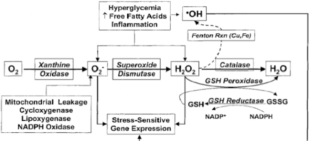
Fig. 1. Endogenous stimuli leading to ROS generation and activation of stress- sensitive gene expression. The endogenous antioxidant enzymes including GSH, superoxide dismutase, GSH peroxidase, and catalase function to maintain redox equilibrium. However, in situations such as chronic hy- perglycemia, the compensatory response is inadequate, leading to both ROS formation and activation of stress- and redox-sensitive gene expres- sion (e.g., via the redox-sensitive transcription factor NF-κB). Catalase is localized primarily in peroxisomes, whereas GSH peroxidase is the major peroxidase in mitochondria [Derived from Ref. 5].
turn, attack, denature or modify various structural and functional molecules15, 16). For instance peroxyni- trite can react with tyrosine or cystein residues of the proteins producing nitrotyrosine or nitrocystein which are considered as footprint of ROS interaction with NO17).
2) Oxidative stress and stress-activated signaling pathways
Recent evidence suggests that common stress-acti- vated signaling pathways such as nuclear factor-κB (NF-κB), p38 MAPK, NH2-terminal Jun kinases/
stress-activated protein kinases (JNK/SAPK) are ac- tivated by oxidative stress and underlies the devel- opment of late diabetic complications5). Among these, transcription factor NF-κB is one major intracellular target of hyperglycemia and oxidative stress. The consequence of these signaling pathways is the pro- duction of gene products, such as VEGF and others, which cause cellular damage and are ultimately re- sponsible for the long-term complications of diabetes.
3) Formation of glycoxidative advanced glycation end products (AGEs)
Possible mediators of untoward effects of hypergly- cemia include AGEs known to accumulate in diabetic subjects. AGEs comprise a variety of molecular structures, such as Nε-(carboxymethyl)lysine (CML), pentosidine, and pyrraline18). They are generated by the Maillard reaction through nonenzymatic glycation of protein amino groups and characterized by differ- ent formation mechanisms18). Among them, CML and pentosidine requires both glycation and oxidation for their formation (thereby termed glycoxidation)19).
Supporting the role of glycoxidative AGE in the pathogenesis of diabetic nephropathy, it was recently demonstrated that among AGE, glycoxidation pro- ducts such as CML and pentosidine, accumulate in expanded mesangial matrix and nodular lesions in DN, in colocalization with malondialdehyde-lysine (MDA-lysine), a lipoxidation product, whereas pyrra- line, another AGE structure whose deposition is rath- er independent from oxidative stress, was not found
within diabetic glomeruli20). Because CML, pentosi- dine, and MDA-lysine are all formed under oxidative stress by carbonyl amine chemistry between protein amino group and carbonyl compounds, their colocali- zation suggests a local oxidative stress and increased protein carbonyl modification in diabetic glomerular lesions.
4) Oxidative stress as a common linking mechanism of hyperglycemia induced damage
It has been suggested that following four pathways are mainly involved in the pathogenesis of long-term diabetic complications : ① increased polyol pathway flux; ② increased AGE formation; ③ activation of protein kinase C (PKC) isoforms; ④ increased hexo- samine pathway flux21). Recent study suggested that excess ROS produced during mitochondrial respiration is a unifying hypothesis linking these seemingly in- dependent four mechanisms22).
Potential mechanisms by which hyperglycemia-in- duced ROS (superoxide) overproduction activates four pathways of hyperglycaemic damage are as follows (Fig. 2). Excess superoxide partially inhibits the gly- colytic enzyme glyceraldehyde-3-phosphate dehydro- genase (GAPDH), thereby diverting upstream me- tabolites from glycolysis into pathways of glucose overutilization. This results in increased flux of dihy- droxyacetone phosphate (DHAP) to diacylglycerol, an activator of PKC, and of triose phosphates to methy- lglyoxal, the main intracellular AGE precursor. In- creased flux of fructose-6-phosphate to UDP-N-ace- tylglucosamine increases modification of proteins by O-linked N-acetylglucosamine (GlcNAc) and in- creased glucose flux through the polyol pathway consumes NADPH and depletes GSH as described below.
2. Sources of oxygen radicals in diabetes mellitus For hyperglycemia, increases in oxidant productions are due to multiple processes. Simply, glucose can undergo non-enzymatic reactions forming gluco-oxi-
dants and glycated products (AGEs), which them- selves can be pro-oxidants23, 24). In addition, high glu- cose-induced ROS production in renal mesangial cell can be effectively blocked by inhibition of PKC, NADPH oxidase, and mitochondrial electron transfer chain complex, suggesting that PKC, NADPH oxi- dase, and mitochondrial metabolism all play a role in high glucose-induced ROS generation25).
Among those possibilities, recent focus has been on mitochondrial metabolism and activation of NADPH oxidases24). Suggestions have been made that most glucose-induced oxidants are derived from glycolysis and mitochondrial oxidative phosphorylation with the productions of superoxide21). Electron trans- fer through mitochondrial enzyme complexes I, III and IV generates a proton gradient that drives ATP synthase (complex V). When the electrochemical po- tential difference generated by this proton gradient is high, the life of superoxide-generating electron trans- port intermediates is prolonged resulting in leakage of superoxide.
There appears to be a threshold value above which superoxide production is markedly increased.
Hyperglycemia increases the proton gradient above this threshold value as a result of overproduction of
electron donors by the Krebs cycle, thereby increas- ing superoxide production21).
Metabolism of high glucose levels can activate NADPH oxidase in the vascular cells independent of mitochondrial metabolisms26). One mechanism that can increase NADPH oxidase activity is the activation of PKC, which is elevated by glucose-induced elevation of diacylglycerol26). It has been also established that angiotensin II stimulates vascular superoxide forma- tion through activation of NADPH oxidase27) and angiotensin-converting enzyme (ACE) inhibition atten- uates the oxidative stress in the diabetic kidney28).
These studies intimate that hyperglycemia and angiotensin II induced oxygen radical formation me- diated by NADPH oxidase play a role in the oxida- tive stress of the diabetic renal microvasculature.
Among other possibilities, it has been suggested that activation of the polyol pathway could be another source of oxidative stress21). In polyol path- way reduction of glucose to sorbitol by NADPH con- sumes NADPH. As NADPH is required for regener- ating reduced GSH, this could induce or exacerbate intracellular oxidative stress.
Koo et al.29) showed that insulin therapy in strep- tozotocin diabetic rat resulted in a significant increase Fig. 2. Potential mechanism by which hyperglycemia-induced mitochondrial
superoxide overproduction activates four pathways of hyperglycemic damage.
in renal NOS expression. This is consistent with the known effect of insulin on NO production and endo- thelial NOS gene expression in endothelial cells via activation of phosphatidylinositol-3 kinase30). In their study29), tissue nitrotyrosine abundance, which is a marker of highly toxic and reactive nitrogen species was paradoxically increased in insulin treated diabetic rat (Fig. 3). This paradoxical result was partially ex- plained by the upregulation of NOS isoforms in all tested tissues and the expected rise in NO production capacity.
This coupled with the residual oxidative stress with incomplete glycemia control in insulin-treated animals can account for the observed elevation of tissue nitrotyrosine abundance. In this regard, it has been suggested that inadequate glucose control with insulin treatment could be another mechanism of oxi- dative stress in diabetic kidney29).
Hypertension
Oxidative stress in the vasculature has been asso- ciated with human essential hypertension, pre-eclamp- sia and several hypertensive animal models, including the spontaneously hypertensive rat (SHR), angiotensin II-induced hypertension, Dahl salt-sensitive hyperten-
sion, lead-induced hypertension, obesity-induced hy- pertension, mineralocorticoid hypertension and hyper- glycemia-induced hypertension3, 31). Most studies indi- cate that antioxidant treatment lowers blood pressure and improves endothelial function in large conduit vessels3, 31). Considering the major role of kidney in the pathogenesis of hypertension, above studies strongly suggested that increased oxidative stress is important in the renal vascular dysfunction in hyper- tension.
1. Oxygen radicals reduce NO function in hypertension
SHR has increased blood pressure and renal vas- cular resistance and an enhanced tubuloglomerular feedback response. Acute and long-term studies sug- gest that oxygen radicals may play an important role in these characteristics. The SOD mimetic TEMPOL (2,2,6,6-tetramethyl-1-piperidinoxyl) normalizes the blood pressure, renal vascular resistance and renal excretion of the oxidative stress marker 8-iso pros- taglandin F2α in the SHR32, 33). Furthermore, TEMPOL increases the basal luminal diameter of in vitro per- fused afferent arterioles of juxtamedullary nephrons in SHR but has no effect in the Wistar-Kyoto rat (WKY)34). These studies suggest that oxygen radicals may contribute to the increased blood pressure and renal vasculature resistance in the SHR.
1) Redox regulation of the afferent arteriole and tubuloglomerular feedback
One of the potential mechanisms for the acute re- duction in blood pressure and renal vascular resis- tance in SHR treated with antioxidants, is via en- hancing NO action in the renal vasculature or in the juxtaglomerular apparatus. NOS type 1 or neuronal NOS is expressed in the macula densa. Renal macula densa derived NO counteracts afferent arteriolar vaso- constriction mediated by the tubuloglomerular feed- back mechanism and thus control intraglomerular pressure and sodium and water excretion35, 36). Sys- temic inhibition of NOS with Nω-nitro-L-arginine Fig. 3. Representative Western blots and correspond-
ing group data illustrating nitrotyrosine abun- dance of kidney cortex in the normal control rats fed regular diet (N=6), untreated diabetic rats (DM, N=6), diabetic rats treated with once-daily ultralente insulin alone (DM+I, N=5) or the combination of insulin and vitamin E and C-fortified diet (DM+I+EC, N=5).
*P<0.005 versus control and DM+I groups;
†P<0.05 versus other groups [Derived from Ref. 29].
methyl ester (L-NAME, NOS inhibitor) blocks the acute anti-hypertensive actions of TEMPOL in SHR
32). This study implies that NO-mediated vasodilation may be restored after scavenging of oxygen radicals in the SHR. The afferent arteriole and macula densa cell both contain a full complement of components of NADPH oxidase that generates superoxide35, 37). Sev- eral recent studies have shown extensive interaction between superoxide and NO in macula densa to regu- late afferent arteriolar tone mediated by the tubulo- glomerular feedback response35, 38). Superoxide in the macula densa and afferent arterioles will inactivate NO and enhance preglomerular resistance in animal models of oxidative stress35). As an increase in affer- ent arteriolar resistance can precede hypertension, oxidative stress could be important in determining the long-term blood pressure and thereby contribute to hypertension.
2) Role of oxidative stress in the angiotensin II induced hypertension
In recent studies, Welch and Wilcox39) showed that two weeks of treatment with the selective angioten- sin AT1 receptor antagonist candesartan in SHR re- stored a normal tubuloglomerular feedback response to NOS inhibitor 7-nitroindazole but had no effect on the response to 7-nitroindazole during TEMPOL mi- croperfusion. These data suggest that angiotensin II stimulation of oxygen radical formation in the jux- taglomerular apparatus, mediated via AT1 receptors, has an important role in elevating oxygen radicals and thereby diminishing NO signaling in the juxta- glomerular apparatus of SHR.
Angiotensin II-induced stimulation of oxygen radi- cal formation, which can diminish NO action, is not specific to the SHR. Nishiyama et al.40) demonstrated that the increased blood pressure and renal vascular resistance in angiotensin II-infused hypertensive rats was ameliorated by TEMPOL. NOS inhibition mark- edly attenuated the hemodynamic response to TEMPOL. These studies strongly implicate angioten- sin II as a major source of oxygen radical formation
in forms of hypertension that are dependent on or sensitive to angiotensin II. More importantly, Higash et al.41) showed that angiotensin II mediated excessive oxidative stress is involved, at least in part, in im- paired endothelium-dependent vasodilatation in pa- tients with renovascular hypertension. In their study
41), correction of renal artery stenosis decreases oxi- dative stress with normalization of endothelial dys- function. These results further support the role of oxidative stress in angiotensin II dependent hyper- tension.
2. Dys-regulation of antioxidant defenses in hypertension
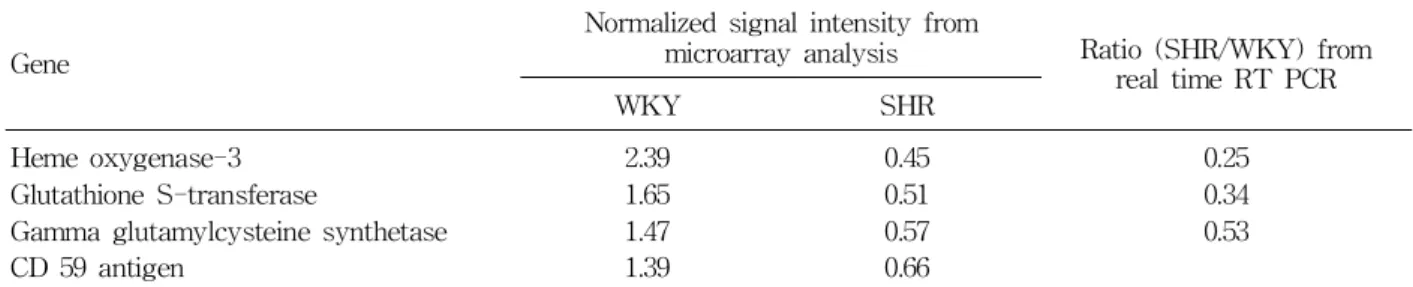
In study using DNA microarray analysis to explore differentially-expressed genes in kidneys between SHR and WKY rat, several genes involved in anti- oxidant system such as gamma glutamylcysteine syn- thetase, GSH S-transferase and heme oxygenase 3 genes were down-regulated in SHR as compared to WKY rat42).
The changes in the expression of these 3 genes were confirmed by real time RT-PCR (Table 1).
Gamma glutamylcysteine synthetase is the rate-limit- ing enzyme for GSH synthesis. GSH S-transferase adds GSH to electrophiles with a variety of chemical structures and protects cells from oxidative stress.
Thus, down-regulations of gamma glutamylcysteine synthetase and GSH S-transferase can lead to GSH deficiency and defective protection against oxidative injury. These events can, in turn contribute to oxida- tive stress and hypertension in SHR. The latter prop- osition is supported by the result of previous study in which induction of oxidative stress by GSH deple- tion caused severe hypertension in genetically normal rats43). Heme oxygenase which exists in constitutive (heme oxygenase-2 and heme oxygenase-3) and in- ducible (heme oxygenase-1) isoforms degrades pro- oxidant heme to produce carbon monoxide and bili- verdin, which is a potent antixidant44). Endogenous carbon monoxide is a potent vasodilator and pos-
sesses anti-inflammatory and anti-apoptotic proper- ties. Therefore, down-regulation of constitutive heme oxygenase 3 isoform as shown in this study could potentially contribute to oxidative stress, inflammation and hypertension in SHR.
3. Oxygen radicals and inflammation in hypertension
Another possible mechanism for the renal protec- tive actions of antioxidants in hypertension is through a reduction in the immune and inflammatory re- sponses. ROS can act as second messengers for sev- eral transcription factors, including NF-κB, which plays a critical role in the activation of multiple genes that contribute to the inflammatory response and end-organ damage3). In the angiotensin II-depen- dent hypertension45) and deoxycorticosterone (DOCA) salt hypertension46) animal models, NF-κB activation is increased in the kidney and renal monocyte/macro- phage infiltration is increased. In association with a reduction in vascular oxygen radical formation, TEMPOL also reduces the blood pressure, NF-κB activation and monocyte/macrophage infiltration in the kidneys of DOCA salt hypertensive rats46).
The transient administration of angiotensin II or NOS inhibitor, L-NAME to rats result in salt-sensi- tive hypertension that persists after removal of the inciting stimulus, and this is associated with the in- filtration of immunocompetent cells and tissue injury in the renal interstitium47, 48). Many of the mononu- clear cells, including some T cells, were shown to express angiotensin II, and there was also local gen-
eration of oxidants47, 48). Tubulointerstitial injury re- sulting from angiotensin II infusion was significantly reduced by anti-inflammatory treatment using myco- phenolate mofetil, as were proliferative activity, T- cell infiltration and activation, superoxide-producing cells, and urinary excretion of marker of oxidative stress, MDA.
Similar results was obtained in SHR, in which my- cophenolate mofetil treatment reduce blood pressure, oxidative stress and inflammatory cell infiltration in kidney49). In DNA microarray analysis of SHR, the complement regulatory membrane protein CD59 gene was down-regulated in SHR as compared to WKY rat42) (Table 1). Because the complement regulatory membrane protein, CD59, restricts membrane attack complex formation, it is likely that down-regulation of CD59 gene can potentially facilitate infiltration of immunocompetent cells in the kidney and, thereby, aggravate hypertension, tissue injury and vascular complication in SHR. These results signify that some of the renal vascular damage in hypertension may be due to the proinflammatory actions of oxygen radicals and inflammation with oxidative stress could be a possible mechanism of hypertension.
Conclusions
Oxidative stress is increased with diabetes mellitus and hypertension. In diabetes, hyperglycemia-induced activation of PKC, NADPH oxidase and mitochondrial metabolism play major role in ROS generation. ROS induced inactivation of NO and induction of stress-
Table 1. Comparison of Expression of mRNA Involved in Antioxidant and Anti-inflammatory System between SHR and WKY Rat
Gene
Normalized signal intensity from
microarray analysis Ratio (SHR/WKY) from real time RT PCR
WKY SHR
Heme oxygenase-3 Glutathione S-transferase
Gamma glutamylcysteine synthetase CD 59 antigen
2.39 1.65 1.47 1.39
0.45 0.51 0.57 0.66
0.25 0.34 0.53
activated signaling pathway seems to be important mechanisms for the chronic diabetic complications in cluding diabetic nephropathy. Recent studies suggest that increased production of superoxide by the mito- chondrial electron transport chain is a causal link between elevated glucose and each of the four main pathways responsible for hyperglycaemic damage i.e.
increased polyol pathway flux; increased AGE forma- tion; activation of PKC; increased hexosamine path- way flux. In experimental hypertension, antioxidant defenses systems are down regulated at least in SHR, an animal model of human essential hyperten- sion. ROS induced inactivation of NO seems to con- tribute to the increased basal vascular tone and sensitivity of the vasculature to angiotensin II and enhanced tubuloglomerular feedback shown in hyper- tensive animal model. It is also suggested that some of the renal vascular damage in hypertension may be due to the proinflammatory actions of oxygen radicals and inflammation with oxidative stress could be another possible mechanism of hypertension. Treat- ment with pharmacological agents that mimic SOD, such as TEMPOL, reduces vascular dysfunction in the kidney during diabetes. In animal models of hy- pertension, TEMPOL decreases markers of oxidative stress, improves renal vascular function and reduces blood pressure. These studies provide a rationale for the development of new pharmacological agents that target oxygen radicals for the treatment of renal dys- function in hypertension and diabetes mellitus.
References
1) Rosenfeld ME : Inflammation, lipids, and free radi- cals : lessons learned from the atherogenic process.
Semin Reprod Endocrinol 16:249-261, 1998
2) Young IS, Woodside JV : Antioxidants in health and disease. J Clin Pathol 54:176-186, 2001
3) Schnackenberg CG : Oxygen radicals in cardiovascu- lar-renal disease. Curr Opin Pharmacol 2:121-125, 2002
4) Allen RG, Tresini M : Oxidative stress and gene regulation. Free Radic Biol Med 28:463-499, 2000 5) Evans JL, Goldfine ID, Maddux BA, Grodsky GM :
Oxidative stress and stress-activated signaling pathways : A unifying hypothesis of type 2 diabetes.
Endocr Rev 23:599-622, 2002
6) De Vriese AS, Verbeuren TJ, Van de Voorde J, Lameire NH, Vanhoutte PM : Endothelial dysfunction in diabetes. Br J Pharmacol 130:963-974, 2000 7) Bank N, Aynedjian HS : Role of EDRF (nitric oxide)
in diabetic renal hyperfiltration. Kidney Int 43:
1306-1312. 1993
8) Gryglewski RJ, Palmer RMJ, Moncada S : Superox- ide anion is involved in the breakdown of endothe- lium-derived relaxing factor. Nature 320:454-456, 1986
9) Ishi N, Patel KP, Lane PH, Taylor T, Bian K, Mu- rad F, Pollock JS, Carmines PK : Nitric oxide syn- thesis and oxidative stress in the renal cortex of rats with diabetes mellitus. J Am Soc Nephrol 12:1630-1639, 2001
10) Ohishi K, Carmines PK : Superoxide dismutase re- stores the influence of nitric oxide on renal arter- ioles in diabetes mellitus. J Am Soc Nephrol 5:
1559-1566, 1995
11) Dai F, Diederich A, Skopec J, Diederich D : Diabe- tes-induced endothelial dysfunction in streptozoto- cin-treated rats : role of prostaglandin endoperoxides and free radicals. J Am Soc Nephrol 4:1327-1336, 1993
12) Schoonmaker GC, Fallet RW, Carmines PK : Super- oxide anion curbs nitric oxide modulation of affer- ent arteriolar ANG II responsiveness in diabetes mellitus. Am J Physiol 278:F302-F309, 2000 13) Squadrito GL, Pryor WA : The formation of peroxy-
nitrite in vivo from nitric oxide and superoxide.
Chem Biol Interact 96:203-206, 1995
14) van der Vliet A, Eiserich JP, O'Neill CA, Halliwell B, Cross CE : Tyrosine modification by reactive ni- trogen species : a closer look. Arch Biochem Bio- phys 319:341-349, 1995
15) Beckman JS, Beckman TW, Chen J, Marshall PA, Freeman BA : Apparent hydroxyl radical production by peroxynitrite : implications for endothelial injury from nitric oxide and superoxide. Proc Natl Acad Sci USA 87:1620-1624, 1990
16) Graham A, Hogg N, Kalyanaraman B, O'Leary V, Darley-Usmar V, Moncada S : Peroxynitrite modifi- cation of low-density lipoprotein leads to recogni- tion by the macrophage scavenger receptor. FEBS Lett 330:181-185, 1993
17) Ceriello A, Mercuri F, Quagliaro L, Assaloni R, Motz E, Tonutti L, Taboga C : Detection of nitro- tyrosine in the diabetic plasma : evidence of oxida- tive stress. Diabetologia 44:834-838, 2001
18) Singh R, Barden A, Mori T, Beilin L : Advanced glycation end-products : a review. Diabetologia 44:
129-146, 2001
19) Horie K, Miyata T, Maeda K, Miyata S, Sugiyama S, Sakai H, van Ypersole de Strihou C, Monnier VM, Witztum JL, Kurokawa K : Immunohistochemi- cal colocalization of glycoxidation products and lipid peroxidation products in diabetic renal glomerular lesions. Implication for glycoxidative stress in the pathogenesis of diabetic nephropathy. J Clin Invest 100:2995-3004, 1997
20) Suzuki D, Miyata T, Saotome N, Horie K, Inagi R, Yasuda Y, Uchida K, Izuhara Y, Yagame M, Sakai H, Kurokawa K : Immunohistochemical evidence for an increased oxidative stress and carbonyl modifica- tion of proteins in diabetic glomerular lesions. J Am Soc Nephrol 10:822-832, 1999
21) Brownlee M : Biochemistry and molecular cell biol- ogy of diabetic complications. Nature 414:813-820, 2001
22) Nishikawa T, Edelstein D, Du XL, Yamagishi S, Matsumura T, Kaneda Y, Yorek MA, Beebe D, Oates PJ, Hammes HP, Giardino I, Brownlee M : Normalizing mitochondrial superoxide production blocks three pathways of hyperglycaemic damage.
Nature 404:787-790, 2000
23) Baynes JW, Thorpe SR : Role of oxidative stress in diabetic complications : A new perspective on an old paradigm. Diabetes 48:1-9, 1999
24) Kuroki T, Isshiki K, King GL : Oxidative Stress : The lead or supporting actor in the pathogenesis of diabetic complications. J Am Soc Nephrol 14:S216- 220, 2003
25) Lee HB, Yu MR, Yang Y, Jiang Z, Ha H : Reactive oxygen species-regulated signaling pathways in dia- betic nephropathy. J Am Soc Nephrol 14:S241-245, 2003
26) Inoguchi T, Li P, Umeda F, Yu HY, Kakimoto M, Imamura M, Aoki T, Etoh T, Hashimoto T, Naruse M, Sano H, Utsumi H, Nawata H : High glucose level and free fatty acid stimulate reactive oxygen species production through protein kinase C-depen- dent activation of NAD(P)H oxidase in cultured vascular cells. Diabetes 49:1939-1945, 2000
27) Lassegue B, Sorescu D, Szocs K, Yin Q, Akers M, Zhang Y, Grant SL, Lambeth JD, riendling KK : Novel gp91 (phox) homologues in vascular smooth muscle cells : nox1 mediates angiotensin II-induced superoxide formation and redox-sensitive signaling pathways. Circ Res 88:888-894, 2001
28) Kedziora-Kornatowska KJ, Luciak M, Paszkowski J : Lipid peroxidation and activities of antioxidant enzymes in the diabetic kidney : effect of treatment with angiotensin convertase inhibitors. IUBMB Life 49:303-307, 2000
29) Koo JR, Vaziri ND : Effects of diabetes, insulin and antioxidants on NO synthase abundance and NO in- teraction with reactive oxygen species. Kidney Int
63:195-201, 2003
30) Kuboki K, Jiang ZY, Takahara N, Ha SW, Igarashi M, Yamauchi T, Feener EP, Herbert TP, Rhodes CJ, King GL : Regulation of endothelial constitutive nitric oxide synthase gene expression in endothelial cells and in vivo : a specific vascular action of in- sulin. Circulation 101:676-681, 2000
31) Koo JR, Ni Z, Oviesi F, Vaziri ND : Antioxidant therapy potentiates antihypertensive action of insu- lin in diabetic rats. Clin Exp Hypertens 24:333-344, 2002
32) Schnackenberg CG, Welch WJ, Wilcox CS : Normal- ization of blood pressure and renal vascular resis- tance in SHR with a membrane-permeable super- oxide dismutase mimetic : role of nitric oxide.
Hypertension 32:59-64, 1998
33) Schnackenberg CG, Wilcox CS : Two-week adminis- tration of tempol attenuates both hypertension and renal excretion of 8-iso prostaglandin F2α. Hyper- tension 33:424-428, 1999
34) Ichihara A, Hayaski M, Hirota N, Saruta T : Super- oxide inhibits neuronal nitric oxide synthase influ- ences on afferent arterioles in spontaneously hyper- tensive rats. Hypertension 37:630-634, 2001
35) Wilcox CS : Redox regulation of the afferent arteri- ole and tubuloglomerular feedback. Acta Physiol Scand 179:217-223, 2003
36) Thorup C, Erik A, Persson G : Macula densa de- rived nitric oxide in regulation of glomerular cap- illary pressure. Kidney Int 49:430-436, 1996
37) Wilcox CS : Reactive oxygen species : roles in blood pressure and kidney function. Curr Hypertens Rep 4:160-166, 2002
38) Welch WJ, Tojo A, Wilcox CS : Roles of NO and oxygen radicals in tubuloglomerular feedback in SHR. Am J Physiol Renal Physiol 278:F769-776, 2000
39) Welch WJ, Wilcox CS : AT1 receptor antagonist combats oxidative stress and restores nitric oxide signaling in the SHR. Kidney Int 59:1257-1263, 2001
40) Nishiyama A, Fukui T, Fujisawa Y, Rahman M, Tian R, Kimura S, Abe Y : Systemic and regional hemodynamic responses to tempol in angiotensin II- infused rats. Hypertension 37:77-83, 2001
41) Higashi Y, Sasaki S, Nakagawa K, Matsuura H, Oshima T, Chayama K : Endothelial function and oxidative stress in renovascular hypertension. N Engl J Med 346:1954-1962, 2002
42) Koo JR, Liang KH, Vaziri ND : Identification of dif- ferentially expressed genes in kidneys of spontane- ous hypertensive rats (SHR) using DNA microarray analysis (abstract). J Am Soc Nephrol 13:A337, 2002
43) Vaziri ND, Wang XQ, Oveisi F, Rad B : Induction
of oxidative stress by glutathione depletion causes hypertension in normal rats. Hypertension 36:142- 146, 2000
44) Agarwal A, Nick HS. Renal response to tissue in- jury : lessons from heme oxygenase-1 gene ablation and expression. J Am Soc Nephrol 11:965-973, 2000
45) Muller DN, Dechend R, Mervaala EM, Park JK, Schmidt F, Fiebeler A, Theuer J, Breu V, Ganten D, Haller H, Luft FC : NF-κB inhibition ameliorates angiotensin II-induced inflammatory damage in rats.
Hypertension 35:193-201, 2000
46) Beswick RA, Zhang H, Marable D, Catravas JD, Hill WD, Webb RC : Long-term antioxidant admin- istration attenuates mineralocorticoid hypertension and renal inflammatory response. Hypertension 37:
781-786, 2001
47) Quiroz, Y, Pons H, Gordon KI, Rincon J, Chavez M,
Parra G, Herrera-Acosta J, Gomez-Garre D, Largo R, Egido J, Johnson RJ, Rodriguez-Iturbe B : My- cophenolate mofetil prevents the salt-sensitive hy- pertension resulting from short-term nitric oxide synthesis inhibition. Am J Physiol Renal Physiol 281:F38-F47, 2001
48) Rodriguez-Iturbe, B, Pons H, Quiroz Y, Gordon K, Rincon J, Chavez M, Parra G, Herrera-Acosta J, Gomez-Garre D, Largo R, Egido J, Johnson RJ : Mycophenolate mofetil prevents salt-sensitive hyper- tension resulting from angiotensin II exposure. Kid- ney Int 59:2222-2232, 2001
49) Rodriguez-Iturbe B, Quiroz Y, Nava M, Bonet L, Chavez M, Herrera-Acosta J, Johnson RJ, Pons HA : Reduction of renal immune cell infiltration re- sults in blood pressure control in genetically hyper- tensive rats. Am J Physiol Renal Physiol 282:
F191-201, 2002