https://doi.org/10.4174/astr.2019.96.6.296 Annals of Surgical Treatment and Research
Impact of tumor sidedness on survival and recurrence patterns in colon cancer patients
Jong Min Lee, Yoon Dae Han, Min Soo Cho, Hyuk Hur, Byung Soh Min, Kang Young Lee, Nam Kyu Kim
Division of Colorectal Surgery, Department of Surgery, Yonsei University College of Medicine, Seoul, Korea
INTRODUCTION
Evidence gathered over the past two decades indicates that colon cancer sidedness is an emerging prognostic factor. Right- sided colon cancer (RCC) shows distinct clinicopathological characteristics and long-term patient outcomes compared with left-sided colon cancer (LCC). Multiple studies have identified differential tumor biology as the reason for differences in patient prognoses for these 2 diseases [1-5]. RCC is more
common in women and the elderly and these tumors are marked by a higher prevalence of poorly differentiated histology, microsatellite instability (MSI-H), and CpG island methylation than seen in LCC tumors. In addition, RCC is fre- quently associated with driver gene mutations in KRAS and BRAF [6-8].
Generally, RCC is associated with worse survival outcomes than LCC. However, reports regarding the prognostic role of primary tumor location with respect to colon cancer stage are
Received November 22, 2018, Revised February 15, 2019, Accepted April 1, 2019
Corresponding Author: Nam Kyu Kim
Division of Colon and Rectal Surgery, Department of Surgery, Severance Hospital, Yonsei University College of Medicine, 50-1 Yonsei-ro, Seodaemun-gu, Seoul 03722, Korea
Tel: +82-2-2228-2100, Fax: +82-2-313-8289 E-mail: namkyuk@yuhs.ac
ORCID code: https://orcid.org/0000-0003-0639-5632
Copyright ⓒ 2019, the Korean Surgical Society
cc Annals of Surgical Treatment and Research is an Open Access Journal. All articles are distributed under the terms of the Creative Commons Attribution Non- Commercial License (http://creativecommons.org/licenses/by-nc/4.0/) which permits unrestricted non-commercial use, distribution, and reproduction in any medium, provided the original work is properly cited.
Purpose: Previous studies have reported conflicting results regarding the prognostic value of tumor sidedness in colon cancer. We investigated the oncologic impact of tumor location and examined whether recurrence patterns were related to tumor sidedness in colon cancer patients.
Methods: We identified stage I–III colon adenocarcinoma patients from a prospective colorectal cancer registry at Severance Hospital, Seoul, Korea, who underwent complete mesocolic excision between 2005 and 2012. Adjusted hazard ratios (HRs) and 95% confidence intervals (CI) for predictors of cancer-specific survival (CSS), recurrence-free survival (RFS), and cumulative recurrence at specific anatomic sites were examined using Cox proportional hazard regression analysis.
Results: Overall, 1,912 patients, 1,077 (56.3%) with left-sided colon cancer (LCC), and 835 (43.7%) with right-sided colon cancer (RCC), at a median follow-up of 59 months, were eligible and included in the study. In univariate analysis, similar 5-year CSS and RFS were observed for LCC and RCC in the total patient population, and when stratified by stage for stage I and II patients. For stage III patients, an adjusted Cox regression analysis indicated that RCC patients had a higher risk of cancer-specific mortality (HR, 1.75; 95% CI, 1.07–2.86; P = 0.024) and recurrence (HR, 1.78; 95% CI, 1.22–2.60; P = 0.003).
Furthermore, RCC was an independent predictor of peritoneal recurrence (HR, 1.86; 95% CI, 1.05–3.29; P = 0.031) in stage III patients.
Conclusion: RCC correlated with worse CSS and RFS than LCC. In stage III patients, RCC correlated with increased risk of peritoneal recurrence. The reasons for these differences remain to be investigated.
[Ann Surg Treat Res 2019;96(6):296-304]
Key Words: Colonic neoplasm, Right, Left, Neoplasm recurrence, local, Survival, Treatment outcome
conflicting [3,9]. For example, Warschkow et al. [3] showed that cancer-specific survival (CSS) was better in RCC patients than in LCC patients (adjusted hazard ratio [HR] for RCC, 0.90;
95% confidence interval [CI], 0.87–0.93; P < 0.001). In stage III patients, RCC and LCC had similar prognoses, whereas prognosis was better for stage I and II RCC patients than LCC patients after propensity score matching [3]. Further, neither the Warschkow study nor most prior studies addressed the question of whether cancer recurrence patterns are related to tumor laterality [10]. Therefore, in-depth investigations of survival and recurrence patterns between RCC and LCC are warranted. In this study, we examined both the prognostic role of tumor sidedness in stage I–III colon cancer and patterns of recurrence with respect to tumor location.
METHODS
The study protocol was approved by the Institutional Review Board of the Human Research Protection Center, Severance Hospital, Seoul, South Korea (approval number: 4-2018-0682).
Informed consent was waived due to the retrospective analysis of this study.
Patients
Consecutive patients from a colorectal cancer registry who underwent surgical resection for colon cancer from November 2005 to October 2012 at Severance Hospital, Yonsei University College of Medicine, Seoul, South Korea were selected for this study. Analysis of the data was performed in March 2018.
Patient samples were staged according to the American Joint Commission on cancer stage I, II, III colon adenocarcinoma, based on pathology. Tumor locations were assigned through the patients’ medical records. Tumors proximal to the hepatic flexure were classified as RCC, and tumors located on the splenic flexure, descending and sigmoid colon were classified as LCC. The embryological reference point dividing RCC and LCC was the distal one third of the transverse colon. Because dividing the transverse colon into thirds can be ambiguous, transverse colon cancer was excluded from the study. Patients with tumors originating from the appendix (n = 35) and rectosigmoid junction (n = 247) or who received palliative resection (n = 174), resection for synchronous colon cancer (n = 80), or who had distant metastases (n = 441) were also excluded.
Surgical technique
As previously described, all patients underwent surgical resections based on the principle of complete mesocolic excision with central vascular ligation (CME with CVL) [11]. Briefly, en bloc resection included the tumor and its surrounding soft tissue enveloped by intact visceral fascia. Supplying vessels
were ligated at their origin to achieve CVL. Specimens included pericolic, intermediate, and principal lymph nodes for adequate lymphadenectomy. If the tumor was suspected to have invaded or threatened the CME plane on the retroperitoneal side in the ascending or descending colon, we deepened our dissection to include perinephric fat tissues in the en bloc specimen.
Outcome measurements
Primary outcomes measured in this study were CSS, recur- rence-free survival (RFS), and cumulative recurrence rate by specific anatomic site. CSS was defined as the length of time from the date of curative resection to the date of death from colon cancer. Patients who died of other causes were censored.
RFS was defined as the length of time the patient survived without any evidence of colon cancer following primary tumor resection. Cumulative incidence of recurrence was defined as the probability that colon cancer recurrence had occurred before the given time. Patients who died without recurrence were censored.
Statistical analysis
Comparison of all variables between RCC and LCC was conducted using a chi-square test for categorical variables, and either Mann-Whitney’s U test or independent t-test for continuous variables. The frequency of recurrence by site and stage was determined using rates and relative risk with a 95%
confidence interval (CI). For long-term outcomes, Kaplan-Meier curves were drawn and log-rank tests were used to compare RCC and LCC. Multivariate Cox regression was performed to assess the effect of tumor location after adjusting for covariates.
All tests were 2-sided, and P-values <0.05 were considered statistically significant. Statistical analysis was performed using SPSS software, version 20.0 (IBM Corp.).
RESULTS
Clinicopathological characteristics
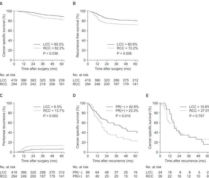
Of the 1912 patients included in the study, 835 (43.7%) underwent right colectomy and 56.3% (n = 1,077) underwent left colectomies or sigmoid colectomy. RCC was more common in older patients and female patients. Other clinical features such as body mass index (BMI) and American Society of Anesthesiologists (ASA) physical status classification (III, IV) did not show significant differences between RCC and LCC.
Prognostic nutritional index (PNI), an immunological and nutritional marker related to short- and long-term outcomes, was better in LCC patients [12,13].
Histological grade was higher, tumor size was larger, and invasion was deeper in RCC, whereas metastatic nodal status and the rate of lymphovascular invasion (LVI) were similar. RCC specimens contained more regional lymph nodes than did LCC
specimens (Table 1).
Surgical characteristics and perioperative outcomes
Minimally invasive surgery was used more frequently in LCC than RCC. Minimally invasive surgeries were more commonly converted to open surgeries with RCC, and RCC surgeries had
longer operation times and more significant blood loss than LCC surgeries. Although the proportion of patients undergoing multivisceral resection was similar between groups, nine cases (1.1%) of RCC, but none of LCC, required duodenum resection to achieve complete R0 resection. The proportion of patients who underwent adjuvant chemotherapy was comparable between
Table 1. Clinicopathological characteristics of all included patients
Characteristic LCC (n = 1,077) RCC (n = 835) P-value
Age (yr) 62 (53–70) 63 (56–71) <0.001
Male sex 654 (60.7) 461 (55.2) 0.015
Body mass index (kg/m2) 23.3 (21.3–25.4) 23.2 (21.4–25.3) 0.339
ASA PS classification ≥ III 189 (17.5) 151 (18.1) 0.762
PNIa) < 50 317 (29.4) 359 (43.0) <0.001
Obstruction 36 (3.3) 24 (2.9) 0.560
Tumor size > 5 cm 267 (24.8) 327 (39.2) <0.001
AJCC stage <0.001
I 279 (25.9) 174 (20.8)
II 378 (35.1) 365 (43.8)
III 420 (39.0) 295 (35.3)
T stage 0.001
1 177 (16.4) 105 (12.6)
2 153 (14.2) 97 (11.6)
3 645 (59.9) 515 (61.7)
4 102 (9.5) 118 (14.1)
N stage 0.231
0 657 (61.0) 541 (64.8)
1 291 (27.0) 206 (24.7)
2 129 (12.0) 88 (10.5)
No. of examined LNs 16 (11–23) 27 (19–37) <0.001
Harvested LN < 12 285 (26.5) 48 (5.7) <0.001
LVI 247 (23.9) 196 (24.2) 0.884
Histologic grade <0.001
G1, 2 1,026 (95.3) 727 (87.1)
G3, 4 51 (4.7) 108 (12.9)
Values are presented as median (interquartile range) or number (%).
LCC, left-sided colon cancer; RCC, right-sided colon cancer; ASA PS, American Society of Anesthesiologists physical status; PNI, prognostic nutritional index; AJCC, American Joint Committee on Cancer; LN, lymph node; LVI, lymphovascular invasion.
a)PNI = 10 × serum albumin (g/dL) + 0.005 × total lymphocyte count (cells/mm3).
Table 2. Surgical characteristics and perioperative outcomes
Variable LCC (n = 1,077) RCC (n = 835) P-value
MIS 815 (75.7) 555 (66.5) <0.001
Surgical duration (min) 192 (153–242) 207 (171–254) <0.001
EBL ≥ 200 mL 181 (16.8) 201 (24.1) <0.001
Open conversion 13/815 (1.6) 25/555 (4.5) 0.001
Multivisceral resection 36 (3.3) 32 (3.8) 0.566
30-Day mortality 1 (0.1) 1 (0.1) >0.999
30-Day morbidity 91 (9.3) 124 (16.2) <0.001
Hospital stay (day) 8 (6–11) 9 (7–12) <0.001
Adjuvant chemotherapy 630 (58.5) 491 (58.9) 0.868
Values are presented as number (%) or median (interquartile range).
LCC, left-sided colon cancer; RCC, right-sided colon cancer; MIS, minimally invasive surgery; EBL, estimated blood loss.
the 2 groups (Table 2).
Survival and recurrence
Differences in 5-year CSS between RCC and LCC for all stages combined (91.9% vs. 94.7%, P = 0.110), and for stage I (100% vs. 99.6%, P = 0.273), and stage II (95.8% vs. 97.1%, P = 0.739) cancers were not observed in unadjusted survival analyses. Further, unadjusted survival curves demonstrated no significant differences in the 5-year RFS between RCC and LCC for all stages combined (85.6% vs. 88.0%, P = 0.079), and for stage I (98.3% vs. 97.0%, P = 0.325), and stage II (88.9%
vs. 90.2%, P = 0.681) cancers. However, for stage III patients, unadjusted Cox regression showed an increased risk for both cancer-specific mortality (HR, 1.53; 95% CI, 1.02–2.30; P =
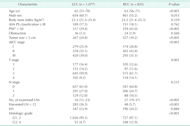
0.037) and recurrence (HR, 1.56; 95% CI, 1.13–2.14; P =0.006) for RCC compared with LCC (Fig. 1A, B). Subsequent multivariable analysis for CSS and RFS were performed with possible confounders including age, sex, BMI, ASA physical status classification, PNI, obstruction, tumor size, histologic grade, LVI, adjuvant chemotherapy, number of examined lymph nodes, estimated blood loss (EBL), T stage, and number of positive LN in stage III. After adjustment, RCC patients had a higher risk of cancer-specific mortality (HR, 1.75; 95% CI, 1.07–2.86; P = 0.024) and recurrence (HR, 1.78; 95% CI, 1.22–2.60; P = 0.003) (Table 3).
When all recurrences were stratified by stage and site, the recurrence rate for all sites was higher only in stage III RCC (27.0% vs. 18.6%, P = 0.012), and peritoneal recurrence (PR) was approximately twice as frequent in RCC (12.5%) than LCC
A B
No. at risk
12 24 60
0 100
80
60
40
20
Cancerspecificsurvival(%)
Time after surgery (mo)
LCC RCC
419 294
386 279
363 242
325 218
LCC = 89.2%
RCC = 82.2%
P = 0.036 0
36 48
309 206
239 161
No. at risk
12 24 60
0 100
80
60
40
20
Recurrencefreesurvival(%)
Time after surgery (mo)
LCC RCC
419 294
366 246
320 200
288 187
LCC = 80.9%
RCC = 72.2%
P = 0.006 0
36 48
275 179
212 141
C D
No. at risk
12 24 60
0 100
80
60
40
recurrencePeritoneal(%) 20
Time after surgery (mo)
LCC RCC
419 294
366 246
320 200
288 187
LCC = 6.5%
RCC = 13.7%
P = 0.002
0
36 48
275 179
212 141
No. at risk
12 24 60
0 100
80
60
40
20
Cancerspecificsurvival(%)
Time after recurrence (mo)
PR( ) PR(+)
88 61
64 40
48 25
37 20
PR( ) = 42.8%
PR(+) = 23.3%
P = 0.010
0
36 48
25 15
19 10
E
No. at risk
12 24 60
0 100
80
60
40
20
Cancerspecificsurvival(%)
Time afterrecurrence(mo)
LCC RCC
24 36
18 22
9 16
8 12
LCC = 15.8%
RCC = 27.0%
P = 0.757
0
36 48
5 10
2 8
Fig. 1. Kaplan-Meier curves for survival analysis within stage III colon cancer patients. Cancer specific survival (CSS) (A), recurrence-free survival (RFS) (B), peritoneal recurrence (PR) (C), CSS from recurrence to death (CSS2) according to presence of peritoneal recurrence (D), and CSS2 in patients with peritoneal recurrence (E). LCC, left-sided colon cancer; RCC, right-sided colon cancer.
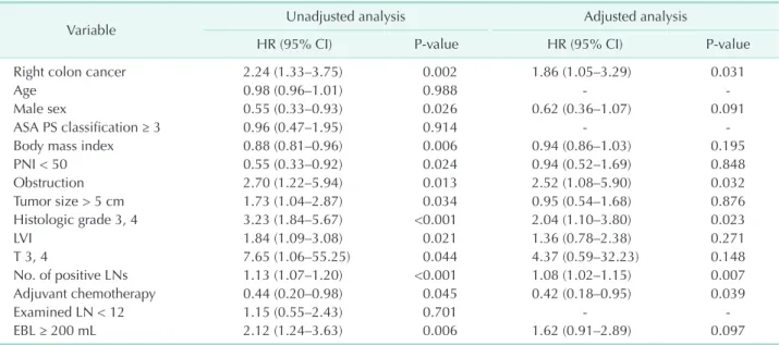
(6%) (Table 4). Five-year cumulative incidence of PR showed significant difference between RCC and LCC in stage III colon cancer (13.7% vs. 6.5%; P = 0.002) (Fig. 1C). A subsequent Cox proportional hazard regression for PR was conducted including all clinical and pathological variables. The locational impact of the tumor on PR in stage III patients persisted following ad- justed analysis (Table 5).
CSS after recurrence (CSS2), was defined as the period be- tween the date that recurrence was initially recognized and the date of either death from colon cancer or the last follow-
up. The Kaplan-Meier curve and log rank test for stage III colon cancer revealed that patients with PR had significantly worse CSS2 than those without PR (Fig. 1D). Further comparison of CSS2 between RCC and LCC showed no differences in stage III patients with PR (Fig. 1E). In multivariable Cox regression analysis of stage III patients, peritoneal metastases exhibited worse results for CSS2 (HR, 1.55; 95% CI, 1.03–2.34; P = 0.035), after adjusting metastases from other sites (Table 6).
Table 3. Multivariate analysis for cancer specific- and recurrence free survival in patients stratified by stage
All stages (n = 1,912) Stage I (n = 453) Stage II (n = 743) Stage III (n = 715) HRa) (95% CI) P-value HRa) (95% CI) P-value HRa) (95% CI) P-value HRa) (95% CI) P-value CSS 1.06 (0.69–1.62) 0.781 0.50 (0.0–99.9)b) 0.998 0.61 (0.27–1.39) 0.248 1.75 (1.07–2.86) 0.024 RFS 1.12 (0.81–1.54) 0.476 1.50 (0.16–13.4) 0.713 0.78 (0.45–1.35) 0.388 1.78 (1.22–2.60) 0.003 Covariates: age, sex, BMI, ASA PS classification, PNI, obstruction, tumor size, AJCC stages, histologic grade, LVI, adjuvant chemotherapy, No. of examined lymph nodes, EBL.
HR, hazard ratio; CI, confidence interval; CSS, cancer specific survival; RFS, recurrence free survival; BMI, body mass index; ASA PS, American Society of Anethesiologists physical status; PNI, prognostic nutritional index; AJCC, American Joint Committee on Cancer;
LVI, lymphovacular invasion; EBL, estimated blood loss.
a)Comparing right colon cancer with left colon cancer. b)95% confidence interval of stage I was very wide due to only one event was noted during follow-up.
Table 4. Details of recurrence during follow-up
Variable LCC RCC Relative risk (95% CI) P-value
Follow-up duration (mo) 68.1 (53.9–86.9) 67.7 (52.0–85.5) - 0.724
Recurrences in all patients 119 (11.0) 115 (13.8) 1.28 (0.97–1.69) 0.072
Recurrences by stage and site
Stage I 2 (1.3) 6 (2.5) 1.92 (0.38–9.67) 0.417
Liver 1 (0.6) 2 (0.7) 1.27 (0.11–14.14) 0.844
Lung 0 (0) 2 (0.7) - 0.259
Bone 0 (0) 2 (0.7) - 0.526
Peritoneal 0 (0) 1 (0.4) - 0.999
Extraregional LN 0 (0) 1 (0.4) - 0.999
Locoregional 1 (0.6) 0 (0) - 0.384
Stage II 38 (10.1) 36 (9.8) 0.97 (0.60–1.57) 0.921
Liver 16 (4.2) 11 (3.0) 0.70 (0.32–1.53) 0.371
Lung 10 (2.6) 12 (3.3) 1.24 (0.53–2.92) 0.610
Bone 0 (0) 1 (0.3) - 0.492
Peritoneal 12 (3.2) 13 (3.6) 1.12 (0.50–2.49) 0.775
Extraregional LN 2 (0.8) 4 (1.1) 1.38 (0.30–6.21) 0.721
Locoregional 6 (1.6) 4 (1.1) 0.68 (0.19–2.44) 0.753
Stage III 78 (18.6) 79 (27.0) 1.65 (1.15–2.37) 0.012
Liver 24 (5.7) 20 (6.8) 1.20 (0.65–2.21) 0.559
Lung 22 (5.2) 18 (6.1) 1.17 (0.61–2.23) 0.621
Bone 1 (0.2) 2 (0.7) 2.86 (0.25–31.68) 0.370
Peritoneal 25 (6.0) 37 (12.5) 2.26 (1.33–3.85) 0.002
Extra-regional LN 12 (2.9) 13 (4.4) 1.56 (0.70–3.48) 0.267
Locoregional 7 (1.7) 10 (3.4) 2.07 (0.77–5.50) 0.137
Values are presented as median (interquartile range) or number (%).
LCC, left-sided colon cancer; RCC, right-sided colon cancer; CI, confidence interval; LN, lymph node.
Table 5. Multivariable analysisa) for peritoneal recurrence in stage III colon cancer patients
Variable Unadjusted analysis Adjusted analysis
HR (95% CI) P-value HR (95% CI) P-value
Right colon cancer 2.24 (1.33–3.75) 0.002 1.86 (1.05–3.29) 0.031
Age 0.98 (0.96–1.01) 0.988 - -
Male sex 0.55 (0.33–0.93) 0.026 0.62 (0.36–1.07) 0.091
ASA PS classification ≥ 3 0.96 (0.47–1.95) 0.914 - -
Body mass index 0.88 (0.81–0.96) 0.006 0.94 (0.86–1.03) 0.195
PNI < 50 0.55 (0.33–0.92) 0.024 0.94 (0.52–1.69) 0.848
Obstruction 2.70 (1.22–5.94) 0.013 2.52 (1.08–5.90) 0.032
Tumor size > 5 cm 1.73 (1.04–2.87) 0.034 0.95 (0.54–1.68) 0.876
Histologic grade 3, 4 3.23 (1.84–5.67) <0.001 2.04 (1.10–3.80) 0.023
LVI 1.84 (1.09–3.08) 0.021 1.36 (0.78–2.38) 0.271
T 3, 4 7.65 (1.06–55.25) 0.044 4.37 (0.59–32.23) 0.148
No. of positive LNs 1.13 (1.07–1.20) <0.001 1.08 (1.02–1.15) 0.007
Adjuvant chemotherapy 0.44 (0.20–0.98) 0.045 0.42 (0.18–0.95) 0.039
Examined LN < 12 1.15 (0.55–2.43) 0.701 - -
EBL ≥ 200 mL 2.12 (1.24–3.63) 0.006 1.62 (0.91–2.89) 0.097
HR, hazard ratio; CI, confidence interval; ASA PS, American Society of Anethesiologists physical status; PNI, prognostic nutritional index; LVI, lymphovascular invasion; LN, lymph node; EBL, estimated blood loss.
a)After all variables that showed P ≥ 0.2 in univariate analysis were removed, multivariable Cox regression analysis was performed using the enter method.
Table 6. Multivariable analysisa) for cancer specific survival after recurrence in stage III patients
Unadjusted analysis Adjusted analysis
HR (95% CI) P-value HR (95% CI) P-value
Right colon cancer 1.45 (0.98–2.15) 0.061 1.21 (0.79–1.83) 0.371
Age 1.00 (0.98–1.01) 0.900 - -
Male sex 1.04 (0.69–1.56) 0.852 - -
ASA PS classification ≥ 3 1.24 (0.75–2.07) 0.388 - -
Body mass index 1.01 (0.95–1.08) 0.611 - -
PNI < 50 1.12 (0.74–1.68) 0.586 - -
Obstruction 1.37 (0.21–4.26) 0.524 - -
Tumor size > 5 cm 0.98 (0.65–1.49) 0.949 - -
Histologic grade 3, 4 1.86 (1.17–2.96) 0.009 1.34 (0.82–2.20) 0.238
LVI 1.26 (0.83–1.91) 0.263
T3, 4 0.96 (0.30–3.05) 0.954
No. of positive LN 1.06 (0.97–1.16) 0.065 1.03 (0.89–1.24) 0.174
Adjuvant chemotherapy 1.30 (0.65–2.60) 0.443 - -
Examined LN < 12 0.76 (0.42–1.36) 0.361 - -
EBL ≥ 200 mL 1.19 (0.77–1.85) 0.423 - -
Liver metastases 0.88 (0.55–1.39) 0.598 - -
Lung metastases 0.90 (0.56–1.44) 0.673 - -
Bone metastases 1.51 (0.37–6.19) 0.564 - -
Extra regional LN metastases 1.74 (1.04–2.89) 0.033 1.44 (0.86–2.43) 0.163
Locoregional metastases 0.96 (0.51–1.82) 0.922 - -
Peritoneal metastases 1.70 (1.13–2.56) 0.011 1.55 (1.03–2.34) 0.035
HR, hazard ratio; CI, confidence interval; ASA PS, American Society of Anethesiologists physical status; PNI, prognostic nutritional index; LVI, lymphovascular invasion; LN, lymph node; EBL, estimated blood loss.
a)After all variables that showed P ≥ 0.2 in univariate analysis were removed, multivariable Cox regression analysis was performed using the enter method.
DISCUSSION
Consistent with prior studies, we found that stage III RCC patients had worse prognoses than stage III LCC patients.
However, we did not detect significant differences in CSS or RFS between RCC and LCC in either our total patient population or in patients with stage I or stage II disease. Other studies have reported conflicting findings at different cancer stages, although the majority showed worse prognosis for stage III RCC patients [1,2]. The most likely explanation for not uncovering differences in the survival of stage I and II patients in this study is a lack of statistical power due to our relatively small sample size. Other studies, for example those that rely on the SEER (Surveillance, Epidemiology, and End Results Program), have data for tens of thousands of patients. Another possible reason for recovering differences only in stage III patients is that the radical lymphadenectomies performed in this study effectively removed remnant tumor cells from stage I and II patients, but not from stage III RCC patients, resulting in worse oncologic outcomes for this group. In this context, the extensive lymphadenectomies performed in this study may have minimized the influence of tumor sidedness on stage I and II patient outcomes, precluding sidedness as a useful prognostic factor for these patients. The median number of regional lymph nodes removed during surgery in this study was 27 for RCC and 16 for LCC, comparable with prospective studies in which surgeons performed CME [14]. In line with more complete resections impacting outcome, Ishihara et al. [15]
showed that tumor location was not a significant predictor of CSS in patients with R0 resection (both primary and metastatic tumors), whereas RCC was associated with worse prognosis in patients with palliative resection. They speculated that
“radicality of surgery may mitigate the difference in biological aggressiveness between RCC and LCC” [15]. This indicates that surgical completeness or quality may be particularly important for advanced RCC.
Another key finding of this study is that RCC significantly raises the risk of PR in stage III patients (HR, 1.86; 95% CI, 1.05–3.29; P = 0.031). Peritoneal metastasis is generally thought to result from direct implantation of cancer cells via serosal invasion, extravasation of cancer cells from perforated or obstructed bowel, and leakage of tumor cells from severed lymphatics or veins [16]. Our data showed the risk of PR was higher in stage III RCC after adjusting for known risk factors such as age, T stage, number of metastatic nodes, obstruction, and adjuvant chemotherapy [17].
The cause for frequent occurrence of peritoneal metastasis in RCC is not clear, but it is thought to be related to tumor biology.
Mutations in BRAF and microsatellite instability (MSI) are more common in RCC than LCC [18,19]. Tran et al. [20] compared colon tumors with and without BRAF mutations and found
higher rates of peritoneal metastases (46% vs. 24%, P = 0.001), and poorer overall survival (10.4 months vs. 34.7 months, P <
0.001) in patients with mutant tumors. Further, tumors with BRAF mutations frequently display adverse histological features such as lymphatic invasion, perineural invasion, high tumor budding, and mucinous and signet-ring cell histology [19].
Mismatch repair (MMR) genes are also frequently mutated in colon cancer. MMR deficiency can be assessed by examining MSI as a proxy for compromised MMR. However, few studies have examined cancer recurrence patterns with respect to MMR deficiency. Kim et al. [21] reported peritoneal metastases were more common in MSI-high tumors, compared with MSI- low/microsatellite stable tumors, in stages I–III colorectal cancer (40.0% vs. 12.3%, P = 0.003). They also demonstrated that MSI- high status was a predictor of worse overall survival in patients with recurrence (HR, 1.36; P = 0.035). Because BRAF mutation status was not available for this study, we examined MMR status with respect to tumor location. We noted that MSI-high status was significantly more common in RCC than LCC (12.4%
vs. 6.3%; P = 0.021) (Supplementary Table 1), supporting the idea that tumor biology might underlie the frequent PRs seen in stage III RCC patients.
Peritoneal metastasis is a powerful negative prognostic factor in colorectal cancer. The prognosis for patients with isol- ated peritoneal metastases is equally as poor for patients with multiple-organ metastases [22,23]. In this study, patients with PR had a worse prognosis with respect to CSS2 than those with other types of recurrences, and PR was a predictor of CSS2 after adjusting for other site recurrences in stage III patients.
Survival after PR was similar for both RCC and LCC patients.
We also found no difference in the rate of curative resection as a treatment for recurrence in LCC and RCC patients (50.0%
for LCC and 40.3% for RCC; P = 0.383) (Supplementary Table 2). The presence of PR seems to be a major contributor to the differences in survival of patients with stage III RCC and LCC.
Mutations in KRAS that confer tumors resistant to anti- epidermal growth factor receptor (EGFR) antibody-based therapies are likely to contribute to the lower survival rates of RCC patients. Patients with KRAS mutations are precluded from anti-EGFR antibody-based treatments for metastatic colorectal cancer (mCRC). However, even in the absence of KRAS mutations, RCC patients may have worse outcomes. A recent meta-analysis of 6 randomized trials compared chemotherapy coupled with anti-EGFR antibody treatment (experimental arm) to chemotherapy alone and to chemotherapy coupled with bevacizumab (an anti-vascular endothelial growth factor (VEGF) antibody) treatment (control arms) in mCRC patients with wild- type RAS. Overall survival and progression-free survival were improved in LCC patients, but not RCC patients, treated with anti-EGFR antibody [24]. This suggests that tumor location may be a predictor of anti-EGFR antibody therapy efficacy.
This study has several limitations. First, despite efforts to collect complete and accurate data, a retrospective review of patients from a single institution may diminish the reliability and generality of the results. For instance, primary tumor location may be reported differently by different surgeons, especially in cases of rectosigmoid junction tumors. However, a strength of this study is that treatment variability was likely minimized as treatments were performed at a single institution over a relatively short time period (7 years). Second, molecular profiling data for tumors was limited. BRAF and KRAS status was not available, which prevented testing the hypothesis that BRAF and KRAS mutations result in a worse prognosis for stage III RCC patients.
Despite our current understanding of how molecular profiles vary according to tumor location, the chief molecules and pathways that lead to better and worse outcomes for LCC and RCC, respectively, are still unknown. Thus, studies of LCC and RCC patient populations bearing defined tumor genotypes will be required to reveal the how tumor location predicts patient survival.
In conclusion, right-sided colon tumors have worse CSS and
RFS, mainly due to the higher risk of PR in stage III colon cancer patients. This suggests that primary tumor location might serve as a biomarker for predicting peritoneal recurrence. The reason for this observation may be due to differences in LCC and RCC tumor biology that remain to be uncovered.
CONFLICTS OF INTEREST
No potential conflict of interest relevant to this article was reported.
ACKNOWLEDGEMENTS
Financial support for English proofreading was provided by Yonsei University College of Medicine.
SUPPLEMENTARY MATERIAL
Supplementary Tables 1–2 can be found via https://www.astr.
or.kr/src/sm/astr-96-296-s001.pdf.
REFERENCES
1. Meguid RA, Slidell MB, Wolfgang CL, Chang DC, Ahuja N. Is there a difference in survival between right- versus left- sided colon cancers? Ann Surg Oncol 2008;15:2388-94.
2. Weiss JM, Pfau PR, O’Connor ES, King J, LoConte N, Kennedy G, et al. Mortality by stage for right- versus left-sided colon cancer: analysis of surveillance, epidemi- ology, and end results--Medicare data. J Clin Oncol 2011;29:4401-9.
3. Warschkow R, Sulz MC, Marti L, Taran- tino I, Schmied BM, Cerny T, et al. Better sur vival in right-sided versus left-sided stage I - III colon cancer patients. BMC Can cer 2016;16:554.
4. Benedix F, Kube R, Meyer F, Schmidt U, Gastinger I, Lippert H, et al. Comparison of 17,641 patients with right- and left- sided colon cancer: differences in epi- demi ology, perioperative course, histo- logy, and survival. Dis Colon Rectum 2010;53:57-64.
5. Lim DR, Kuk JK, K im T, Shin EJ.
Comparison of oncological outcomes of right-sided colon cancer versus left-sided colon cancer after curative resection:
Which side is better outcome? Medicine (Baltimore) 2017;96:e8241.
6. Missiaglia E, Jacobs B, D’Ario G, Di Narzo AF, Soneson C, Budinska E, et al. Distal and proximal colon cancers differ in terms of molecular, pathological, and clin ic al features. Ann Oncol 2014;25:1995- 2001.
7. Nawa T, Kato J, Kawamoto H, Okada H, Yamamoto H, Kohno H, et al. Differences between right- and left-sided colon cancer in patient characteristics, cancer morphology and histology. J Gastroenterol Hepatol 2008;23:418-23.
8. Yang SY, Cho MS, Kim NK. Difference between right-sided and left-sided colo- rec tal cancers: from embryology to mole- cular subtype. Expert Rev Anticancer Ther 2018;18:351-8.
9. Moritani K, Hasegawa H, Okabayashi K, Ishii Y, Endo T, Kitagawa Y. Difference in
the recurrence rate between right- and left-sided colon cancer: a 17-year experi- ence at a single institution. Surg Today 2014;44:1685-91.
10. Ishihara S, Murono K, Sasaki K, Yasuda K, Otani K, Nishikawa T, et al. Impact of primary tumor location on postoperative recur rence and subsequent prognosis in nonmetastatic colon cancers: a multi- center retrospective study using a pro- pensity score analysis. Ann Surg 2018;
267:917-21.
11. Cho MS, Baek SJ, Hur H, Soh Min B, Baik SH, Kyu Kim N. Modified complete mesocolic excision with central vascular ligation for the treatment of right-sided colon cancer: long-term outcomes and prog nostic factors. Ann Surg 2015;261:708- 15.
12. Noh GT, Han J, Cho MS, Hur H, Min BS, Lee KY, et al. Impact of the prognostic nutritional index on the recovery and long-term oncologic outcome of patients with colorectal cancer. J Cancer Res Clin
Oncol 2017;143:1235-42.
13. Tokunaga R, Sakamoto Y, Nakagawa S, Miyamoto Y, Yoshida N, Oki E, et al. Prog- nostic nutritional index predicts severe complications, recurrence, and poor prognosis in patients with colorectal can- cer undergoing primary tumor resection.
Dis Colon Rectum 2015;58:1048-57.
14. Kontovounisios C, Kinross J, Tan E, Brown G, Rasheed S, Tekkis P. Complete meso colic excision in colorectal cancer: a syste matic review. Colorectal Dis 2015;17:
7-16.
15. Ishihara S, Nishikawa T, Tanaka T, Tanaka J, Kiyomatsu T, Kawai K, et al. Prognostic impact of tumor location in stage IV colon cancer: a propensity score analysis in a multicenter study. Int J Surg 2014;12:925- 30.
16. Dawson LE, Russell AH, Tong D, Wisbeck WM. Adenocarcinoma of the sigmoid colon: sites of initial dissemination and clinical patterns of recurrence following surgery alone. J Surg Oncol 1983;22:95-9.
17. Segelman J, Granath F, Holm T, Machado
M, Mahteme H, Martling A. Incidence, prevalence and risk factors for peritoneal carcinomatosis from colorectal cancer. Br J Surg 2012;99:699-705.
18. Nitsche U, Stogbauer F, Späth C, Haller B, Wilhelm D, Friess H, et al. Right sided colon cancer as a distinct histopatho- logical subtype with reduced prognosis.
Dig Surg 2016;33:157-63.
19. Sinicrope FA, Mahoney MR, Yoon HH, Smyrk TC, Thibodeau SN, Goldberg RM, et al. Analysis of molecular markers by anatomic tumor site in stage III Colon Carcinomas from Adjuvant Chemotherapy Trial NCCTG N0147 (Alliance). Clin Cancer Res 2015;21:5294-304.
20. Tran B, Kopetz S, Tie J, Gibbs P, Jiang ZQ, Lieu CH, et al. Impact of BRAF muta tion and microsatellite instability on the pat- tern of metastatic spread and prog nosis in metastatic colorectal cancer. Can cer 2011;117:4623-32.
21. Kim CG, Ahn JB, Jung M, Beom SH, Kim C, Kim JH, et al. Effects of microsatellite instability on recurrence patterns and
out comes in colorectal cancers. Br J Can- cer 2016;115:25-33.
22. Arakawa K, Kawai K, Ishihara S, Hata K, Nozawa H, Oba K, et al. Prognostic signi ficance of peritoneal metastasis in stage IV colorectal cancer patients with R0 resection: a multicenter, retrospective study. Dis Colon Rectum 2017;60:1041-9.
23. Franko J, Shi Q, Meyers JP, Maughan TS, Adams RA, Seymour MT, et al. Prognosis of patients with peritoneal metastatic colorectal cancer given systemic therapy:
an analysis of individual patient data from prospective randomised trials from the Analysis and Research in Cancers of the Digestive System (ARCAD) database.
Lancet Oncol 2016;17:1709-19.
24. Arnold D, Lueza B, Douillard JY, Peeters M, Lenz HJ, Venook A, et al. Prognostic and predictive value of primary tumour side in patients with RAS wild-type meta static colorectal cancer treated with chemo- therapy and EGFR directed antibodies in six randomized trials. Ann Oncol 2017;28:1713-29.