Letter to the Editor
498 Ann Dermatol
Received August 9, 2012, Revised November 19, 2012, Accepted for publication December 5, 2012
Corresponding author: Moon-Kyun Cho, Department of Dermatology, Soonchunhyang University Seoul Hospital, 59 Daesagwan-ro, Yong- san-gu, Seoul 140-743, Korea. Tel: 82-2-709-9368, Fax: 82-2-709- 9374, E-mail: [email protected]
This is an Open Access article distributed under the terms of the Creative Commons Attribution Non-Commercial License (http://
creativecommons.org/licenses/by-nc/3.0) which permits unrestricted non-commercial use, distribution, and reproduction in any medium, provided the original work is properly cited.
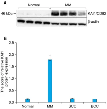
Fig. 1. (A) KAI1/CD82 protein was expressed on malignant melanoma by western blot analysis. (B) The score of relative KAI1/CD82 protein expression. Normal: normal skin, MM:
malignant melanoma, SCC: squamous cell carcinoma, BCC: basal cell carcinoma.
http://dx.doi.org/10.5021/ad.2013.25.4.498
Elevated KAI1 Protein Expression Identified in Malignant Melanoma
Moon-Kyun Cho
Department of Dermatology, Soonchunhyang University College of Medicine, Seoul, Korea
Dear Editor:
KAI1, also known as CD82, is a member of the TM4SF protein family. KAI1 was first discovered during a study on T-cell activation. While recent studies have proposed that TM4SF affects the behavior of cancer cell growth, the exact mechanism has not been described. KAI1/CD82 is found in high levels in the spleen, lung, liver, placenta, kidney and prostate, in moderate levels in the pancreas, skeletal muscle, and thymus, and in low levels in the brain, heart, ovary, stomach, and uterus. However, It was not reported that KAI1/CD82 protein was expressed in normal skin tissue.
Although there have been many studies on the role in the regulation of cell growth, cell-to-cell adhesion and moti- lity, KAI1 and the TM4 family function remains unclear.
An association between KAI1/CD82 and cancer progres- sion was discovered during a study conducted to identify metastasis suppressor genes.
KAI1 expression is reduced in a large number of cancers.
Dong et al.1 reported reduced RNA expression of KAI1 during prostate cancer metastasis by northern blot analy- sis. Transfecting metastatic prostate cancer cells into nude mice resulted in suppression of lung metastasis by KAI1.
Tests using reverse transcriptase-polymerase chain reac- tion and immunohistochemistry have reported reduced levels of CD82 mRNA and protein in thyroid cancer2. A
study by Hinoda et al.3 used immunohistochemical tests to assess the association between KAI1 and the progres- sion of gastric cancer. Northern blot analysis conducted by Guo et al.4 showed similar levels of KAI1 mRNA expression in both normal and cancerous esophagus and similar levels in tissue samples with or without metastasis.
Therefore, metastasis in esophageal and gastric cancer is independent of KAI14. However, a recent study has revea- led increased expression of KAI1 protein in chromophobe renal cell carcinoma5. In light of this, we explored the expression of KAI1 in skin cancers in order to determine whether it downregulates or upregulates the expression of
Letter to the Editor
Vol. 25 No. 4, 2013 499 Fig. 2. Representative immunohistochemistry staining for KAI1/CD82 protein expression in paraffin-embedded malignant melanoma (A: strongly positive immunostaining, ×200), squamous cell carcinoma (B: very weakly positive immunostaining, ×200), basal cell carcinoma (C: very weakly positive immunostaining, ×200) and normal skin (D: no immunostaining, ×100).
skin tumors. The Institutional Review Board of Seoul Soonchunhyang University Hospital reviewed and appro- ved this research protocol, which involved the use of tissue samples.
We experimentally assessed whether loss of KAl1 protein expression in metastatic cancer is a common event in malignant skin cancers or if it occurs only in some tumor types. Based on the aforementioned finding, we chose basal cell carcinoma (BCC), squamous cell carcinoma (SCC) and malignant melanoma (MM) and used western blot analysis in order to determine if KAI1 expression suppresses all cancer types or certain cancer types.
We evaluated the expression of KAI1 protein in MM, SCC and BCC using western blot and immunohistochemistry.
In this study, KAI1, which is a marker of metastatic potentiality in breast cancer, was measured in BCC, SCC and MM using western blot and the immunohistologic method. The results show that KAI1 was hardly expressed in 6 cases of BCC. In 6 cases of SCC, KAI1 was hardly
expressed. Loss of KAI1 protein expression including SCC, BCC in our study corresponded to previous studies results6-8. In the 6 cases of MM, the expression of KAI1 was strongly positive (Fig. 1A). The amount of expression in western blotting is displayed graphically (Fig. 1B). The expression patterns of KAI1 protein in MM were similar with those by Yusenko and Kovacs5. However, different patterns were shown in other types of cancer including prostate, gastric, colon, cervix, breast, bladder, lung, pancreas, liver, and thyroid cancers. An immunohis- tochemical study showed that the staining pattern of KAI1 in normal human skin tissues mirrored those of western blot analysis (Fig. 2).
To the best of our knowledge, KAI1 expression in cutaneous BCCs, SCCs and MMs has not yet been studied.
KAI1 protein expression has been known to be associated with a higher grade of malignant skin tissue. However, the exact function and mechanism of KAI1 during tumor genesis have not been confirmed. While KAI1 is suppres-
Letter to the Editor
500 Ann Dermatol
Received October 19, 2012, Revised December 1, 2012, Accepted for publication December 5, 2012
Corresponding author: Dong-Youn Lee, Department of Dermatology, Samsung Medical Center, 81 Irwon-ro, Gangnam-gu, Seoul 135-710, Korea. Tel:
82-2-3410-3543, Fax: 82-2-3410-3869, E-mail: dylee@ skku.edu
This is an Open Access article distributed under the terms of the Creative Commons Attribution Non-Commercial License (http://
creativecommons.org/licenses/by-nc/3.0) which permits unrestricted non-commercial use, distribution, and reproduction in any medium, provided the original work is properly cited.
sed in specific types of cancer, KAI1 is over expressed in MM. Interestingly, KAI1 was not expressed in cutaneous BCCs, SCCs and normal tissues. More work is needed to characterize the pathway through which KAI1 is over- expressed in MM skin tumor.
ACKNOWLEDGMENT
This work was supported by the Soonchunhyang Univer- sity Research Fund.
REFERENCES
1. Dong JT, Isaacs WB, Barrett JC, Isaacs JT. Genomic organi- zation of the human KAI1 metastasis-suppressor gene. Geno- mics 1997;41:25-32.
2. Chen Z, Mustafa T, Trojanowicz B, Brauckhoff M, Gimm O, Schmutzler C, et al. CD82, and CD63 in thyroid cancer. Int J Mol Med 2004;14:517-527.
3. Hinoda Y, Adachi Y, Takaoka A, Mitsuuchi H, Satoh Y, Itoh F, et al. Decreased expression of the metastasis suppressor
gene KAI1 in gastric cancer. Cancer Lett 1998;129:229-234.
4. Guo XZ, Friess H, Maurer C, Berberat P, Tang WH, Zim- mermann A, et al. KAI1 is unchanged in metastatic and nonmetastatic esophageal and gastric cancers. Cancer Res 1998;58:753-758.
5. Yusenko MV, Kovacs G. Identifying CD82 (KAI1) as a marker for human chromophobe renal cell carcinoma. Histopatho- logy 2009;55:687-695.
6. Geradts J, Maynard R, Birrer MJ, Hendricks D, Abbondanzo SL, Fong KM, et al. Frequent loss of KAI1 expression in squamous and lymphoid neoplasms. An immunohistoche- mical study of archival tissues. Am J Pathol 1999;154:1665- 1671.
7. Farhadieh RD, Smee R, Ow K, Yang JL, Russell PJ, Crouch R, et al. Down-regulation of KAI1/CD82 protein expression in oral cancer correlates with reduced disease free survival and overall patient survival. Cancer Lett 2004;213:91-98.
8. Okochi H, Kato M, Nashiro K, Yoshie O, Miyazono K, Furue M. Expression of tetra-spans transmembrane family (CD9, CD37, CD53, CD63, CD81 and CD82) in normal and neoplastic human keratinocytes: an association of CD9 with alpha 3 beta 1 integrin. Br J Dermatol 1997;137:856-863.
http://dx.doi.org/10.5021/ad.2013.25.4.500
Lichenoid Drug Eruption after Low-Dose Imatinib Mesylate Treatment
Jae-Hyung Lee, Jong-Yoon Chung, Mi-Young Jung, Cho Rok Kim, Ji-Ho Park, Ji-Hye Park
1, Jong-Hee Lee, Joo-Heung Lee, Jun-Mo Yang, Dong-Youn Lee
Department of Dermatology, Samsung Medical Center, Sungkyunkwan University School of Medicine,
1Department of Dermatology, Kangbuk Samsung Hospital, Sungkyunkwan University School of Medicine, Seoul, Korea
Dear Editor:
Imatinib mesylate, a selective antitumor tyrosine kinase inhibitor, has been approved as the first-line therapy for gastrointestinal stromal cell tumor and chronic myeloid leukemia. Lichenoid drug eruption (LDE) is known to
cause uncommon adverse cutaneous reactions of imatinib mesylate, usually appearing with 400 mg or more of imatinib mesylate per day1,2. Here, we present a case of severe LDE after low-dose imatinib mesylate treatment for gastrointestinal stromal cell tumor.