Low Grade Fibromyxoid Sarcoma in Thigh
Bong-Jin Lee, MD, Woo-Sung Park, MD, Jong-Mun Jin, MD, Chang-Won Ha, MD*, Sang-Hoon Lee, MD
Department of Orthopaedic Surgery, *Pathology, Cheju Halla General Hospital, Jeju, Korea
A low grade fibromyxoid sarcoma is a rare soft tissue tumor that has a tendency to develop in the deep soft tissue of young adults and the potential for local recurrence or distant metastasis. There have been several case reports and sporadic reports in the literature. However, only 1 case has been reported in Korea but without a follow-up result. We describe a 54-year-old female patient with a low-grade fi bromyxoid sarcoma of the thigh that had been growing slowly for 34 years. A marginal resection of this tumor was performed. Currently, the patient is doing well without evidence of local recurrence or distant metastasis at 5 years after surgery.
Keywords: Low grade fi bromyxoid sarcoma, Thigh
Case Report
Clinics in Orthopedic Surgery 2009;1:240-243 • doi:10.4055/cios.2009.1.4.240Copyrights © 2009 by Th e Korean Orthopaedic Association Clinics in Orthopedic Surgery • pISSN 2005-291X eISSN 2005-4408 Received May 3, 2008; Accepted September 29, 2008
Correspondence to: Woo-Sung Park, MD
Department of Orthopedic Surgery, Cheju Halla General Hospital, 1963-2 Yeon-dong, Jeju 690-170, Korea
Tel: +82-64-740-5030, Fax: +82-64-743-3110 E-mail: [email protected]
A low grade fi bromyxoid fi brosarcoma is a recently recog- nized, uncommon soft tissue neoplasm. Th e condition was reported originally by Evans in 1987, who subsequently described ten additional cases in 1993. Since then, a few sporadic case reports and series have been reported.1-7) We describe the MRI and histology findings associated with the clinical results of a 5 year follow-up in a 54-year- old female patient with a low-grade fi bromyxoid sarcoma and discuss the diff erential diagnosis with a review of the relevant literature.
CASE REPORT
A 54-year-old female complained of a slowly growing mass in her thigh over a 34 year period. She has a two year history of hemodialysis due to chronic renal failure. A physical examination demonstrated a small melon sized mass on the anterior side of the distal thigh. Th e mass was not tender but rubbery hard and mobile.
The plain radiographs revealed a huge soft tissue mass with irregular calcification in the distal thigh.
On MRI, the tumor was 17 × 11 × 9 cm in size located under the quadriceps muscle. Th e tumor matrix was par- tially calcified and relatively well defined with irregular low signal intensity in the T1 weighted image and heterogeneous low and high signal in the T2 weighted image. There was no abnormal signal of the underlying bony cortex and bone marrow (Fig. 1).
Grossly, the excised tumor was a well circumscribed, lobulated round and firm mass. The cut surface was yellowish-white and fi brous to myxoid without any areas of hemorrhage or necrosis.
Th e optical microscopy examination demonstrated a mass with sharp demarcation, a nodular growth pat- tern and intervening hypocellular collagenous stroma (Fig. 2A). The tumor showed a biphasic pattern with fibrous and myxoid areas. The cells were small, bland, regular, fibroblastic spindle cells with minimal nuclear pleomorphism, low to moderate cellularity, and swirling, whorled growth (Fig. 2B). Th e background matrix ranged from fi bromyxoid to densely fi brous (Fig. 2C).
Immunohistochemically, the tumor cells were dif- fusely and strongly positive for vimentin, but were negative for cytokeratin, smooth muscle actin, S-100 protein and neuron specific enolase. The tumor was identical im- munohistochemically to a low grade fi bromyxoid sarcoma with strong positivity to vimentin (Fig. 2D).
Five years aft er the excision, the patient is alive with no evidence of local recurrence or distant metastasis. Th e
241
Lee et al. Low Grade Fibromyxoid Sarcoma in Th igh Clinics in Orthopedic Surgery • Vol. 1, No. 4, 2009 • www.ecios.org
ranges of knee and hip joint motion were normal and the strength of the quadriceps muscle was good (Fig. 3).
DISCUSSION
Low grade fibromyxoid sarcoma is a bland soft tissue tumor histologically. The tumor has aggressive behavior in that a complete excision does not necessarily prevent the development of local recurrence or distant metastasis many years later.
Among the 27 cases reviewed, 16 showed local recurrences and 9 had lung metastases.1-7) The interval to local recurrence varied from 2 to 13 years (median, 4 years). The interval to the lung metastasis ranged from zero (metastases at presentation) to 45 years (median, 5 years).4,5) Moreover, 1 case became dedifferentiated at 30 years after surgery.4) Therefore, it was believed that a
minimum 5 year follow-up will be needed to discuss the results of this tumor.
A review of the literature revealed the mean age at the first operation to be 33.2 years (range, 6 to 65 years) with 66.7% being male. Frequent sites were the thigh, shoulder, inguinal area and chest wall (Table 1). The preoperative duration varied from 3 days to 22 years and that of the current case (34 years) was the longest.1-7)
The main pathologies to be considered in a differ- ential diagnosis include desmoid fibromatosis, peripheral nerve sheath tumor, myxoid liposarcoma, spindle cell liposarcoma and low grade myxofi brosarcoma.3-6)
Desmoid fi bromatosis is poorly defi ned and shows a greater degree of cellularity, prominent fascicular prolif- eration, and more interstitial collagen fibers. Low grade fibromyxoid sarcoma can be distinguished readily from a nerve sheath tumor by the absence of the S-100 protein Fig. 1. On MRI, the tumor is a 17 × 11 × 9 cm sized mass located under the quadriceps muscle. The tumor matrix is partially calcifi ed and relatively well defi ned one having irregular low signal intensity in the T1 weighted image (A) and heterogeneous low signal and high signal in the T2 weighted image (B-D).
242
Lee et al. Low Grade Fibromyxoid Sarcoma in Th igh Clinics in Orthopedic Surgery • Vol. 1, No. 4, 2009 • www.ecios.org
Fig. 2. Optical microscopy examination demonstrates a mass with sharp demarcation, nodular growth pattern and intervening hypocellular collagenous stroma (A: Hematoxylin and eosin stain, ×100). The tumor shows a biphasic pattern with fi brous and myxoid areas, minimal nuclear pleomorphism, low to moderate cellularity, and a swirling, whorled growth (B: Hematoxylin and eosin stain, ×200). The background matrix ranges from fi bromyxoid to densely fi brous (C: Hematoxylin and eosin stain, ×400). Immunohistochemically, the tumor cells are diffusely and strongly positive for vimentin (D:
Immunohistochemical stain for vimentin, ×1400).
or an association with any nerve, and a lesser degree of waviness of the individual nuclei than a nerve sheath tumor.
Low grade fi bromyxoid sarcoma lacks lipoblast and arborizing vascular vessels, which are characteristic of myxoid liposarcomas. The absence of adipose elements excludes the possibility of spindle cell liposarcoma.
Low grade myxofi brosarcoma diff ers from low grade fibromyxoid sarcoma in that it generally occurs in older adults, is always predominantly myxoid and composed of fusiform cells with hyperchromatic atypical nuclei, and does not metastasize.
Low grade fi bromyxoid sarcoma is a distinct neoplasm with a tendency to recur or metastasize. Histological ly, it is
characterized by the presence of bland spindle cells with a mainly whorled pattern of growth, set in alternating areas with a myxoid or fibrous stroma. Immunohistochemical studies allow a discrimination of this entity from other benign and malignant lesions.
We report a patient with of low grade fi bromyxoid sarcoma, who was treated with a marginal resection, and had no sign of recurrence or metastasis during a 5 year follow-up. However, close observation will be needed for several decades.
ACKNOWLEDGEMENTS
The authors wish to thank Professor Yong-Koo Park,
243
Lee et al. Low Grade Fibromyxoid Sarcoma in Th igh Clinics in Orthopedic Surgery • Vol. 1, No. 4, 2009 • www.ecios.org
Author Location
Evans (1993)4)
Goodlad (1995)6)
Devaney (1990)1)
Fukunaga (1996)5)
Dvornik (1997)2)
Lee (2004)7)Total
Thigh 2 3 1 6
Shoulder 3 1 4
Inguinal
area 2 1 1 4
Chest wall 1 3 4
Buttock 1 1 2
Neck 1 1 2
Popliteal 1 1
Axilla 1 1
Perineum 1 1
Psoas
muscle 1 1
Mesentery 1 1
Total 12 11 1 1 1 1 27
Table 1. The Location of Low Grade Fibromyxoid Sarcomas
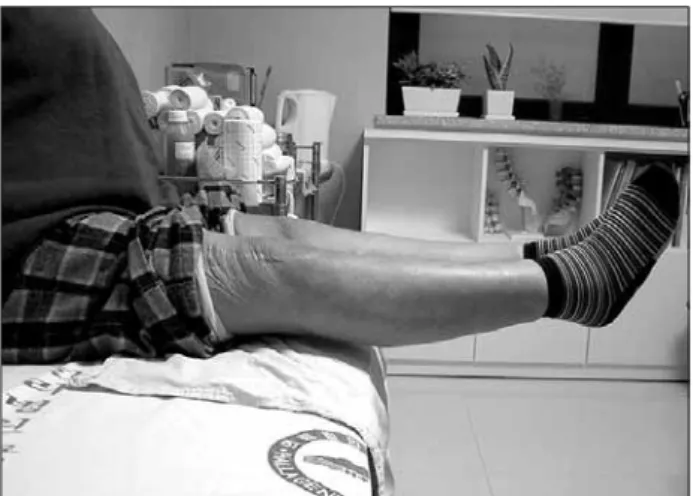
Fig. 3. The patient is still alive with no evidence of local recurrence and distant metastasis fi ve years after excision. The range of motion of the knee joint is full and the strength of the quadriceps muscle is normal.
REFERENCES
1. Devaney DM, Dervan P, O'Neill S, Carney D, Leader M.
Low-grade fibromyxoid sarcoma. Histopathology. 1990;
17(5):463-5.
2. Dvornik G, Barbareschi M, Gallotta P, Dalla Palma P. Low grade fibromyxoid sarcoma. Histopathology. 1997;30(3):
274-6.
3. Evans HL. Low-grade fibromyxoid sarcoma: a report of two metastasizing neoplasms having a deceptively benign appearance. Am J Clin Pathol. 1987;88(5):615-9.
4. Evans HL. Low-grade fi bromyxoid sarcoma: a report of 12
cases. Am J Surg Pathol. 1993;17(6):595-600.
5. Fukunaga M, Ushigome S, Fukunaga N. Low-grade fi bromyxoid sarcoma. Virchows Arch. 1996;429(4-5):301-3.
6. Goodlad JR, Mentzel T, Fletcher CD. Low grade fi bromyxoid sarcoma: clinicopathological analysis of eleven new cases in support of a distinct entity. Histopathology. 1995;26(3):229- 37.
7. Lee SS, Song C, Sun DH, Moon MS. Low grade fi bromyxoid sarcoma in shoulder: one case report. J Korean Bone Joint Tumor Soc. 2004;10(2):130-3.
Kyung Hee University, who confi rmed the diagnosis of this case. None of the authors received any financial support for this study.