D I A B E T E S & M E T A B O L I S M J O U R N A L
This is an Open Access article distributed under the terms of the Creative Commons At- tribution Non-Commercial License (http://creativecommons.org/licenses/by-nc/3.0/) which permits unrestricted non-commercial use, distribution, and reproduction in any medium, provided the original work is properly cited.
Effects of Spironolactone and Losartan on Diabetic Nephropathy in a Type 2 Diabetic Rat Model
Mi Young Lee1,*, Myoung Sook Shim2,*, Bo Hwan Kim1, Soon Won Hong3, Ran Choi1, Eun Young Lee4, Soo Min Nam5, Gun Woo Kim1, Jang Yel Shin1, Young Goo Shin1, Choon Hee Chung1
1Department of Internal Medicine, Yonsei University Wonju College of Medicine, Wonju,
2Department of Internal Medicine, Gangneung Asan Hospital, University of Ulsan College of Medicine, Gangneung,
3Department of Pathology, Yonsei University College of Medicine, Seoul,
4Department of Internal Medicine, Soonchunhyang University College of Medicine, Cheonan,
5Department of Internal Medicine, Sun General Hospital, Daejeon, Korea
Background: While there is an evidence that the anti-inflammatory properties of spironolactone can attenuate proteinuria in type 2 diabetes, its effects on vascular endothelial growth factor (VEGF) expression in diabetic nephropathy have not been clear- ly defined. In this study, we examined the effects of spironolactone, losartan, and a combination of these two drugs on albumin- uria, renal VEGF expression, and inflammatory and oxidative stress markers in a type 2 diabetic rat model.
Methods: Thirty-three Otsuka-Long-Evans-Tokushima-Fatty (OLETF) rats were divided into four groups and treated with dif- ferent medication regimens from weeks 25 to 50; OLETF diabetic controls (n=5), spironolactone-treated (n=10), losartan-treat- ed (n=9), and combination of spironolactone- and losartan-treated (n=9).
Results: At week 50, the albumin-to-creatinine ratio was significantly decreased in the losartan and combination groups com- pared to the control OLETF group. No decrease was detected in the spironolactone group. There was a significant reduction in renal VEGF, transforming growth factor (TGF)-β, and type IV collagen mRNA levels in the spironolactone- and combination regimen-treated groups. Twenty-four hour urine monocyte chemotactic protein-1 levels were comparable in all four groups but did show a decreasing trend in the losartan and combination regimen groups. Twenty-four hour urine malondialdehyde levels were significantly decreased in the spironolactone- and combination regimen-treated groups.
Conclusion: These results suggest that losartan alone and a combined regimen of spironolactone and losartan could ameliorate albuninuria by reducing renal VEGF expression. Also, simultaneous treatment with spironolactone and losartan may have pro- tective effects against diabetic nephropathy by decreasing TGF-β and type IV collagen expression and by reducing oxidative stress in a type 2 diabetic rat model.
Keywords: Diabetic nephropathy; Losartan; Spironolactone; Vascular endothelial growth factor
Corresponding author: Choon Hee Chung
Department of Internal Medicine, Yonsei University Wonju College of Medicine, 162 Ilsan-dong, Wonju 220-701, Korea
E-mail: [email protected]
Received: Jul. 1, 2010; Accepted: Oct. 26, 2010
INTRODUCTION
It is well known that the renin-angiotensin-aldosterone (RAA) system is an important pathway of progression in cardiovas- cular disease, diabetic nephropathy, and chronic renal disease through a mechanism of inflammation, fibrosis, and necrosis [1,2]. For this reason, angiotensin converting enzyme inhibi-
tor (ACEI) and angiotensin receptor blocker (ARB) are effec- tive in the treatment of chronic heart failure and diabetic ne- phropathy [3-6]. However, long term use of RAA system block- ade therapies have been shown to result in increased plasma aldosterone levels in 40% of patients with diabetic nephropa- thy and 20% of patients with chronic renal failure. This condi- tion is referred to as the aldosterone escape phenomenon [7].
pISSN 2233-6079 · eISSN 2233-6087
The proteinuria-reducing effects of ACEI and ARB are also decreased in these patients [8]. It is expected that a reduction in aldosterone levels alone could be a crucial treatment target in diabetic nephropathy.
Recent data suggests that an aldosterone receptor blocker could reduce proteinuria by decreasing various growth factors [9,10]. It has also been reported that aldosterone receptor block- er treatment can reduce proteinuria in patients with chronic renal disease who did not respond to ACEI therapy [11].
Vascular endothelial growth factor (VEGF), a strong angio- genic factor, is known to play a major role in the neovascular- ization of atherosclerotic plaques and solid cancers [12,13], and in the progression of diabetic nephropathy [14]. Although it is reported that ARB treatment protects against diabetic nephrop- athy by reducing VEGF [15,16], there is no data demonstrat- ing the effect of aldosterone receptor blocker therapy on renal VEGF expression.
In this study, we investigated the effects of losartan, spirono- lactone, and a combination of these two drugs on albuminuria and renal VEGF expression in a type 2 diabetic rat model.
METHODS
Experimental rats
Thirty-three male Otsuka-Long-Evans-Tokushima-Fatty rats (OLETF rats; Otsuka Pharmaceutical, Tokushima, Japan) were divided into four groups and received different medication regimens: spironolactone (50 mg/kg/day), losartan (20 mg/kg/
day), or a combination of the two from weeks 25 to 50. The first group consisted of untreated diabetic OLETF controls (CO; n=5), the second group was spironolactone-treated dia- betic rats (SPR; n=10), the third group was losartan-treated diabetic rats (LO; n=9) and the fourth group was spironolac- tone and losartan combination-treated diabetic rats (COM;
n=10). All groups had access to standard rat chow and drink- ing water, ad libitum. This research protocol was approved by the animal ethics committee of the Yonsei University Wonju College of Medicine (Wonju, Korea).
Basic parameters
Body weights and blood glucose levels (Surestep; Lifescan Inc., Milpitas, CA, USA) were recorded at weeks 15, 30, and 50.
Blood pressure was measured using tail-cuff plethysmography at weeks 30 and 50. Twenty-four hour urines were collected at weeks 15, 30, and 50 for measuring and urine protein levels
(Roche Molecular Biochemicals, Indianapolis, IN, USA) and albumin-creatinine ratio (ACR) values (by ELISA; Shibayagi, Shibukawa, Japan) were determined.
Kidney extraction
At week 50, all experimental rats were sacrificed under anes- thesia by intraperitoneal injection of Zoletil® (30 mg/kg). One kidney was rapidly fixed in 4% paraformaldehyde for 24 hours and paraffin-embedded for immunohistochemical study and histologic examination. The other kidney was flash frozen in liquid nitrogen for subsequent protein and RNA extraction.
Frozen kidneys were stored at -70°C until analyzed by Western blot and real time reverse transcription-polymerase chain reac- tion (RT-PCR) for VEGF, transforming growth factor (TGF)-β, and collagen type IV.
Histologic examination of kidney
Paraffin-embedded kidney tissues were cut into 7 μm sections and stained with periodic acid-Schiff (PAS). A glomerular ma- trix index (GMI) score was measured and the degree of sclero- sis for each glomerulus was graded from 0 to 4 as follows [17]:
grade 0: normal; grade 1: mild sclerosis (less than 25% of glom- erulus); grade 2: moderate sclerosis (25 to 50% of glomerulus);
grade 3: moderate-severe sclerosis (50 to 75% of glomerulus);
grade 4: severe sclerosis (75 to 100% of glomerulus). Ten glom- eruli were scored in each kidney section.
Immunohistochemical staining of VEGF
Tissue sections were affixed onto slides and deparaffinized for immunohistochemical staining. The slides were then trans- ferred to a 10-mmol/L citrate buffer solution (pH 6.0), washed with distilled water and blocked with 0.05% H2O2-methanol for 15 minutes. A 1:1,000 dilution of anti-VEGF monoclonal antibody (Santa Cruz Biotechnology Inc., Santa Cruz, CA, USA) was applied and slides were incubated at room tempera- ture. A biotinylated secondary antibody from a rat ABC stain- ing kit (Santa Cruz Biotechnology Inc.) was used followed by incubation in a horseradish peroxidase Avidin-Biotin Com- plex solution (ABC reagent). The slides were then incubated in a peroxidase substrate containing 0.05% 3,3ʹ-diaminoben- zidine tetrahydrochloride (DAB). Stained sections were viewed with a light microscope equipped with a charge-coupled de- vice camera (Pulnix America Inc., Sunnyvale, CA, USA) and glomerular VEGF optical densities were measured by image analysis.
Real time RT-PCR
Total RNA was extracted from frozen kidney tissues using TRIzol® LS (GIBCO-BRL, Grand Island, NY, USA) and then reverse transcribed into cDNA using oligo-(dT) primers (Pro- mega, Madison, WI, USA). Real time RT-PCR was performed using a SYBR Green RT-PCR kit (Qiagen, Valencia, CA, USA) and analyzed with a Rotor-Gene RG-3000 cycler (Corbett Re- search, Mortlake, NSW, Australia). Primer oligonucleotide se- quences for VEGF, TGF-β, collagen type IV and GAPDH are as follows:
VEGF forward: 5ʹ-GTATATCTTCAAGCCGTCCTGT- GTG-3ʹ, VEGF reverse: 5ʹ-GATCCGCATGATCTGCA TAGTGAC-3ʹ, TGF-β forward: 5ʹ-TGAGTGGCTGT CTTTTGACG-3ʹ, TGF- β reverse: 5ʹ-TGGGACTGAT CCCATTGATT-3ʹ, Collagen type IV forward: 5ʹ-CCA GGATTCCAAGGTCAGAA-3ʹ, Collagen type IV reverse:
5ʹ-CCCTGGTTCTCCTTTGATGA-3ʹ, GAPDH forward:
5ʹ-TCAGGTCATCACTATCGGCAATG-3ʹ, GAPDH re- verse: 5ʹ-GGAATTGAATGTAGTTTCATGGATGC-3ʹ Real-time RT-PCR amplification was performed with one cycle at 95°C for 10 minutes followed by 40 cycles that consist- ed of denaturation for 15 seconds at 94°C, annealing for 30 seconds at 58°C and extension for 30 seconds at 72°C. After rinse and melting processes, fluorescent products were detect- ed during the 92°C extension cycle. The cycle threshold (∆Ct= CtVEGF-Ctβ-actin) of each sample was calculated and the relative change ratio determined using an mRNA ratio of VEGF/
β-actin.
Western blot analysis
The cortex of each kidney was homogenized in RIPA buffer, incubated on ice for 20 minutes and centrifuged at 15,000 rpm to remove cellular debris. 10 µg samples of protein lysate were electrophoresed on 10% SDS-PAGE gels at 100 V and trans- ferred onto polyvinylidene fluoride (PVDF) membranes for 1 hour at 280 mA in a Tris-based buffer. Non-specific binding sites were blocked with a 5% non-fat dried milk solution for 1 hour. Membranes were incubated overnight with anti-rat β-actin antibody (1:2,000 dilution; Cell Signaling Technology Inc., Beverly, MA, USA) or VEGF antibody (1:1,000 dilution;
R&D System, Minneapolis, MN, USA) followed by incubation with anti-rabbit IgG or anti-goat IgG HRP antibodies. Signal was visualized using the ELC Western Blotting Analysis Sys- tem (Amersham Biosciences, Buckinghamshire, UK).
Monocyte chemotactic protein-1 (MCP-1) and malondialdehyde (MDA)
Twenty-four hour urine MCP-1 levels were measured by quan- titative sandwich ELISA (Biosource Inc., Camarillo, CA, USA) and used as an inflammatory marker. Urine MDA levels were measured by a rapid and sensitive fluorometric HPLC method and used as an oxidative stress marker [18].
Statistical analyses
All data are presented as mean±standard deviation. The data were analyzed statistically using one-way ANOVA and the Tukey test (multiple comparisons). All analyses were performed using a Windows-based, SPSS statistical package version 12.0 (SPSS Inc., Chicago, IL, USA). P<0.05 was considered to be statistically significant.
RESULTS
Basic parameters of experimental rats
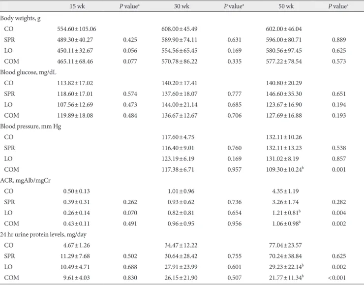
There were no significant differences in body weights and plas- ma glucose levels at each time point. However, blood pressure levels of the COM group at week 50 were significantly lower than that of the CO group (Table 1). At weeks 15 and 30, there were no differences in 24-hour urine protein levels and ACR among all groups. At week 50, the LO and COM groups showed significant decreases in 24-hour urine protein levels and ACR, but the SPR group did not show reduced proteinuria and ACR when compared to the CO group (Table 1).
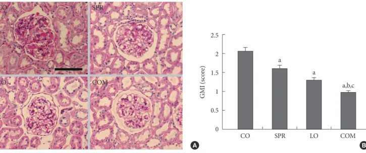
PAS staining of glomeruli and GMI score
Glomerular mesangial expansion was observed in the CO group compared to the other groups in PAS staining of glom- eruli (Fig. 1A). The GMI scores were significantly decreased in all medicated groups compared to the CO group. Also, the COM group showed a marked decrease in GMI scores com- pared to the SPR and LO groups (Fig. 1B).
Analysis of renal VEGF expression
Glomerular VEGF immunohistology revealed darker staining in the CO group compared to all of the medication treated groups (data not shown). The optical density of immunohisto- chemical staining for VEGF in the LO and COM groups was significantly decreased compared to the CO group. The SPR group showed no appreciable difference when compared with the CO group (Fig. 2A). Real time RT-PCR analysis revealed
Table 1. Clinical characteristics of experimental rats
15 wk P valuea 30 wk P valuea 50 wk P valuea
Body weights, g
CO 554.60±105.06 608.00±45.49 602.00±46.04
SPR 489.30±40.27 0.425 589.90±74.11 0.631 596.00±80.71 0.889
LO 450.11±32.67 0.056 554.56±65.45 0.169 580.56±97.45 0.625
COM 465.11±68.46 0.077 570.78±86.22 0.335 577.22±78.54 0.573
Blood glucose, mg/dL
CO 113.82±17.02 140.20±17.41 140.80±20.29
SPR 118.60±17.01 0.574 137.60±18.07 0.777 146.60±35.30 0.651
LO 107.56±12.69 0.473 144.00±21.14 0.685 123.67±16.90 0.194
COM 119.89±18.08 0.484 136.67±12.67 0.706 127.69±16.88 0.193
Blood pressure, mm Hg
CO 117.60±4.75 132.11±10.26
SPR 116.40±9.01 0.760 132.11±13.23 0.538
LO 123.19±6.19 0.169 131.02±8.19 0.857
COM 117.38±6.71 0.957 109.30±10.24b 0.001
ACR, mgAlb/mgCr
CO 0.50±0.13 1.01±0.96 4.35±1.19
SPR 0.39±0.31 0.262 0.93±0.62 0.736 3.26±1.74 0.282
LO 0.26±0.14 0.070 0.82±0.81 0.654 1.21±0.81b 0.004
COM 0.43±0.11 0.491 0.96±0.95 0.956 1.06±0.98b 0.002
24 hr urine protein levels, mg/day
CO 4.67±1.26 34.47±12.22 77.04±23.57
SPR 11.29±7.68 0.502 30.64±28.42 0.755 70.24±38.84 0.625
LO 10.49±4.71 0.688 27.91±23.99 0.601 29.23±22.14b 0.002
COM 9.61±4.03 0.830 26.15±21.90 0.507 21.77±11.34b <0.001
Data are presented by mean±standard deviation.
CO, control Otsuka-Long-Evans-Tokushima-Fatty (OLETF) group; SPR, spironolactone-treated OLETF group; LO, losartan-treated OLETF group; COM, spironolactone- and losartan-treated OLETF group; ACR, albumin-creatinine ratio.
aP value vs. CO group, bP<0.05 compared with CO group.
that VEGF mRNA expression was 0.35-fold in the SPR group, 0.67-fold in the LO group, and 0.22-fold in the COM group when compared to the CO group (Fig. 2B). Western blot anal- ysis, however, did not show any significant differences be- tween the groups (Fig. 2C).
Expression of TGF-β, type IV collagen
Real time RT-PCR analysis showed a significant decrease in TGF-β and type IV collagen mRNA expression in the SPR and COM groups compared to the CO group (Fig. 3). The SPR and COM groups also showed lower TGF-β mRNA expression than the LO group (Fig. 3).
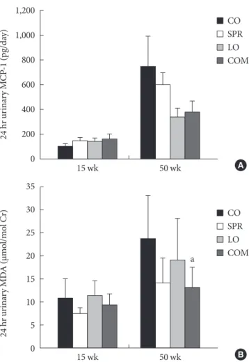
MCP-1 and MDA
At weeks 15 and 50, 24-hour urine MCP-1 levels were similar in all four groups but exhibited a decreasing trend in the LO and COM groups (Fig. 4A). At week 50, MDA levels were sig- nificantly decreased in the COM group compared to the CO group (Fig. 4B).
DISCUSSION
Our data shows that losartan (ARB) alone or a combination treatment consisting of losartan and spironolactone (aldoste- rone receptor blocker) can reduce proteinuria by reducing re-
Fig. 2. Analysis of renal vascular endothelial growth factor (VEGF) expression. (A) Optical densities of immunohistochemi- cal staining for VEGF. (B) VEGF mRNA expression by real-time RT-PCR, and (C) Western blot for renal VEGF expression. Opti- cal densities of glomerular VEGF in the LO and COM groups were significantly lower than those of the CO and SPR groups.
Quantitative analysis revealed that expression of VEGF mRNA was 0.35-fold in the SPR group, 0.67-fold in the LO group, and 0.22-fold in the COM group when compared with the CO group.
Western blot did not show any differences among groups. CO, control Otsuka-Long-Evans-Tokushima-Fatty (OLETF) group;
SPR, spironolactone-treated OLETF group; LO, losartan-treated OLETF group; COM, spironolactone- and losartan-treated OLETF group. aP<0.05 compared with CO group.
Fig. 1. PAS staining of glomeruli (A) and glomerular matrix indices (GMI) (B). PAS staining of glomeruli showed marked me- sangial expansion and sclerosis in the CO group compared to other groups. Compared with the CO group, GMI scores were sig- nificantly decreased in the SPR, LO, and COM groups. CO, control Otsuka-Long-Evans-Tokushima-Fatty (OLETF) group; SPR, spironolactone-treated OLETF group; LO, losartan-treated OLETF group; COM, spironolactone- and losartan-treated OLETF group. aP<0.05 compared with CO group, bP<0.05 compared with SPR group, cP<0.05 compared with LO group. Scale bar, 100 μm, ×400.
CO SPR
LO COM
A B
CO SPR LO COM
2.5 2 1.5 1 0.5 0
GMI (score) a
a
a,b,c
CO SPR LO COM C
1 0.9 0.8 0.7 0.6 0.5 0.4 0.3 0.2 0.1 0
VEGF/beta actin ratio
CO SPR LO COM A
180 160 140 120 100 80 60 40 20 0
VEGF optical density
a a
CO SPR LO COM B
1.2 1 0.8 0.6 0.4 0.2 VEGF/GAPDH (relative changes) 0
a
a
nal VEGF expression in a type 2 diabetic rat model.
Diabetic nephropathy is the most common cause of end- stage renal disease (ESRD). Since diabetic ESRD patients are more prone to cardiovascular mortality than other ESRD pa- tients, early identification of diabetic nephropathy and prompt renoprotective treatment are critical for the prevention of end organ damage from diabetic nephropathy [19].
It is well known that therapies with ACEI and ARB can re- tard the progression of diabetic nephropathy, chronic heart failure and chronic renal failure by reducing blood pressure and by decreasing inflammatory and sclerosing effects [3-6].
Recent data indicates that spironolactone, an aldosterone re- ceptor blocker, can reduce proteinuria by providing anti-in- flammatory protection and by decreasing oxidative stress [10, 20]. In our study, losartan- and the combination regimen-treat- ed rat groups showed significant decreases in 24-hour urine protein levels and ACR while the spironolactone monotherapy group did not show these significant reductions. This discrep- ancy may be explained by the differences in treatment initia- tion time periods. Recent studies reported that treatment with aldosterone receptor blockers in the early stages of nephropa- thy may reduce proteinuria but has no beneficial effects at later stages of the disease [21,22]. Since we administered spirono- lactone treatment as late as week 25, its protective actions
against diabetic nephropathy could have been inadequate compared to other studies that initiated treatment at earlier stages [10]. Another possible explanation is the different ef- fects of spironolactone on hemodynamics and the fibrosis process. We observed decreased mesangial expansion and sclerosis in both the SPR and COM groups, which is consis- tent with results from previous studies [23,24]. However, there were no differences in blood pressure between the groups. It could be explained that spironolactone can act on diabetic ne- phropathy by non-hemodynamic mechanisms [25].
1.2 1 0.8 0.6 0.4 0.2 0
Relative changes
TGF-β a,b a,b
Type IV collagen a,b
a,b
CO SPR LO COM
Fig. 3. Transforming growth factor (TGF)-β and collagen type IV mRNA expression by real time RT-PCR. Compared with the CO and LO groups, TGF-β and collagen type IV mRNA expression of the SPR and COM groups were significantly de- creased. CO, control Otsuka-Long-Evans-Tokushima-Fatty (OLETF) group; SPR, spironolactone-treated OLETF group;
LO, losartan-treated OLETF group; COM, spironolactone- and losartan-treated OLETF group. aP<0.05 compared with CO group, bP<0.05 compared with LO group.
1,200 1,000 800 600 400 200 0
24 hr urinary MCP-1 (pg/day)
15 wk 50 wk
CO SPR LO COM
A 35
30 25 20 15 10 5 0
24 hr urinary MDA (μmol/mol Cr)
15 wk 50 wk
CO SPR LO a COM
B
Fig. 4. Twenty-four hour urinary monocyte chemotactic pro- tein-1 (MCP-1) (A) and malondialdehyde (MDA) (B) levels.
MCP-1 levels were not different among the four groups, but showed a decreasing trend in the LO and COM groups. MDA levels were significantly decreased in the COM group com- pared to the CO group. CO, control Otsuka-Long-Evans- Tokushima-Fatty (OLETF) group; SPR, spironolactone-treat- ed OLETF group; LO, losartan-treated OLETF group; COM, spironolactone- and losartan-treated OLETF group. aP<0.05 compared with CO group.
In diabetic nephropathy, VEGF expression may be increased by various growth factors including platelet derived growth factor and the accumulation of hyperglycemia-induced, ad- vanced glycation end products [26,27]. Recent studies re- vealed that ACEI and ARB can decrease proteinuria by reduc- ing VEGF expression in type 2 diabetic rats [15,28]. Our data also suggests that a combination therapy with losartan and spironolactone may affect diabetic nephropathy by reducing VEGF expression despite incidental, disparate results between VEGF mRNA expression, immunohistochemical staining, and Western blot analysis. The discrepancies between mRNA expression and protein levels may be due to the interpretation of our data, which was in part based on inconsistent tissue sampling. While we could analyze individual glomeruli by im- munohistochemical staining, we could not isolate glomeruli from tubules and interstitial tissue when extracting total RNA and protein for real time RT-PCR and Western blot analysis.
Activation of the renin-angiotensin-aldosterone (RAA) sys- tem causes increased production of reactive oxygen species through activation of the NADPH oxidase enzymatic complex in numerous tissues, including the kidney [29,30]. Angioten- sin and other cytokines can also cause diabetic nephropathy through inflammatory mechanisms [29,30]. Blockade of the RAA system may have protective effects against diabetic ne- phropathy through anti-oxidative and anti-inflammatory mechanisms [20]. In our study, only a combination therapy of losartan and spironolactone reduced MDA levels. MCP-1 lev- els showed a slight decreasing trend in the combination regi- men treated group but this was not statistically significant.
These results indirectly explain that spironolactone and losar- tan combination therapy may be an effective treatment regi- men for diabetic nephropathy by reducing oxidative stress.
In conclusion, we suggest that ARB therapy in combination with ARB and aldosterone receptor blocker therapies may have protective effects against diabetic nephropathy by reducing VEGF expression. We also propose that combination treat- ments could reduce proteinuria by anti-oxidative mechanisms and by decreasing TGF-β and type IV collagen expression.
ACKNOWLEDGMENT
This study was supported in part by a research fund from The Korean Society of Hypertension (2006).
REFERENCES
1. Brown NJ, Vaughan DE, Fogo AB. Aldosterone and PAI-1: im- plications for renal injury. J Nephrol 2002;15:230-5.
2. Stier CT Jr, Chander PN, Rocha R. Aldosterone as a mediator in cardiovascular injury. Cardiol Rev 2002;10:97-107.
3. Pfeffer MA, Braunwald E, Moye LA, Basta L, Brown EJ Jr, Cuddy TE, Davis BR, Geltman EM, Goldman S, Flaker GC, Klein M, Lamas GA, Packer M, Rouleau JL, Rutherford J, Wertheimer JH, Morton Hawkins C; SAVE Investigators. Ef- fect of captopril on mortality and morbidity in patients with left ventricular dysfunction after myocardial infarction. Re- sults of the survival and ventricular enlargement trial. The SAVE Investigators. N Engl J Med 1992;327:669-77.
4. Schieffer B, Wirger A, Meybrunn M, Seitz S, Holtz J, Riede UN, Drexler H. Comparative effects of chronic angiotensin-con- verting enzyme inhibition and angiotensin II type 1 receptor blockade on cardiac remodeling after myocardial infarction in the rat. Circulation 1994;89:2273-82.
5. Brenner BM, Cooper ME, de Zeeuw D, Keane WF, Mitch WE, Parving HH, Remuzzi G, Snapinn SM, Zhang Z, Shahinfar S;
RENAAL Study Investigators. Effects of losartan on renal and cardiovascular outcomes in patients with type 2 diabetes and nephropathy. N Engl J Med 2001;345:861-9.
6. Parving HH, Lehnert H, Brochner-Mortensen J, Gomis R, An- dersen S, Arner P; Irbesartan in Patients with Type 2 Diabetes and Microalbuminuria Study Group. The effect of irbesartan on the development of diabetic nephropathy in patients with type 2 diabetes. N Engl J Med 2001;345:870-8.
7. Cicoira M, Zanolla L, Franceschini L, Rossi A, Golia G, Zeni P, Caruso B, Zardini P. Relation of aldosterone “escape” despite angiotensin-converting enzyme inhibitor administration to impaired exercise capacity in chronic congestive heart failure secondary to ischemic or idiopathic dilated cardiomyopathy.
Am J Cardiol 2002;89:403-7.
8. Sato A, Hayashi K, Naruse M, Saruta T. Effectiveness of aldo- sterone blockade in patients with diabetic nephropathy. Hy- pertension 2003;41:64-8.
9. Guo C, Martinez-Vasquez D, Mendez GP, Toniolo MF, Yao TM, Oestreicher EM, Kikuchi T, Lapointe N, Pojoga L, Wil- liams GH, Ricchiuti V, Adler GK. Mineralocorticoid receptor antagonist reduces renal injury in rodent models of types 1 and 2 diabetes mellitus. Endocrinology 2006;147:5363-73.
10. Han KH, Kang YS, Han SY, Jee YH, Lee MH, Han JY, Kim HK, Kim YS, Cha DR. Spironolactone ameliorates renal injury and
connective tissue growth factor expression in type II diabetic rats. Kidney Int 2006;70:111-20.
11. Sato A, Hayashi K, Saruta T. Antiproteinuric effects of miner- alocorticoid receptor blockade in patients with chronic renal disease. Am J Hypertens 2005;18:44-9.
12. Jain RK. Tumor angiogenesis and accessibility: role of vascular endothelial growth factor. Semin Oncol 2002;29(6 Suppl 16):3-9.
13. Moehler TM, Ho AD, Goldschmidt H, Barlogie B. Angiogene- sis in hematologic malignancies. Crit Rev Oncol Hematol 2003;
45:227-44.
14. The EUCLID Study Group. Randomised placebo-controlled trial of lisinopril in normotensive patients with insulin-depen- dent diabetes and normoalbuminuria or microalbuminuria.
Lancet 1997;349:1787-92.
15. Lee EY, Shim MS, Kim MJ, Hong SY, Shin YG, Chung CH.
Angiotensin II receptor blocker attenuates overexpression of vascular endothelial growth factor in diabetic podocytes. Exp Mol Med 2004;36:65-70.
16. Nagisa Y, Shintani A, Nakagawa S. The angiotensin II receptor antagonist candesartan cilexetil (TCV-116) ameliorates retinal disorders in rats. Diabetologia 2001;44:883-8.
17. Tojo A, Kimura K, Nanba S, Matsuoka H, Sugimoto T. Varia- tions in renal arteriolar diameter in deoxycorticosterone ace- tate-salt hypertensive rats. A microvascular cast study. Vir- chows Arch A Pathol Anat Histopathol 1990;417:389-93.
18. Korchazhkina O, Exley C, Andrew Spencer S. Measurement by reversed-phase high-performance liquid chromatography of malondialdehyde in normal human urine following deriva- tisation with 2,4-dinitrophenylhydrazine. J Chromatogr B An- alyt Technol Biomed Life Sci 2003;794:353-62.
19. Remuzzi G, Schieppati A, Ruggenenti P. Clinical practice. Ne- phropathy in patients with type 2 diabetes. N Engl J Med 2002;
346:1145-51.
20. Han SY, Kim CH, Kim HS, Jee YH, Song HK, Lee MH, Han KH, Kim HK, Kang YS, Han JY, Kim YS, Cha DR. Spironolac- tone prevents diabetic nephropathy through an anti-inflam- matory mechanism in type 2 diabetic rats. J Am Soc Nephrol 2006;17:1362-72.
21. Nakhoul F, Khankin E, Yaccob A, Kawachi H, Karram T, Awaad H, Nakhoul N, Hoffman A, Abassi Z. Eplerenone potentiates the antiproteinuric effects of enalapril in experimental nephrot- ic syndrome. Am J Physiol Renal Physiol 2008;294:F628-37.
22. Piecha G, Koleganova N, Gross ML, Geldyyev A, Adamczak
M, Ritz E. Regression of glomerulosclerosis in subtotally ne- phrectomized rats: effects of monotherapy with losartan, spi- ronolactone, and their combination. Am J Physiol Renal Physi- ol 2008;295:F137-44.
23. Perez-Rojas J, Blanco JA, Cruz C, Trujillo J, Vaidya VS, Uribe N, Bonventre JV, Gamba G, Bobadilla NA. Mineralocorticoid receptor blockade confers renoprotection in preexisting chron- ic cyclosporine nephrotoxicity. Am J Physiol Renal Physiol 2007;292:F131-9.
24. Bobadilla NA, Gamba G. New insights into the pathophysiol- ogy of cyclosporine nephrotoxicity: a role of aldosterone. Am J Physiol Renal Physiol 2007;293:F2-9.
25. Kelly DJ, Aaltonen P, Cox AJ, Rumble JR, Langham R, Panagi- otopoulos S, Jerums G, Holthofer H, Gilbert RE. Expression of the slit-diaphragm protein, nephrin, in experimental diabetic nephropathy: differing effects of anti-proteinuric therapies.
Nephrol Dial Transplant 2002;17:1327-32.
26. Williams B, Baker AQ, Gallacher B, Lodwick D. Angiotensin II increases vascular permeability factor gene expression by human vascular smooth muscle cells. Hypertension 1995;25:913-7.
27. Wendt TM, Tanji N, Guo J, Kislinger TR, Qu W, Lu Y, Bucci- arelli LG, Rong LL, Moser B, Markowitz GS, Stein G, Bierhaus A, Liliensiek B, Arnold B, Nawroth PP, Stern DM, D’Agati VD, Schmidt AM. RAGE drives the development of glomerulo- sclerosis and implicates podocyte activation in the pathogene- sis of diabetic nephropathy. Am J Pathol 2003;162:1123-37.
28. Kakizawa H, Itoh Y, Imamura S, Matsumoto T, Ishiwata Y, Ono Y, Yamamoto K, Kato T, Hayakawa N, Oda N, Goto Y, Goto Y, Nagasaka A, Senda T, Itoh M. Possible role of VEGF in the progression of kidney disease in streptozotocin (STZ)-in- duced diabetic rats: effects of an ACE inhibitor and an angio- tensin II receptor antagonist. Horm Metab Res 2004;36:458-64.
29. Blendea MC, Jacobs D, Stump CS, McFarlane SI, Ogrin C, Bahtyiar G, Stas S, Kumar P, Sha Q, Ferrario CM, Sowers JR.
Abrogation of oxidative stress improves insulin sensitivity in the Ren-2 rat model of tissue angiotensin II overexpression.
Am J Physiol Endocrinol Metab 2005;288:E353-9.
30. Kelly DJ, Wilkinson-Berka JL, Ricardo SD, Cox AJ, Gilbert RE.
Progression of tubulointerstitial injury by osteopontin-induced macrophage recruitment in advanced diabetic nephropathy of transgenic (mRen-2)27 rats. Nephrol Dial Transplant 2002;17:
985-91.