서 론
당뇨병은 심부전을 일으키는 중요한 위험인자로 간주되 고 있으며, Framingham Heart Study 결과에서 나타났듯이 당뇨병환자는 연령보정 대조군에 비해 남자에서는 2배, 여 자에서는 5배 심부전 발생이 증가한다1). 당뇨병성 심근병증 (diabetic cardiomyopathy)은 고혈압이나 관상동맥질환 여 부와 무관하게 당뇨병환자에서 발생하는 심실기능저하로 정 의할 수 있으며2,3) 심장초음파로 확장기 심실기능저하 (diastolic cardiac dysfunction)와 심근비대를 관찰할 수 있 다. 또한 당뇨병성 심근병증은 기능적 이상과 함께 심근세 포 (cardiomyocyte)의 염증 반응, 세포 사멸 및 심근의 섬유 화로 인한 형태학적 이상을 동반한다4,5). 당뇨병성 심근병증 의 발생 기전으로 몇 가지 가설이 제안되었으며 이는 (1) 칼 슘 항상성의 이상으로 인한 심근 수축력의 저하, (2) 레닌- 안지오텐신계의 활성화로 인한 심근세포 사멸과 심근의 섬 유화, (3) 산화 스트레스를 통한 심근세포 사멸, (4) 지방산 산화의 증가로 인한 심근효율 (cardiac efficiency)의 감소, (5) 미토콘드리아 기능이상 등으로 요약할 수 있다3). 본 종
설에서는 이 가운데 최근 많은 연구가 이루어지고 있는 심 장의 기질 대사 (substrate metabolism)의 변화에 따른 미토 콘드리아 기능이상에 대해 정리하고자 한다.
기질 대사의 변화; 포도당 산화의 감소와 지방산 이용 증가
수축과 이완을 반복하는 사람의 심장은 매일 3.5~5 kg의 ATP를 생산하고 소비하는데 이를 위해서 지속적인 영양소와 산소 공급이 필수적이다. 이에 따라 심근세포는 세포 내 ATP 생성 소기관인 미토콘드리아를 풍부하게 갖고 있으며 이는 전체 세포 용적의 40%에 달한다. 생리적으로 심장은 지방산 의 산화를 통해 60~70%의 ATP를 생성하며, 포도당과 유산 으로부터 30~40%의 ATP를 생산한다. 한편 인슐린저항성을 동반한 비만이나 당뇨병이 발생하면 심장은 좀 더 많은 지방 산을 ATP 생성에 사용하는 한편 포도당 산화는 감소된다6,7). 4주령의 db/db 생쥐와 ob/ob 생쥐 심장에서 이미 지방산 산 화의 증가와 포도당 산화 감소를 관찰할 수 있다6). 이와 더불 어 심근세포에서의 지방산 섭취 증가에 따른 심근의 지질 축
당뇨병성 심근병증과 미토콘드리아 기능 이상
중앙대학교 의과대학 내과학교실
안지현․김재택
Mitochondrial Dysfunction in Diabetic Cardiomyopathy
Jihyun Ahn, Jaetaek Kim
Division of Endocrinology and Metabolism, Department of Internal Medicine, Chung-Ang University College of Medicine
Abstract
Metabolic syndrome and diabetes are associated with increased risk of cardiac dysfunction independently of underlying coronary artery disease. The underlying pathogenesis is partially understood but accumulating evidence suggests that alterations of cardiac energy metabolism might contribute to the development of contractile dysfunction. Recent findings suggest that myocardial mitochondrial dysfunction may play an important role in the pathogenesis of cardiac contractile dysfunction in type 2 diabetes. This review is focused on evaluating mechanisms for the mitochondrial abnormalities that may be involved in the development and progression of cardiac dysfunction in diabetes. (KOREAN DIABETES J 32:467-473, 2008) Key Words: Cardiac Dysfunction, Diabetes, Energy Metabolism, Mitochondria
적이 일어나 지질독성 (lipotoxicity)이 발생하며 이에 따른 부산물인 세라마이드 (ceramide) 생성으로 인해 심근세포의 아포프토시스 (apoptosis)를 초래한다8-10).
당뇨병 심장에서 기질 대사의 변화가 일어나는 기전으로 심 근세포에서의 인슐린 신호전달 장애, 심근세포로 지방산 전달 의 증가와 함께 peroxisome proliferator-activated receptor-α (PPAR-α) 전사 신호전달계의 활성화가 중요하다11-13). 리간드 활성화 전사인자 (ligand-activated transcriptional factor)인 PPAR-α는 생체 내 리간드인 지방산과 결합, 활성화되어 retinoid X 수용체와 복합체를 형성하여 지방산 대사를 조 절하는 단백질의 유전자 프로모터 부위에 결합한다. 따라서 PPAR-α는 전사 수준 (transcriptional level)에서 지방산 대사 를 조절하게 되며, 지방산 대사가 활발히 일어나는 심장에서 많은 양의 PPAR-α가 발현되는 것을 관찰할 수 있다14). 실제 로 PPAR-α는 심장의 지방산 대사 각 단계에서 담당 유전자 를 조절하는데, PPAR-α 녹아웃 (PPAR-α-/-) 생쥐는 심근세 포로의 지방산 섭취를 담당하는 CD36의 표현, 미토콘드리 아로 지방산을 운반하는 carnitine palmitoyl transferase-1, β-산화에 관련된 acyl CoA dehydrogenase와 hydroxy acyl CoA dehydrogenase 유전자가 모두 감소되어 있음이 알려
졌다15,16). 한편 PPAR-α를 과발현시킨 생쥐는 제2형 당뇨병
모델 생쥐와 비슷한 심장의 표현형을 나타내는데12) 이 생쥐 의 심근에서 pyruvate dehydrogenase kinase 4 (PDK4)가 증가되어 있으며 이는 pyruvate dehydrogenase (PDH)를 억 제하여 포도당 산화를 감소시킨다. 하지만 당뇨병 심장에서 지방산 산화의 증가의 기전을 PPAR-α 신호전달계의 활성 화로만 설명할 수는 없다. 최근 db/db 생쥐와 ob/ob 생쥐 심 장을 분석한 보고에서 어린 연령 (4주령)에서는 PPAR-α와 그 조절유전자의 변화가 없었으며, 15주령이 되어야 비로소 증가함을 관찰하였다6). 따라서 4주령에서 관찰된 지방산 산 화의 증가는 PPAR-α에 비의존적인 다른 기전이 있음을 시 사하며, PPAR-α를 통한 신호전달은 오랫동안 당뇨병 상태 가 지속될 때 활성화되어 증가된 지방산 산화를 유지시키는 것으로 이해된다.
증가된 심근 산소소비량 (Myocardial Oxygen Consumption, MVO
2)에 따른
심근효율의 감소
포도당 산화의 경우 산소 한 분자 당 2.58 ATP를 생산하 지만, 지방산 산화의 경우 산소 한 분자를 소비하여 2.33 ATP 밖에 생산하지 못한다. 심장의 스트레스 상황 즉, 저산
소증과 허혈 상태에서는 포도당 산화를 증가시켜 심장을 보 호한다. 따라서 당뇨병 심장에서 발생하는 지방산 산화의 증가는 동일한 ATP 생성을 위해 더 많은 산소가 필요하고 결국 심근 산소소비량 증가를 초래하게 된다17-19). 심근 산소 소비량 증가는 심근효율 (심장의 부하능/심근 산소소비량)의 감소로 이어지며 이러한 심장 상태는 제한된 산소를 소비하 여 ATP를 생성해야 하는 혈역학적인 스트레스 즉, 허혈/재 관류 상황에 매우 취약하여 심기능저하로 이어질 수 있다20). 제1형 당뇨병환자를 대상으로 시행한 양전자 방출 단층촬영 술 (positron emission tomography) 연구에서 심근의 지방산 이용의 증가, 포도당 산화 감소 및 심근 산소소비량의 증가 가 확인되었으며21,22), db/db 생쥐에 허혈 자극을 가했을 때 저하된 심기능과 심근효율은 재관류 시 인슐린과 포도당 용 액을 사용하였을 때 모두 회복되었다23). 당뇨병으로 인한 심근 산소소비량 증가와 심근효율의 감소 기전은 아직 정확 히 알려지지 않았지만 최근 유력한 가설로 미토콘드리아 uncoupling이 제시되었다.
미토콘드리아 Uncoupling
지방산의 β-산화 또는 포도당의 해당과정을 통해 생성된 acetyl-CoA는 미토콘드리아에서 tricarboxylic acid (TCA) 회로를 거치면서 전자수송체인 NADH와 FADH2를 만든다.
미토콘드리아 내벽에는 전자전달환 (electron transport chain) 이 존재하며 복합체 I (complex I)에서 복합체 IV (complex IV)을 통해 단계적으로 전자가 산소 분자로 전달되는 과정 에서 수소 이온이 미토콘드리아 기질 밖으로 나오게 되면서 막 전위차가 형성되며 수소 이온이 F0/F1-ATP synthase (complex V)에 의해 미토콘드리아 기질로 다시 흘러들어갈 때 ADP에서 ATP가 생성되는데 이 과정을 산화 인산화 (oxidative phosphorylation, OXPHOS)라고 한다. 따라서 정상 적으로 미토콘드리아에서의 산소소비량과 ATP 생성은 정확히 짝지어진다(coupling of oxidative phosphorylation). 하지만 일부의 수소 이온은 기질로 유입될 때 ATP 생성을 동반하 지 않으며 (uncoupled respiration), 이 경우 수소 이온은 uncoupling protein (UCP)으로 알려진 미토콘드리아 내막의 통로를 통해 유입된다. 현재까지 UCP 아형은 5가지가 밝 혀져 있다 (UCP1~UCP5)24). UCP1은 갈색지방에서 열발생 에 중요한 역할을 담당하며25) 심장은 UCP2와 UCP3를 표 현하는데 그 기능에 대해서는 아직 충분히 알려져 있지 않
다26,27). 정상적인 미토콘드리아 대사과정에서 일부의 전자
는 유출 (leak)되어 산소를 완전히 환원시키지 못해
superoxide와 같은 활성산소족 (reactive oxygen species, ROS)을 만들게 된다. 이와 같이 미토콘드리아에서의 산화 과정 중 발생한 ROS는 UCP2와 UCP3을 활성화하여 미세 한 uncoupling을 일으켜 ATP 생성에는 영향을 미치지 않으 며 ROS를 해독 (detoxification)시켜 미토콘드리아를 보호할 것으로 생각된다28). 하지만 지방산 산화의 증가로 인해 전 달되는 전자의 양이 많아져 미토콘드리아 내막의 막전위가 일정 역치 이상으로 증가하면 ROS 생성이 증가한다. UCP 단백질은 ROS와 지질 과산화 (lipid peroxidation) 과정의 중 간 생성물인 4-hydroxy-2-nonenal (4-HNE) 등에 의해 활성화
된다29,30). 과다 생성된 ROS는 UCP를 통해 미토콘드리아
uncoupling을 유도하여 미토콘드리아에서의 산소소비량은 증가하지만 ATP 생성은 이에 비례하여 증가하지 못한다.
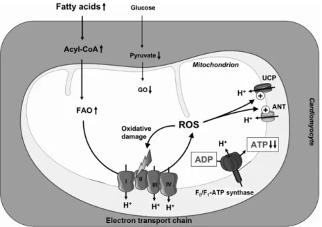
또한 ROS는 OXPHOS에 관여되는 단백질에 직접적인 산 화스트레스를 가하여 ATP 생성에 장애를 초래한다 (Fig.
1). 고지방식이 또는 스트렙토조토신 유발 당뇨병 쥐의 심 장에 UCP 표현이 증가되어 있으며31-33) 사람에서 혈중 지방 산 농도는 심장의 UCP2와 UCP3 표현과 상관관계를 보였 다34). UCP2와 UCP3는 PPAR-α 전사 경로에 의해 표현이
증가한다. UCP2과 UCP3 단백질 표현은 PPAR-α-/- 생쥐의 심장에서 감소되어 있고, UCP3 단백질 표현은 스트렙토조 토신 유발 당뇨병 생쥐에서 증가되었음이 관찰되었다. db/db 생쥐의 심장에서 UCP3 표현은 변화가 없었지만 활성도가 증가되었으며, ex vivo에서 심장을 포도당과 palmitate로 관 류하였을 때 포도당 관류 시와 비교하여 ADP-자극 산소소비 량이 증가하였다35). 반면 UCP3는 포도당을 주된 기질로 사 용하는 허혈성 심장 (ischemic heart)에서 감소되어 있다36). 결국 당뇨병 심장에서 지속적인 UCP 활성화는 심근효율을 감소시켜 심기능저하를 초래할 수 있다. 향후 심장에서 UCP2와 UCP3각각의 역할과 지방산 산화/ROS 생성과 UCP 상호작용에 대해 추가적인 연구가 필요하다. 미토콘드 리아에서 에너지 효율을 감소시키는 또 다른 단백질로 adenine nucleotide translocator (ANT)를 들 수 있다. 설치 류에서 ANT는 두 개의 아형 (ANT1, ANT2)이 있으며 심 장과 골격근에서 표현된다7). 최근 Boudina 등37)은 db/db 생 쥐 심장의 미토콘드리아에서 수소 이온의 기질 내 유입의 부분을 ANT가 담당한다고 보고하였다.
Fig. 1. The proposed mechanism of decreased cardiac efficiency in diabetic heart. Increased serum fatty acid levels and cardiac insulin resistance may lead to increased fatty acid oxidation (FAO) and decreased glucose oxidation (GO). The resulting increase in reducing equivalent delivery to the respiratory chain may increase mitochondrial generation of reactive oxygen species (ROS), which may induce mitochondrial uncoupling by activating uncoupling proteins (UCP) and adenine nucleotide translocator (ANT). This may result in increased mitochondrial oxygen consumption, thereby increasing FAO even further. Increased oxygen consumption in the absence of increased cardiac work caused by a relative energy deficit reduces cardiac efficiency. On the other hand, ROS may also induce oxidative stress to mitochondrial proteins involved in oxidative phosphorylation, thereby reducing ATP synthesis (adapted from reference 44).
미토콘드리아 생성 (Biogenesis)
기존의 미토콘드리아가 성장하거나 자신의 DNA 분열을 통해 증식하는 것을 미토콘드리아 생성으로 정의할 수 있다.
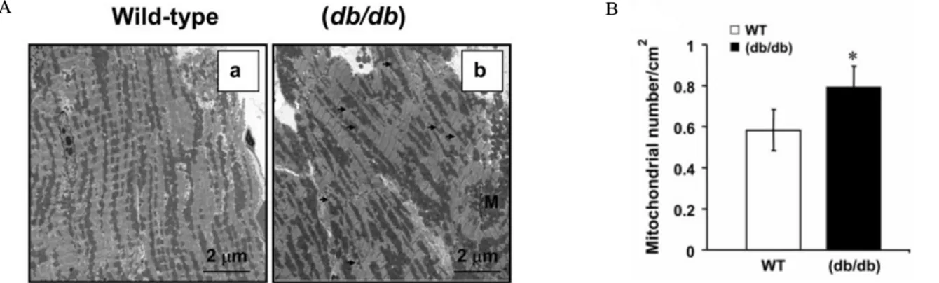
제1형과 제2형 당뇨병 동물모델 심장에서 미토콘드리아 증 식이 관찰되었다38,39). 미토콘드리아 생성의 주요한 조절인 자로 peroxisome proliferator-activated receptor gamma co-activator (PGC-1)을 들 수 있다. PGC-1은 전사인자와의 결합을 통해 전사인자의 전사 활성도를 증가시킨다. PGC-1 의 아형으로 PGC-1α, PGC-β 및 PGC-1-related coactivator 가 밝혀졌으며 PGC-1α와 PGC-1β는 미토콘드리아가 풍부 한 갈색지방과 심장에서 높은 표현을 보인다40,41). PGC-1α 는 PPAR-α 및 PPAR-β와 결합하여 지방산 산화와 관련된 유전자 표현을 증가시키고, 핵수용체 estrogen receptor related receptor (ERR)와 결합하여 포도당 산화를 감소시킨 다. 또한 전사인자 nuclear respiratory factor 1 (NRF-1)과 결합하여 OXPHOS 관련 유전자의 전사를 증가시키고 미토 콘드리아 증식과 DNA의 전사를 촉진하여 미토콘드리아 생 성을 증가시킨다40). db/db 생쥐와 ob/ob 생쥐의 심장에서 미토콘드리아 용적과 DNA 함량이 증가되었지만37,42,43) PGC-1α 표현은 ob/ob 생쥐 심장에서 증가하지 않았으며, 따라서 미토콘드리아 증식은 PGC-1α 비의존적으로 진행됨 을 시사한다38,40). 또한 db/db 생쥐에서 증가된 미토콘드리 아 생성 (Fig. 2)이 PGC-1α의 표현 증가와 관련이 있었지만 OXPHOS 유전자 표현은 증가하지 않은 것을 볼 때 미토콘 드리아 기능이상으로 인해 미토콘드리아 생성이 보상적으로 증가하는 것으로 추정된다37,44). 앞으로 미토콘드리아 생성이 당뇨병 심장에서 지니는 의미에 대한 추가 연구가 필요하다.
결 론
당뇨병성 심근병증의 발생 기전을 설명하기 위해 그 동 안 다양한 모델이 제시되었으며 최근 심장의 미토콘드리아 기능이상에 대한 많은 증거들이 밝혀지고 있다. 심근세포에 서 미토콘드리아 기능이상은 심장의 ATP생성을 저해하여 며 결국 심실기능저하를 초래한다. 즉 미토콘드리아 기질 이용의 변화, 증가된 ROS와 UCP의 활성화를 통해 미토콘 드리아 기능이상이 발생하는데 향후 각각을 표적 (target)으 로 하여 당뇨병성 심근병증의 새로운 치료제 개발을 기대할 수 있을 것이다.
참 고 문 헌
1. Kannel WB, McGee DL: Diabetes and cardiovascular disease. The Framingham study. JAMA 241:2035-8, 1979
2. Rubler S, Dlugash J, Yuceoglu YZ, Kumral T, Branwood AW, Grishman A: New type of cardiomyopathy associated with diabetic glomerulo- sclerosis. Am J Cardiol 30:595-602, 1972
3. Boudina S, Abel ED: Diabetic cardiomyopathy revisited.
Circulation 115:3213-23, 2007
4. Borezuk A, Factor SM: Pathological alterations of the heart in diabetes mellitus. In Chatham JC, Forder JR, McNeill JH, editors. The heart in diabetes. p. 23-40, Norwell, MA: Kluwer Academic Publishers, 1996 5. Asbun J, Villarreal FJ: The pathogenesis of myocardial
fibrosis in the setting of diabetic cardiomyopathy. J A
B
Fig. 2. Increased cardiac mitochondrial proliferation in db/db mice. A. Representative electron micrographs from wild-type (a) and db/db (b) hearts. Magnification ×2000. B. A quantitative analysis of mitochondrial number in each group (wild type or db/db). * P < 0.05 vs. wild type (adapted from reference 37).
Am Coll Cardiol 47:693-700, 2006
6. Buchanan J, Mazumder PK, Hu P, Chakrabarti G, Roberts MW, Yun UJ, Cooksey RC, Litwin SE, Abel ED: Reduced cardiac efficiency and altered substrate metabolism precedes the onset of hyperglycemia and contractile dysfunction in two mouse models of insulin resistance and obesity. Endocrinology 146:5341-9, 2005
7. Mazumder PK, O'Neill BT, Roberts MW, Buchanan J, Yun UJ, Cooksey RC, Boudina S, Abel ED:
Impaired cardiac efficiency and increased fatty acid oxidation in insulin-resistant ob/ob mouse hearts.
Diabetes 53:2366-74, 2004
8. McGavock JM, Victor RG, Unger RH, Szczepaniak LS: Adiposity of the heart, revisited. Ann Intern Med 144:517-24, 2006
9. Sharma S, Adrogue JV, Golfman L, Uray I, Lemm J, Youker K, Noon GP, Frazier OH, Taegtmeyer H:
Intramyocardial lipid accumulation in the failing human heart resembles the lipotoxic rat heart. FASEB J 18:1692-700, 2004
10. Szczepaniak LS, Dobbins RL, Metzger GJ, Sartoni-D’
Ambrosia G, Arbique D, Vongpatanasin W, Unger R, Victor RG: Myocardial triglycerides and systolic function in humans: in vivo evaluation by localized proton spectroscopy and cardiac imaging. Magn Reson Med 49:417-23, 2003
11. Yang J, Sambandam N, Han X, Gross RW, Courtois M, Kovacs A, Febbraio M, Finck BN, Kelly DP:
CD36 deficiency rescues lipotoxic cardiomyopathy.
Circ Res 100:1208-17, 2007
12. Finck BN, Lehman JJ, Leone TC, Welch MJ, Bennett MJ, Kovacs A, Han X, Gross RW, Kozak R, Lopaschuk GD, Kelly DP: The cardiac phenotype induced by PPARalpha overexpression mimics that caused by diabetes mellitus. J Clin Invest 109:121-30, 2002
13. Finck BN, Han X, Courtois M, Aimond F, Nerbonne JM, Kovacs A, Gross RW, Kelly DP: A critical role for PPARalpha-mediated lipotoxicity in the pathogenesis of diabetic cardiomyopathy: modulation by dietary fat content. Proc Natl Acad Sci U S A 100:1226-31,
2003
14. Finck BN: The role of the peroxisome proliferator -activated receptor alpha pathway in pathological remodeling of the diabetic heart. Curr Opin Clin Nutr Metab Care 7:391-6, 2004
15. Aoyama T, Peters JM, Iritani N, Nakajima T, Furihata K, Hashimoto T, Gonzalez FJ: Altered constitutive expression of fatty acid-metabolizing enzymes in mice lacking the peroxisome proliferator-activated receptor alpha. J Biol Chem 273:5678-84, 1998
16. Lee SS, Pineau T, Drago J, Lee EJ, Owens JW, Kroetz DL, Fernandez-Salguero PM, Westphal H, Gonzalez FJ: Targeted disruption of the alpha isoform of the peroxisome proliferator-activated receptor gene in mice results in abolishment of the pleiotropic effects of peroxisome proliferators. Mol Cell Biol 15:3012-22, 1995
17. Dagenais GR, Jalbert B: Effect of increased free fatty acids on myocardial oxygen extraction and angina threshold during atrial pacing. Circulation 56:315-9, 1977
18. Pearce FJ, Forster J, DeLeeuw G, Williamson JR, Tutwiler GF: Inhibition of fatty acid oxidation and in normal and hypoxic perfused rat hearts by 2-tetradecylglycidic acid. J Mol Cell Cardiol 11:893 -915, 1979
19. Vik-Mo H, Mjos OD: Influence of free fatty acids on myocardial oxygen consumption and ischemic injury.
Am J Cardiol 48:361-5, 1981
20. An D, Rodrigues B: Role of changes in cardiac metabolism in development of diabetic cardiomyopathy.
Am J Physiol Heart Circ Physiol 291:H1489-506, 2006
21. Herrero P, Peterson LR, McGill JB, Matthew S, Lesniak D, Dence C, Gropler RJ: Increased myocardial fatty acid metabolism in patients with type 1 diabetes mellitus. J Am Coll Cardiol 47:598-604, 2006 22. Peterson LR, Herrero P, McGill J, Schechtman KB,
Kisrieva-Ware Z, Lesniak D, Gropler RJ: Fatty acids and insulin modulate myocardial substrate metabolism in humans with type 1 diabetes. Diabetes 57:32-40, 2008
23. Hafstad AD, Khalid AM, How OJ, Larsen TS, Aasum E: Glucose and insulin improve cardiac efficiency and post-ischemic functional recovery in perfused hearts from type 2 diabetic (db/db) mice. Am J Physiol Endocrinol Metab 292:E1288-94, 2007
24. Ledesma A, de Lacoba MG, Rial E: The mitochondrial uncoupling proteins. Genome Biol 3:REVIEWS3015, 2002
25. Nicholls DG, Locke RM: Thermogenic mechanisms in brown fat. Physiol Rev 64:1-64, 1984
26. Boss O, Samec S, Paoloni-Giacobino A, Rossier C, Dulloo A, Seydoux J, Muzzin P, Giacobino JP:
Uncoupling protein-3: a new member of the mitochondrial carrier family with tissue-specific expression. FEBS Lett 408:39-42, 1997
27. Gong DW, He Y, Karas M, Reitman M: Uncoupling protein-3 is a mediator of thermogenesis regulated by thyroid hormone, beta3-adrenergic agonists, and leptin. J Biol Chem 272:24129-32, 1997
28. Brand MD, Esteves TC: Physiological functions of the mitochondrial uncoupling proteins UCP2 and UCP3.
Cell Metab 2:85-93, 2005
29. Lambert AJ, Brand MD: Superoxide production by NADH:ubiquinone oxidoreductase (complex I) depends on the pH gradient across the mitochondrial inner membrane. Biochem J 382:511-7, 2004
30. Nishikawa T, Edelstein D, Du XL, Yamagishi S, Matsumura T, Kaneda Y, Yorek MA, Beebe D, Oates PJ, Hammes HP, Giardino I, Brownlee M: Normalizing mitochondrial superoxide production blocks three pathways of hyperglycaemic damage. Nature 404:787 -90, 2000
31. Young ME, Patil S, Ying J, Depre C, Ahuja HS, Shipley GL, Stepkowski SM, Davies PJ, Taegtmeyer H: Uncoupling protein 3 transcription is regulated by peroxisome proliferator-activated receptor in the adult rodent heart. FASEB J 15:833-45, 2001
32. Chou CJ, Cha MC, Jung DW, Boozer CN, Hashim SA, Pi-Sunyer FX: High-fat diet feeding elevates skeletal muscle uncoupling protein 3 levels but not its activity in rats. Obes Res 9:313-9, 2001
33. Kageyama H, Suga A, Kashiba M, Oka J, Osaka T,
Kashiwa T, Hirano T, Nemoto K, Namba Y, Ricquier D, Giacobino JP, Inoue S: Increased uncoupling protein-2 and -3 gene expressions in skeletal muscle of STZ-induced diabetic rats. FEBS Lett 440:450-3, 1998
34. Murray AJ, Anderson RE, Watson GC, Radda GK, Clarke K: Uncoupling proteins in human heart.
Lancet 364:1786-8, 2004
35. Ho OJ, Aasum E, Severson DL, Chan WY, Essop MF, Larsen TS: Increased myocardial oxygen consumption reduces cardiac efficiency in diabetic mice. Diabetes 55:466-73, 2006
36. Essop MF, Razeghi P, McLeod C, Young ME, Taegtmeyer H, Sack MN: Hypoxia-induced decrease of UCP3 gene expression in rat heart parallels metabolic gene switching but fails to affect mitochondrial respiratory coupling. Biochem Biophys Res Commun 314:561-4, 2004
37. Boudina S, Sena S, Theobald H, Sheng X, Wright JJ, Hu XX, Aziz S, Johnson JI, Bugger H, Zaha VG, Abel ED: Mitochondrial energetics in the heart in obesity related diabetes: direct evidence for increased uncoupled respiration and activation of uncoupling proteins. Diabetes 56:2457-66, 2007
38. Boudina S, Sena S, O'Neill BT, Tathireddy P, Young ME, Abel ED: Reduced mitochondrial oxidative capacity and increased mitochondrial uncoupling impair myocardial energetics in obesity. Circulation 112:2686-95, 2005
39. Shen X, Zheng S, Thongboonkerd V, Xu M, Pierce WM Jr, Klein JB, Epstein PN: Cardiac mitochondrial damage and biogenesis in a chronic model of type 1 diabetes. Am J Physiol Endocrinol Metab 287:E896 -905, 2004
40. Finck BN, Kelly DP: Peroxisome proliferator-activated receptor gamma coactivator-1 (PGC-1) regulatory cascade in cardiac physiology and disease. Circulation 115:2540-8, 2007
41. Ventura-Clapier R, Garnier A, Veksler V: Transcriptional control of mitochondrial biogenesis: the central role of PGC-1alpha. Cardiovasc Res 79:208-17, 2008 42. Duncan JG, Fong JL, Medeiros DM, Finck BN, Kelly
DP: Insulin-resistant heart exhibits a mitochondrial biogenic response driven by the peroxisome proliferator -activated receptor/PGC-1 gene regulatory pathway.
Circulation 115:909-17, 2007
43. Boudina S, Abel ED: Mitochondrial uncoupling: a key
contributor to reduced cardiac efficiency in diabetes.
Physiology (Bethesda) 21:250-8, 2005
44. Bugger H, Abel ED: Molecular mechanisms for myocardial mitochondrial dysfunction in the metabolic syndrome. Clin Sci (Lond) 114:195-210, 2008