가피를 동반하지 않은 쯔쯔가무시 수막뇌염 1례
전남대학교의과대학소아과학교실
양재혁・황인지・송지호・이누리샘・우영종・김영옥
Submitted: 30 May, 2016 Revised: 13 June, 2016 Accepted: 26 June, 2016
Correspondence to Young Ok Kim, MD, PhD
Department of Pediatrics, Chonnam National Univer
sity Medical School, 42, Jebongro, Donggu, Gwangju, 61469, Republic of Korea
Tel: +82622206646, Fax: +82622206646 Email: [email protected]
A Case of Scrub Typhus Meningoencephalitis without Eschar
Scrub typhus caused by Orientia tsutsugamushi manifests as fever, headache, myal
gia and sometimes central nervous system dysfunction after mite bite. Eschar is an important diagnostic factor for scrub typhus but can be absent in about 30% of cases. We report a 6yearold boy with scrub typhus meningoencephalitis, in whom diagnosis was late due to absence of eschar and direct exposure history. He was trans
ferred to our hospital due to altered mental status and seizures following sustained fever. Previously, he had been diagnosed with aseptic meningitis as cerebrospinal fluid examination showed pleocytosis with normal range of glucose and protein. No pathogen had been proven and no abnormalities had been found in brain magnetic resonance imaging. On admission to our hospital, electroencephalography showed diffuse delta slowing with bilateral frontal sharps and scrub typhus antibody was positive, although the patient had no eschar. Family historytaking revealed his grand
mother had been recently treated for scrub typhus and his father helping her farm and often cared for the patient wearing his working pants, as a possible vehicle of infection. Recovery was gradually upon treatment with clarithromycin, intravenous methylprednisolone and immunoglobulin. As scrub typhus can be present without eschar and can be transmitted through contaminated clothes, it needs to be consi
dered for patients presenting with meningoencephalitis in a vulnerable season.
Key Words: Scrub typhus, Meningoencephalitis, Child
Jae Hyuk Yang, MD, In Ji Hwang, MD, Ji Ho song, MD, Noorisaem Rhee, MD, Young Jong Woo, MD, Young Ok Kim, MD, PhD
Department of Pediatrics, Chonnam National University Medical School, Gwangju, Republic of Korea
Copyright © 2016 by The Korean Child Neurology Society
http://www.cns.or.kr
Introduction
Scrub typhus is an acute febrile disease caused by rickettsial infection after the bite of chigger larvae infected with Orientia tsutsugamushi1). It is prevalent in a wide area of eastern Asia and the western pacific region1). Persons doing occu
pational or recreational behavior who come in contact with miteinfested habitats, such as bush and grass, are most susceptible to infected1). Scrub typhus has a wide spectrum of clinical manifestations within 710 days after mite bite, which include fever, rash, eschar, and gastrointestinal, pulmonary, renal, or hematological symptoms1,2). Some patients with scrub typhus can display central nervous system (CNS) dysfunction evident as altered mental status or sensorium, seizure, aseptic meningitis, and encephalitis24).
The classic case description of scrub typhus includes an eschar at the site of
chigger feeding, regional lymphadenopathy, and a maculopapular rash5,6). The presence of an eschar is an important diagnostic sign of scrub typhus, but may not be found in up to 30% of cases5,7,8). We report a patient with scrub typhus meningoencephalitis, in whom diag nosis was late due to absence of eschar and direct exposure history to mites.
Case report
A 6yearold boy who was previously healthy was transferred to our hospital due to stuporous mental state following fever and generalized tonicclonic seizure. Five days before, he had visited another clinic due to a 5day history of fever and subsequent seizures. He was diagnosed with aseptic meningitis as cerebro
spinal fluid (CSF) examination showed only increase of CSF white blood cells (WBCs, 292 cells/mm3) with normal CSF protein, CSF glucose and serum Creactive protein (CRP). No pathogen was isolated and no abnormalities were seen in enhanced brain ma
gnetic resonance imaging (MRI) (Fig. 1). On admission to our hospital, his vital sign was stable without fever. Physical exami
nation revealed an enlarged right cervical lymph node but de
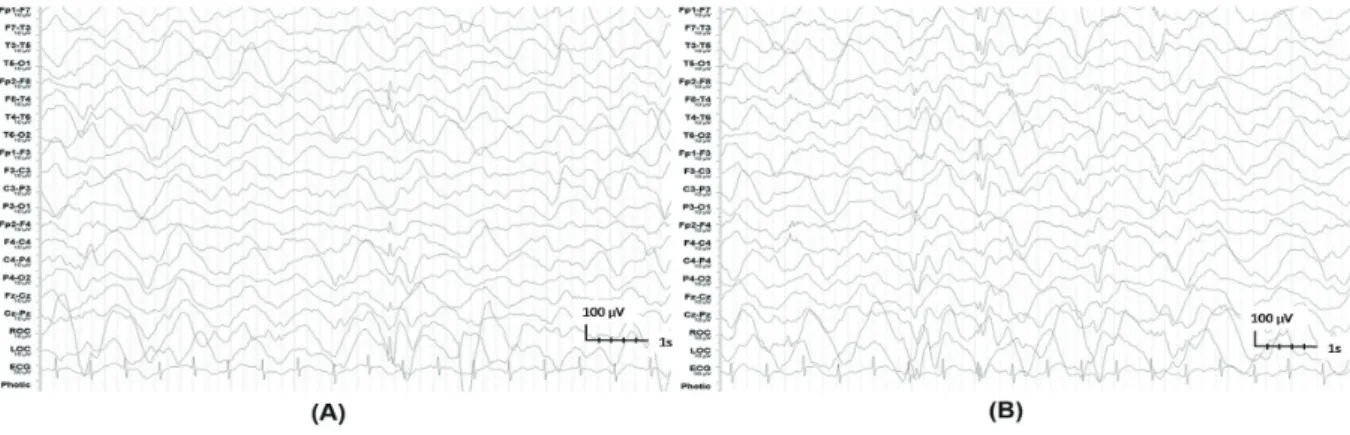
monstrated no skin lesion including rash or eschar. He could open and focus his eyes when experiencing pain, and sometimes uttered inappropriate words. Electroencephalography showed diffuse dysmorphic and slow delta background activities with bilateral independent frontal sharps (Fig. 2).
CSF analysis on admission showed evidence of improvement, with a WBC count of 5/mm3 with normal level of CSF glucose and protein. Laboratory investigations for usual pathogens of meningoencephalitis in the previous clinic and our hospital in
cluded CSF Gram stain, acid fast bacilli stain, and potassium hyd
roxide mount; CSF latex particle agglutination test for bacteria;
CSF multiplex polymerase chain reaction (PCR) test for bacteria (e.g., Streptococcus pneumoniae, Haemophilus influenza, Neis
seria meningitis, Listeria monocytogenes, and group B strepto
coccus) and viruses (e.g., herpes simplex virus type 1, herpes simplex virus type 2, human herpes virus 6, cytomegalovirus, ebsteinbarr virus, and varicella zoster virus); enterovirus PCR test of CSF and stool; antiJapanese encephalitis virus antibody in serum; CSF PCR test for tuberculosis; and bacterial culture of CSF, blood, and urine. All failed to reveal the causative pathogen.
Fig. 1. Result of enhanced brain magnetic resonance imaging. T1-weighted saggital (A), T2-weighted axial (B), diffusion-weighted axial (C),
and T1-weighted axial enhanced (D) images demonstrate no abnormal findings in parenchyma with ab normal signal intensity or enhancement.
Fig. 2. Electroencephalography of the patient on admission showed diffuse dysmorphic and slow delta background activities with
bilateral independent frontal sharps (A and B with sharps from the right and left frontal areas, respectively).
Further serologic tests for fever of unknown origin included:
1) serum antibodies against cytomegalovirus, EpsteinBarr virus, herpes virus, surface antigen test for hepatitis B virus and Myco
plasma pneumoniae; 2) multiplex PCR test for throatdwelling viruses including influenza virus; and 3) rapid immunochroma
tographic test (ICT) for hantaan virus, leptospira and O. tsutsu
gamushi. Although the patient showed no eschar or direct expo
sure history to mites, ICT for O. tsutsugamushi was positive, with an antibody titer in a passive hemagglutination assay (PHA) of 1:80. No abnormal findings were seen in the other tests for other pathogens. Urine analysis, other basic laboratory tests including thyroid function test, chest Xray, and electrocardiography showed no abnormal results.
In family history, his grandmother had complained of fever, rash, and general weakness, and was recently diagnosed as scrub typhus with a clue of eschar on her vulva. She recovered on do
xycycline therapy. Although our patient had no history of expo
sure to the grandmother’s vegetable garden, where his father also worked, his father often cared for his son while wearing his working pants. The clothing was a possible vehicle of infection to our patient. The patient was treated with clarithromycin (30 mg/kg/day for 14 days), intravenous methylprednisolone (15 mg/kg/day for 5 days), and immunoglobulin treatment (1g/kg/
day for 2 days). Generalized tonicclonic seizures recurrently appeared for the first 3 days despite fosphenytoin, valproic acid and topiramate treatment. On day 5 of admission, seizures disap
peared and he improved to an alert mental state. On day 13, he could walk alone and was discharged next day on valproic acid and topiramate.
Discussion
Scrub typhus is a febrile endemic disease in AsianPacific areas including Korea3,7). It is prevalent in autumn and has been reported from June to November in South Korea (from rainy months to the time of harvest)9). It is a clinically important, as it can cause many serious complications including CNS symptoms3). A study of 189 patients with scrub typhus reported that 79 patients (41.8
%) had CNS symptoms3). Among them, headache was the most common (100.0%), followed by altered sensorium (53.2%), neck stiffness (48.1%), and seizure (15.2%)3). Misra et al. reported that among 37 scrub typhus patients, headache occurred in 33 pati
ents (89.2%), altered sensorium in 32 patients (86.5%), neck stiff
ness in 18 patients (48.6%), and seizure in 8 patients (21.6%)4). In addition to these symptoms, ataxia, delirium, cranial nerve palsy, quadriplegia, hypesthesia, motor weakness, and diplopia have
been reported1). Palanivel et al. reported that among 67 pediatric scrub typhus patients, 58.2% had altered sensorium and 28.4%
showed seizures8). However, headache, which is the most com
mon neurologic symptom of patients with scrub typhus, was not investigated in the study8).
Brain or spinal MRI lesions in patients with scrub typhus have been reported only in some patients, which are usually limited to the gray matter1,4,6,10). This might be because scrub typhus in
volves invasion of small vessels1,4,10). Among 25 patients who re
ceived brain MRIs (consisting of scrub typhus meningoencepha
litis, encephalitis, and encephalopathy) in the study of Mirsa et al., only one patient showed an abnormal finding of leptomenin
geal enhancement4). Two case reports have described spinal cord lesions: one in the thoracic spine and another in the cervical spine and the brainstem1,11). Recently, a few cases of scrub typhus were reported with periventricular and/or deep white matter lesion1,10).
CSF examinations in patients with scrub typhus meningitis or meningoencephalitis have revealed the mildtomoderate in
crease of CSF WBC with lymphocyte dominance in addition to mild increase of CSF protein2,3). WBC in CSF usually normalizes in a shorter period2,3). Pai et al. reported that CSF WBC counts were elevated from 5 to 110/mm3 with lymphocyte dominance in 12 of 25 patients with CNS symptoms2). Boorugu et al.’s study of 39 patients with CNS symptoms showed similar results: mean CSF WBC count was 47.3/mm3 (range; 2450/mm3) with lympho
cyte dominance, and mean CSF protein was 90.6 mg/dL (range;
14360 mg/dL)3).
For diagnosis of scrub typhus, the eschar is the single most useful clue5,7). The eschar begins as a small papule, which en
larges, undergoes central necrosis, and eventually acquires a blackened crust with an erythematous halo resembling a cigarette burn6). An eschar usually occurs at sites where skin surfaces meet (e.g., axilla and groin) or where clothes bind (e.g., neck and waist)5,6). However, as it can be absent in 30% of patients with scrub typhus, clinical suspicion and early request for an approp
riate laboratory investigation are very important in a prevalent season5,7). As the levels of hepatic enzymes or alkaline phospha
tase frequently elevated, patients with hepatitis and CNS symp
toms need to be screened for scrub typhus6,7).
The main stay in scrubtyphus diagnostics remains serology5,
6,12). The indirect fluorescent antibody (IFA) test that is the most sensitive method is known as the gold standard5,13). However, the IFA is still expensive, needs several days to get a result, and re
quires experts with considerable training to perform it5,13). There
fore, the alternative diagnostic method of PHA has been used widely for recent two decades; nevertheless it has a lower sen
sitivity, especially in the acute phase5,13). Recently, the ICT was
introduced as a rapid diagnostic test to replace the PHA12). Pre
vious studies found that the sensitivities and specificities of ICT were ranged from 46.3% to 67.9% and 67.9% to 95.1%, respec
tively12).
The drug of choice for scrub typhus treatment is doxycycline and chloramphenicol in adult patients7). However, these are not appropriate for children due to their adverse effects14,15). Doxy
cycline is not recommended in children under 8 years of age as it can result in teeth staining15). Chloramphenicol can cause gray baby syndrome and irreversible bone marrow toxicity of fatal aplastic anemia16). Therefore, macrolides are recommended as alternative antimicrobials for children especially under 8 years of age1416). Several studies showed they were not less effective for treatment of scrub typhus and can be better tolerated in children than doxycycline1416).
Most patients showing neurologic symptomes associated with scrub typhus have good prognosis. Misra et al. reported that all 31 patients with imparied conciousness were completely re
covered within 1 month4). However, rare compilcation such as cerebral infarction, polyneurolathy, and transverse myelitis have been reported in other literature3,11).
As scrub typhus meningoencephalitis can be seen without eschar and can be caused by indirect exposure through conta
minated clothes, serum antibody test targeting O. tsutsugamushi should be considered in patients with meningoencephalitis who seek treatment between June and November in Korea. Our patient with altered mental state, seizure, and prolonged fever showed positive result for antibodies against O. tsutsugamushi, although he had no eschar and no previous history of recrea
tional behavior in contact with miteinfested habitats, such as bush and grass. His CSF pleocytosis was improved in a week and he had recovered in 14 days without sequelae.
요약
쯔쯔가무시병은 O. tsutsugamushi에 감염된 털진드기의 유충에 물려 발생하는 질환으로 발열, 두통, 근육통 등의 증상을 보이며, 신 경학적 증상이 나타나기도 한다. 가피는 쯔쯔가무시 진단에 중요하지 만 30% 정도에서는 관찰되지 않는다. 저자들은 가피 및 직접 노출된 병력이 없어 진단이 늦어졌던, 쯔쯔가무시 수막뇌염 환아 증례를 경 험하였기에 보고하고자 한다. 타병원에서 무균성 뇌수막염 진단을 받 고 5일간 치료한 6세 남자 환자가 지속되는 발열과 이후 발생한 의식 의 쳐짐, 반복되는 경련을 주소로 전원되었다. 타병원에서 시행한 뇌 척수액 검사에서 백혈구 증가(292개/mm3)를 보였으나 척수액 당과 단백은 정상 이었고, 세균 및 바이러스는 검출되지 않았으며, 뇌 자기 공명영상은 정상이었다. 입원 후 시행한 뇌파검사에서 전반적으로 느
린 서파와 양쪽 전두엽 쪽의 예파가 관찰되었고, 불명열에 대해 시행 한 혈액검사에서 쯔쯔가무시 항체가 양성이었다. 환아는 피부 가피 가 없고, 직접적으로 노출된 병력도 없었지만 할머니가 2주 전 쯔쯔 가무시로 진단받아 치료받았고, 자주 환아와 접촉하였던 아버지도 같이 농장 일을 했었다고 했다. 따라서, 아버지의 오염된 옷이 환아에 게 쯔쯔가무시병을 옮긴 매개체로 생각된다. 환아는 clarithromycin, me thylprednisolone, immunoglobulin 치료 후 호전되어 14일째에 퇴원하였다. 쯔쯔가무시병은 가피가 없이 발생하기도 하며, 오염된 옷 이 매개체가 될 수 있으므로 유행하는 시기에 수막뇌염으로 내원하 는 환아에서는 진단에 고려해야 할 것이다.
References
1) Yum KS, Na SJ, Lee KO, Ko JH. Scrub typhus meningo-encepha- litis with focal neurologic signs and associated brain MRI abnor- mal findings: literature review. Clin Neurol Neurosurg 2011;113:
250-3.
2) Pai H, Sohn S, Seong Y, Kee S, Chang WH, Choe KW. Central nervous system involvement in patients with scrub typhus. Clin Infect Dis 1997;24:436-40.
3) Boorugu H, Chrispal A, Gopinath KG, Chandy S, Prakash JJ, Abra- ham AM, et al. Central nervous system involvement in scrub typhus. Trop Doct 2014;44:36-7.
4) Misra UK, Kalita J, Mani VE. Neurological manifestations of scrub typhus. J Neurol Neurosurg Psychiatry 2015;86:761-6.
5) Koh GC, Maude RJ, Paris DH, Newton PN, Blacksell SD. Diagnosis of Scrub Typhus. Am J Trop Med Hyg 2010;82:368-70.
6) Jeong YJ, Kim S, Wook YD, Lee JW, Kim KI, Lee SH. Scrub typhus:
clinical, pathologic, and imaging findings. Radiographics 2007;
27:161-72.
7) Kim DM. Clinical Features and Diagnosis of Scrub Typhus. Infect Chemother 2009;41:315-22.
8) Palanivel S, Nedunchelian K, Poovazhagi V, Raghunadan R, Ramachandran P. Clinical profile of scrub typhus in children.
Indian J Pediatr 2012;79:1459-62.
9) Min YS, Lim HS, Lee K, Jung C, Cheong HK. A study on the epi- demiologic characteristics of scrub typhus in Gyeongsangbuk- do, 1999-2001. Korean J Epidemiol 2005;27:70-9.
10) Heo YJ, Jeong HW. Uncommon manifestations of scrub typhus encephalitis in two cases: Clinical and magnetic resonance imaging findings. J Korean Soc Radiol 2015;73:337-42.
11) Lee KL, Lee JK, Yim YM, Lim OK, Bae KH. Acute transverse my- elitis associated with scrub typhus: case report and a review of literatures. Diagn Microbiol Infect Dis 2008;60:237-9.
12) Lee KD, Moon C, Oh WS, Sohn KM, Kim BN. Diagnosis of scrub typhus: introduction of the immunochromatographic test in Korea. Korean J Intern Med 2014;29:253-5.
13) Brown GW, Shirai A, Rogers C, Groves MG. Diagnostic criteria for scrub typhus: probability values for immunofluorescent
antibody and Proteus OXK agglutinin titers. Am J Trop Med Hyg 1983;32:1101-7.
14) Kim EJ, Lee CY, Oh YG, Yun HS, Kim JD. Four cases of scrub ty- phus treated with azithromycin in children. J Korean Pediatr Soc 2003;46:188-91.
15) Lee KY, Lee HS, Hong JH, Hur JK, Whang KT. Roxithromycin treatment of scrub typhus (tsutsugamushi disease) in children.
Pediatr Infect Dis J 2003;22:130-3.
16) Strickman D, Sheer T, Salata K, Hershey J, Dasch G, Kelly D, et al.
In vitro effectiveness of azithromycin against doxycycline-resis- tant and -susceptible strains of Rickettsia tsutsugamushi, etiologic agent of scrub typhus. Antimicrob Agents Chemother 1995;39:
2406-10.