ABSTRACT
Purpose: Acellular dermal matrix (ADM) supports tissue expanders or implants in implant-based breast reconstruction. The characteristics of ADM tissue are defined by the manufacturing procedure, such as decellularization, preservation, and sterilization, and are directly related to clinical outcomes. This study aimed to compare the properties of a new pre-hydrated-ADM (H-ADM-low) obtained using a decellularization reagent reduction process with a low concentration of detergent with those of radiation-sterilized H-ADM and freeze-dried ADM (FD-ADM).
Methods: ADMs were evaluated in terms of structure, mechanical quality, and cytotoxicity using histochemical staining, tensile strength testing, and in vitro cell viability analysis.
Results: The tissue structure of H-ADM-low (CGDERM ONE-STEP) was similar to that of native skin despite complete decellularization. By contrast, in FD-ADM, the tissue structure was damaged by the freeze-drying process, and radiation-sterilized H-ADM showed a compact fibrillar arrangement. Furthermore, matrix components such as collagen and elastin were preserved in H-ADM-low, whereas a loss of elastin fibers with fragmented distribution was observed in radiation-sterilized H-ADMs. H-ADM-low's tensile strength (58.84 MPa) was significantly greater than that of FD-ADM (38.60 MPa) and comparable with that of radiation- sterilized H-ADMs. The residual detergent content in H-ADM-low (47.45 mg/L) was 2.67-fold lower than that of H-ADM decellularized with a conventional detergent concentration (126.99 mg/mL), and this finding was consistent with the cell viability results (90.7% and 70.7%, respectively), indicating that H-ADM-low has very low cytotoxicity.
Conclusions: H-ADM-low produced through aseptic processes retains the original tissue structure, demonstrates excellent mechanical properties, and does not affect cell viability.
Therefore, this newer H-ADM is suitable for use in implant-based breast reconstruction.
Keywords: Acellular dermis; Biological preservation; Breast implantation; Detergents;
In vitro techniques
Original Article
Received: Oct 15, 2020 Accepted: Nov 28, 2020 Correspondence to Il Jae Lee
Department of Plastic and Reconstructive Surgery, Ajou University School of Medicine, 164 World cup-ro, Yeongtong-gu, Suwon 16499, Korea.
E-mail: i00325@live.co.kr
© 2020 Korean Breast Cancer Society This is an Open Access article distributed under the terms of the Creative Commons Attribution Non-Commercial License (https://
creativecommons.org/licenses/by-nc/4.0/) which permits unrestricted non-commercial use, distribution, and reproduction in any medium, provided the original work is properly cited.
ORCID iDs Ji Young Kim
https://orcid.org/0000-0001-5539-7269 Kyung Min Yang
https://orcid.org/0000-0002-5605-7124 Ji Hyun Youn
https://orcid.org/0000-0003-2939-8457 Heejun Park
https://orcid.org/0000-0001-8250-1690 Hyung Min Hahn
https://orcid.org/0000-0002-4756-1804 Il Jae Lee
https://orcid.org/0000-0002-9478-6969 Conflict of Interest
The authors declare that they have no competing interests.
Ji Young Kim 1, Kyung Min Yang 2, Ji Hyun Youn 3, Heejun Park 3, Hyung Min Hahn 2, Il Jae Lee 2
1Department of Surgery, Ajou University Hospital, Suwon, Korea
2Department of Plastic and Reconstructive Surgery, Ajou University School of Medicine, Suwon, Korea
3CGBio Co. Ltd., Seoul, Korea
In Vitro Analysis of Histology,
Mechanics, and Safety of Radiation-
free Pre-hydrated Human Acellular
Dermal Matrix
Author Contributions
Conceptualization; Lee IJ, Kim JY, Yang KM;
Investigation; Park H; Data curation: Kim JY, Youn JH; Formal analysis: Kim JY, Youn JH;
Writing - original draft: Kim JY, Park H, Youn JH; Writing - review & editing: Yang KM, Hahn HM, Lee IJ.
INTRODUCTION
Acellular dermal matrix (ADM) refers to decellularized tissues obtained from humans, cattle, and pigs, in which cellular components that may induce immune responses have been removed. ADM is composed of the extracellular matrix (ECM), including collagen and growth factors, which upon transplantation into the body should be integrated into the recipient's tissue through re-cellularization and re-vascularization [1,2]. ADM of the bovine and porcine origin is commonly available in sufficient amounts to be used over a large body area;
however, even after the decellularization process, antigens foreign to the human body (e.g., alpha-gal) may remain and induce allergic reactions during transplantation [3,4]. Human- derived ADM overcomes the problem of immunogenicity and maintains the original structure of the dermal tissue, allowing fibroblasts, which play an important role in regeneration, to penetrate the tissue more efficiently. Therefore, ADM of human origin is preferred over that derived from animal sources [5] and is widely used for tissue reconstruction, especially breast reconstruction with tissue expanders and breast silicone implants.
ADM has been used to treat full-thickness burns since 1995 [6] and is now widely utilized in plastic and reconstructive surgery [7]. The most common methods of breast reconstruction after mastectomy are autologous and implant-based tissue restoration approaches. In particular, ADM has been used to support tissue expanders and implants in implant- based breast reconstruction since 2003 [7,8]. ADM has not only supportive effects but also minimizes fibrosis around the implants and reduces inflammatory reactions caused by implantation [9]. When ADM was used in implant-based breast reconstruction, the incidence of spherical constriction was significantly reduced, and the esthetic satisfaction of both surgeons and patients was excellent [10,11].
Currently, ADM used for breast reconstruction can be classified into freeze-dried,
cryopreserved, and pre-hydrated types. Freeze-dried ADM (FD-ADM) represents a sheet that can be safely stored at room temperature (e.g., AlloDerm), and its clinical effects have been demonstrated for many years in various studies [6,8]. However, the drying process can cause a loss of dermal tissue and critical structural components, and the relatively long rehydration (20 minutes or more) required before application makes use of FD-ADM rather inconvenient [12]. To overcome these disadvantages, cryopreserved ADM (CP-ADM) was developed (e.g., CGCRYODERM). CP-ADM is obtained without the drying process and the dermal structure is better preserved than that in FD-ADM. Furthermore, CP-ADM can be used immediately after a 5 minutes thawing step and is soft and flexible [13]. However, the disadvantage of CP- ADM is that it must be stored and transported at temperatures below −40°C. To circumvent the problems arising with the use of these ADM products, pre-hydrated ADM (H-ADM) was developed. H-ADM does not require rehydration or thawing, can be stored at room temperature, and can be immediately applied (e.g., AlloDerm RTU [Ready-To-Use] and DermACELL) [12].
Unlike conventional RTU products, CGDERM ONE-STEP is a new H-ADM product prepared with a low concentration of decellularization reagent and manufactured by the decellularization reagent reduction process under aseptic conditions in good manufacturing practice (GMP)-compliant facilities. The purpose of this study was to compare the structural and mechanical properties and safety of CGDERM ONE-STEP with those of FD-ADM and radiation-sterilized H-ADMs to determine the suitability of this new H-ADM for application in breast reconstruction.
METHODS
ADM products
H-ADM-low (CGDERM ONE-STEP) and FD-ADM (CGDERM) were obtained from CGBIO Co., Ltd. (Hwaseong, Korea). Both products were manufactured using donor skin tissues collected in accordance with the guidelines of the U.S. Food and Drug Administration and American Association of Tissue Banks and decellularized by the manufacturer. All processes related to tissue processing were conducted in a class 100 cleanroom in accordance with ISO 14664 (cleanrooms and associated controlled environments). H-ADM-low was prepared with a low-concentration of decellularization reagent, whereas H-ADM-high was prepared with conventional concentration conditions. Both H-ADMs were packaged and stored in a saline preservation solution. H-ADMs were manufactured in a sterile environment and did not undergo an additional sterilization procedure. After manufacturing, the sterility of H-ADMs was confirmed by a bacterial culture test according to USP <71> Sterility Testing in EONE Laboratories (Incheon, Korea). FD-ADM was packaged and stored in a freeze-dried state. FD-ADM was hydrated in physiological saline for 30 minutes before the experiments.
To assess the changes in tissue characteristics caused by the sterilization process, H-ADM samples were prepared using E-beam (H-ADM-e) and gamma irradiation (H-ADM-g) at 15 kGy each using Greenpia Technology (Yeoju, Korea). To evaluate the decellularization status and tissue structure (hematoxylin and eosin [HE] staining), each of these ADM products was compared with native unprocessed human cellular dermal matrix (native skin), which served as a control material.
Histological examination
To evaluate histological characteristics, all ADM tissues were fixed in 10% formalin, embedded in paraffin, and sectioned at a thickness of 5 μm. Sections were deparaffinized with xylene, hydrated in graded alcohol series (100%, 85%, and 70%), running tap water, and stained. To evaluate the decellularization status and tissue structure, HE staining was performed by incubating tissue sections with Harris HE Y for 5 minutes and 2 minutes, respectively. Tissue collagen content was analyzed by Masson's trichrome (MT) staining performed by incubation in Bouin's solution at 56°C for 1 hour, then in Weiger's hematoxylin for 10 minutes, Biebrich scarlet-acid fuchsin for 15 minutes, phosphomolybdic- phosphotungstic acid solution for 15 minutes, and aniline blue for 10 minutes. Tissue elastin content was evaluated by Verhoeff-Van Gieson (VVG) staining. Tissue sections were incubated in Verhoeff 's solution for 1 hour, 2% ferric chloride for 1 minute, 5% sodium thiosulfate for 1 minute, and Van Gieson's solution for 5 minutes. All reagents were purchased from Sigma- Aldrich (St. Louis, MO, USA). Stained tissue specimens were observed under an optical microscope (Olympus Optical Co., Tokyo, Japan) at × 100 magnification.
Mechanical testing
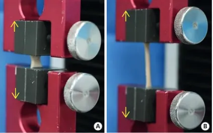
The mechanical properties were analyzed in FD-ADM, H-ADM-low, H-ADM-high, H-ADM-e, and H-ADM-g samples (tissue area of 50 × 10 mm, with an average thickness of 2.35 and 1.82 mm for FD-ADM and H-ADMs, respectively). Each specimen (n = 5 per group) was analyzed in a universal testing machine (UTM; Test One, Siheung, Korea) according to the guidelines (#D638-03) of the American Society for Testing and Materials. Specimens were loaded into tensile grips attached to the UTM and pulled apart at a rate of 100 mm/min until failure (Figure 1). The tensile strength was calculated by dividing the maximum load (N) value by the area of the cross-section (stretch length × thickness) and expressed as megapascal (MPa, 1 N/
mm2 equal to 1 MPa).
Measurement of residual detergent concentration
The amount of detergent (sodium dodecyl sulfate [SDS]) remaining in ADM tissues was analyzed using the methylene blue method [14]. The SDS complex with methylene blue is insoluble in chloroform and formation can be measured based on the optical density (OD) at 650 nm. H-ADM samples (H-ADM-high, H-ADM-low) decellularized using high (0.5%) and low (0.25%) SDS concentrations were soaked in sterile distilled water for 72 ± 2 hours at 37°C.
Then, 1 μL of each eluate was mixed with 99 μL of 0.0125% methylene blue solution (Sigma- Aldrich) and 200 µL chloroform (Sigma-Aldrich), and vortexed for 1 minute. After incubation at room temperature for 30 minutes, 150 μL of the lower layer was transferred to a 96-well plate and OD650 was measured using a spectrophotometer (Molecular Devices, San Jose, CA, USA). The SDS concentration in each ADM sample was calculated based on a linear standard curve (R2 = 0.9956) generated with 10 mg/mL stock solution of SDS (Sigma-Aldrich).
Cytotoxicity assessment
Cytotoxicity was evaluated using the mouse fibroblast cell line L-929 (NCTC clone 929:
CCL-1, American Type Culture Collection ATCC®, Manassas, USA). Cells were cultured in minimum essential medium (MEM; Gibco, Grand Island, USA) supplemented with 100 μg/
mL streptomycin, 100 U/mL penicillin, and 10% heat-inactivated fetal bovine serum at 37°C and 5% CO2.
To compare the cytotoxicity of H-ADM-low and H-ADM-high, 4 g of each tissue was eluted in 20 mL of MEM in an incubator at 37°C ± 1°C for 24 ± 2 hours. L-929 cells were seeded at a density of 1 × 10⁴ cells/100 μL/well in a 96-well-plate and cultured at 37°C and 5% CO2 for 24
± 2 hours. The medium was then removed, and 100 μL of H-ADM eluates, MEM positive, or dimethyl sulfoxide (DMSO), which was toxic to cells (Sigma-Aldrich; negative), was added in triplicate. After incubation at 37°C and 5% CO2 for 24 ± 2 hours, the cells were assessed for viability using the 3-(4,5-dimethylthiazol-2-yl)-2,5-diphenyltetrazolium bromide (MTT) assay.
H-ADM eluates and negative and positive control solutions were removed, and 50 μL of MTT solution (Sigma-Aldrich) was added. After 2 hours of incubation at 37°C and 5% CO2, the OD was measured at 570 nm.
A B
Figure 1. Tensile strength testing. (A) The tissue was fixed in the universal testing machine and (B) stretched in the direction of the yellow arrows.
Statistical analysis
All data are reported as mean ± standard deviation. Statistical analyses were conducted using Microsoft® Excel® software (Microsoft, Redmond, USA). Samples were compared using a paired one-tailed t-test, and p < 0.05 was considered to indicate significant differences.
RESULTS
Evaluation of the decellularized state and structure of ADM samples
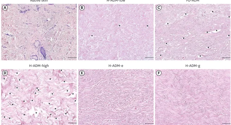
Optical microscopy analysis of HE-stained tissues indicated that H-ADM-low was completely decellularized with no remaining cell debris; furthermore, the overall tissue structure was similar to that of native skin (Figure 2A and B). In contrast, although no remaining cells were detected in FD-ADM, this appeared to contain a large number of voids due to tissue damage, which was not observed in H-ADM-low or native skin (Figure 2C). H-ADM-high decellularized with a conventional detergent concentration showed an amorphous structure instead of a dense fibrous structure, and several voids due to tissue damage were present (Figure 2D).
Both radiation-sterilized H-ADM tissues (H-ADM-e and H-ADM-g) had a condensed structure and compact fibrillar mesh-like morphology (Figure 2E and F). The histological analysis results suggest that H-ADM-low manufactured through the new decellularization reagent reduction process was completely decellularized, had little tissue damage owing to
A B C
D E F
Native skin H-ADM-low FD-ADM
H-ADM-high H-ADM-e H-ADM-g
Figure 2. Evaluation of the structure and decellularization status in ADM samples. Samples were fixed and stained with HE; representative images are shown (magnification × 100; scale bars, 200 μm). (A) Native skin, (B) H-ADM-low, (C) FD-ADM, (D) H-ADM-high, (E) H-ADM-e, and (F) H-ADM-g. The cell nuclei are stained dark-blue or purple and the extracellular material, such as collagen, is stained pink. The stars indicate the location of the void.
ADM = acellular dermal matrix; HE = hematoxylin and eosin; Native skin = unprocessed human cellular dermal matrix; H-ADM-low = hydrated-acellular dermal matrix prepared using low sodium dodecyl sulfate concentration; FD-ADM = freeze-dried acellular dermal matrix; H-ADM-high = hydrated-acellular dermal matrix prepared using conventional high SDS concentration; H-ADM-e = E-beam radiation-sterilized pre-hydrated acellular dermal matrix; H-ADM-g = gamma radiation-sterilized pre-hydrated acellular dermal matrix.
the exclusion of sterilization and freeze-drying steps, and preserved the structure similar to that of native skin.
Evaluation of collagen and elastin components
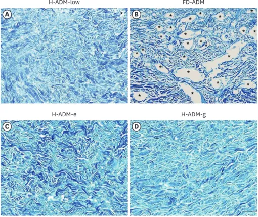
The content and distribution pattern of collagen and elastin, important structural
components of ADMs, were evaluated by MT staining (Figure 3) and VVG staining (Figure 4).
In H-ADM-low, collagen and elastin fibers tended to be uniformly distributed throughout the tissue without structural distortion. However, in the FD-ADM, H-ADM-e, and H-ADM-g samples, the fibers showed structural defects as the tissue was damaged and condensed.
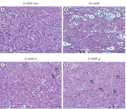
In particular, VVG staining revealed far fewer elastin fibers in FD-ADM and radiation- treated H-ADMs (H-ADM-e and H-ADM-g) than in H-ADM-low and that these fibers had a fragmented distribution pattern (Figure 5). In conclusion, collagen and elastin fibers in H-ADM-low were uniformly distributed and preserved close to the native skin without structural damage, which can be attributed to the fact that the H-ADM-low product did not undergo additional sterilization and freeze-drying processes.
Evaluation of tensile strength
To assess the mechanical properties of the ADM tissues, we measured their tensile strength (Figure 5). The results indicated the tensile strength of H-ADM-low was comparable with that of native skin (58.84 and 52.54 MPa, respectively). In contrast, the tensile strength of
A B
H-ADM-low FD-ADM
C D
H-ADM-e H-ADM-g
Figure 3. Collagen structure in ADM samples. Representative images of tissues stained with MT are shown (magnification × 100; scale bars, 200 μm) (A) H-ADM-low, (B) FD-ADM, (C) H-ADM-e, and (D) H-ADM-g. The collagen fibers are stained blue. The stars indicate the location of the void.
ADM = acellular dermal matrix; MT = Masson's trichrome; H-ADM-low = hydrated-acellular dermal matrix prepared using low sodium dodecyl sulfate concentration; FD-ADM = freeze-dried acellular dermal matrix;
H-ADM-e = E-beam radiation-sterilized pre-hydrated acellular dermal matrix; H-ADM-g = gamma radiation- sterilized pre-hydrated acellular dermal matrix.
A B
H-ADM-low FD-ADM
C D
H-ADM-e H-ADM-g
Figure 4. Elastin structure in ADM samples. Representative images of tissues stained with VVG staining method are shown (magnification × 100; scale bars, 200 μm). (A) H-ADM-low, (B) FD-ADM, (C) H-ADM-e, (D) H-ADM-g.
The elastic fibers and collagen fibers are stained black and red, respectively. The arrows indicate the fragmented elastic fibers and the stars indicate the location of the void.
ADM = acellular dermal matrix; VVG = Verhoeff-van Gieson; H-ADM = pre-hydrated acellular dermal matrix;
H-ADM-low = hydrated-acellular dermal matrix prepared using low sodium dodecyl sulfate concentration;
FD-ADM = freeze-dried acellular dermal matrix; H-ADM-e = E-beam radiation-sterilized pre-hydrated acellular dermal matrix; H-ADM-g = gamma radiation-sterilized pre-hydrated acellular dermal matrix.
0
* *
Tensile strength (MPa)
40 80 100
H-ADM-low FD-ADM
60
20
H-ADM-g H-ADM-e
Native skin
Figure 5. Evaluation of tissue mechanical properties by the tensile strength test. The results are presented as the mean ± standard deviation (n = 5).
Native skin = unprocessed human cellular dermal matrix; FD-ADM = freeze-dried acellular dermal matrix;
H-ADM-low = hydrated-ADM prepared using sodium dodecyl sulfate concentration; H-ADM-e = E-beam radiation- sterilized pre-hydrated acellular dermal matrix; H-ADM-g = gamma radiation-sterilized pre-hydrated acellular dermal matrix.
*p < 0.05.
FD-ADM (38.60 MPa) was significantly lower than that of the H-ADM-low (p = 0.027) and E-beam-irradiated H-ADM-e (54.92 MPa, p = 0.018); this was also lower than that of gamma radiation-treated H-ADM-g (55.33 MPa), although the difference was not significant. These results indicated that the tensile strength of H-ADM-low was similar to that of native skin and significantly higher than that of FD-ADM, whereas sterilization by irradiation did not significantly affect the mechanical properties of H-ADM.
Evaluation of residual SDS content and cytotoxicity
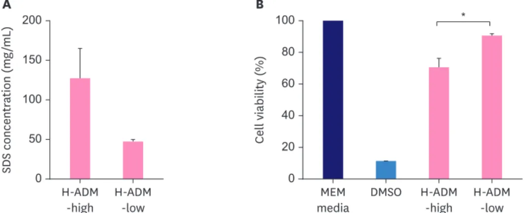
The detergent content in the eluates of H-ADM samples (H-ADM-high and H-ADM-low) decellularized using high and low SDS concentrations was 126.99 ± 38.13 and 47.45 ± 2.49 mg/mL, respectively, indicating a decrease of SDS concentration of 2.67-fold (p = 0.054) in H-ADM-low (Figure 6A) compared with that in H-ADM-high.
To determine whether SDS concentration used for decellularization influenced H-ADM cytotoxicity, L929 cells were incubated with H-ADM extracts. Cell viability with DMSO (which is toxic to cells), H-ADM-high, and H-ADM-low groups was 11.32%, 70.70% ± 5.67%, and 90.65% ± 1.10%, respectively (Figure 6B), compared with that of the MEM control group (100% viability), and the difference between the 2 H-ADM groups was significant (p = 0.014).
Thus, as the cell viability in the H-ADM-low group was over 90%, H-ADM-low decellularized using a low SDS concentration could be considered safe.
DISCUSSION
ADM was first used for breast areas in 2001 and since then has been extensively employed in implant-based breast reconstruction. ADM was used in 61,713 (61%) of 101,658 breast reconstruction cases performed in the USA in 2018 [15]. The application of ADM in implant-
0
*
Cell viability (%)
40 80 100
DMSO MEM
media 60
20
H-ADM -low H-ADM
-high SDS concentration (mg/mL) 0
100 200
150
50
H-ADM -low H-ADM
-high
A B
Figure 6. Residual detergent concentration and cytotoxicity of H-ADM prepared using different concentrations of detergent. (A) H-ADM-high and H-ADM-low samples were decellularized using high and low SDS concentrations, respectively. The residual detergent concentrations (mg/mL) are presented as the mean ± SD (n = 3). (B) L929 cells were treated with MEM media (control), DMSO (which is toxic to cells), and eluates of H-ADM-high and H-ADM-low. The cytotoxicity results (%) are presented as the mean ± SD (n = 3).
H-ADM = pre-hydrated acellular dermal matrix; H-ADM-high = hydrated-acellular dermal matrix prepared using high sodium dodecyl sulfate concentration; H-ADM-low = hydrated-acellular dermal matrix prepared using low sodium dodecyl sulfate concentration; SDS = sodium dodecyl sulfate; MEM = minimum essential medium; DMSO
= dimethyl sulfoxide; FD-ADM = freeze-dried acellular dermal matrix; H-ADM-e = E-beam radiation-sterilized pre- hydrated acellular dermal matrix; H-ADM-g = gamma radiation-sterilized pre-hydrated acellular dermal matrix;
SD = standard deviation.
*p < 0.05.
based breast reconstruction supports tissue expanders and implants, reduces capsular contracture and implant migration, increases fill volume, and improves esthetic outcome [9-11]. However, the use of ADM in implant-based breast reconstruction has been reported to increase the risk of seroma and infection [16]. There have also been conflicting results regarding ADM application after postmastectomy radiation therapy (PMRT), which is known to increase the chances of reconstruction failure and/or capsular contracture after implantation [17], where some studies noted that ADM alleviated such PMRT effects, whereas others indicated that this did not [18,19]. In this respect, standardization of ADM manufacturing procedures and careful evaluation of characteristics and safety may help improve the clinical outcomes of breast reconstruction. In particular, human-derived ADM preparations may show significant variations depending on the tissue donor. Therefore, to ensure predictable outcomes after AMD implantation, it is important to minimize changes that may be introduced during the manufacturing process, including decellularization, preservation, and sterilization. In this study, we investigated whether CGDERM ONE-STEP (H-ADM-low), a new product developed through hydration preservation and decellularization with reduced detergent concentration under aseptic conditions of GMP facilities, had suitable histological and mechanical properties for application in breast reconstruction.
The ADM manufacturing process includes decellularization, tissue preservation, and sterilization. Decellularization refers to the procedure of removing immunogenic cellular components, nucleic acids, membrane lipids, and cytosolic proteins from donor skin samples. If decellularization is not properly performed, the remaining materials may induce inflammatory reactions after transplantation and negatively affect the remodeling process [20]. However, since decellularization causes various degrees of tissue damage, it is important to maintain an optimal balance between complete decellularization and preservation of the original tissue structure. The most common decellularization method involves the use of detergents such as SDS, which is considered an excellent agent for cell removal. When SDS was applied to human skin, porcine small intestinal submucosa, or fibroblast sheets, the efficiency of decellularization increased with concentration, although the overall ECM density, including the glycosaminoglycan content, decreased and the collagen fiber network was damaged [21]. In previous studies, SDS was applied at an average concentration of 0.5% w/v (range 0.1%–1%) [21]. H-ADM-low was produced using SDS at 0.25%, which is half of the average concentration, and HE staining showed that the matrix was completely decellularized, i.e., there were no residual cell debris or other immunogenic components of the native skin. However, if 0.5% SDS was used, the H-ADM structure, including the fibrous matrix, was damaged. These findings suggest that the process of H-ADM-low manufacturing using low detergent concentration is suitable for both decellularization and preservation of the tissue structure.
The presence of contaminant SDS used for decellularization in ADM may cause toxic effects on surrounding cells after transplantation. A previous study showed that at concentrations less than 50 mg/L, the residual detergent in ADM did not cause cytotoxic effects in human endothelial cells [22]. In the current study, the residual SDS content in H-ADM-low decellularized with 0.25% SDS (47.45 mg/L) was below this level, whereas that in H-ADM- high decellularized with 0.5% SDS (126.99 mg/L) was considerably higher than the safe level of 50 mg/L. Accordingly, the former did not significantly affect cell survival (90.65%), whereas the latter decreased this to 70.70%. In conclusion, we suggest that radiation-free H-ADM-low can significantly reduce the potential risk for cytotoxicity by using detergents at lower concentrations.
The method of ADM preservation affects user convenience and tissue structural properties.
H-ADM can be stored at room temperature and used immediately without rehydration or thawing steps, which is very convenient. However, FD-ADM requires freezing/drying, which damages tissue and destroys collagen triple-helix structure by breaking hydrogen bonds, thus decreasing tensile strength [23]. Consistent with these findings, we observed gaps in the structure of FD-ADM and significantly lower tensile strength compared with H-ADM, which was similar in mechanical properties to the native skin. Thus, H-ADM-low, which does not undergo an additional preservation procedure, maintains the original structure and tensile strength of the human dermis.
ADM used for breast reconstruction can sometimes considered as a nidus of infection.
However, there was no significant difference in the rate of complications such as infection or seroma formation between patient groups receiving sterile or aseptically produced ADM for breast reconstruction [24]. H-ADM-low was manufactured under aseptic conditions and has been observed to remain sterile for at least 2 years from the date of production, as evidenced by bacterial culture tests (data not shown). This indicates that there is no difference in sterility between H-ADM-low and samples subjected to additional sterilization procedures.
However, sterilization had a negative effect on the tissue structure of H-ADM. Terminal sterilization is generally performed using ionizing radiation such as E-beam and gamma radiation. Use of E-beam radiation at a low intensity (25 kGy) has been reported to result in crosslinked collagen fiber matrix, which increased the tensile strength and resistance to enzymatic degradation [25]. However, use of high E-beam radiation intensity (50–70 kGy) produced severed collagen fibers in the ADM and damaged their aligned structure [26]. In contrast, the aseptic process can not only preserve the original porous tissue structure but also the matrix protein components such as collagen and elastin [27]. The native structure of matrix proteins is critical for their function as scaffolds for tissue regeneration and integration of cells such as fibroblasts as well as for their activity as signaling molecules that promote cell homing, adhesion, migration, and proliferation [28]. In this study, additional sterilization of H-ADM-low, manufactured through the aseptic process, with E-beam and gamma radiation at 15 kGy did not affect H-ADM tensile strength. However, long elastin fiber strands, which were preserved in the original H-ADM-low, were fragmented in the irradiated tissues, and collagen fibers were crosslinked, showing a compact fibrillar structure.
Overall, instead of the porous network characteristic for the native skin, irradiated H-ADMs (H-ADM-e and H-ADM-g) had a condensed structure, which can negatively affect host cell infiltration, graft incorporation, and remodeling at the ADM transplantation site [29].
Thus, H-ADM-low manufactured through the aseptic process not only has undamaged ECM components such as collagen and elastin but also retains a porous structure similar to that of the native skin, which is favorable for breast reconstruction.
This study has several limitations. First, whereas different cell types are involved in the integration of ADM into host tissues, only a single mouse fibroblast cell line was evaluated in this study. Second, although the study performed qualitative analysis of the histological changes in tissue structure and ECM contents around the ADM, no quantitative analysis was performed. Third, the effect of radiation on the structure and content of ECM was analyzed histologically, but the effect of the applied detergent concentration was not addressed in this study. Despite these limitations, the study demonstrated the effect of the detergent concentration and irradiation applied in the manufacturing process on the structure and contents of H-ADM. This information may be useful for surgeons to help
predict the outcomes when H-ADM is used for soft tissue augmentation or reconstruction.
To fully understand the mechanism underlying post-transplantation integration of the newly developed H-ADM-low, qualitative/quantitative experiments with various cell types and in vivo models are required. In addition, our results demonstrated that decellularization with low concentrations of detergent could lower the cytotoxicity of ADM. The potential cytotoxicity of ADM cannot be excluded as a cause of complications in patients undergoing breast reconstruction. We believe that elimination of the majority of the potential risk factors is important to ensure successful breast reconstruction in patients. Whether the low cytotoxicity of H-ADM-low reduces the incidence of complications should be studied in the future.
REFERENCES
1. Capito AE, Tholpady SS, Agrawal H, Drake DB, Katz AJ. Evaluation of host tissue integration,
revascularization, and cellular infiltration within various dermal substrates. Ann Plast Surg 2012;68:495-500.
PUBMED | CROSSREF
2. Garcia O Jr, Scott JR. Analysis of acellular dermal matrix integration and revascularization following tissue expander breast reconstruction in a clinically relevant large-animal model. Plast Reconstr Surg 2013;131:741e-751e.
PUBMED | CROSSREF
3. Bellows CF, Alder A, Helton WS. Abdominal wall reconstruction using biological tissue grafts: present status and future opportunities. Expert Rev Med Devices 2006;3:657-75.
PUBMED | CROSSREF
4. Galili U. The α-Gal epitope (Galα1-3Galβ1-4GlcNAc-R) in xenotransplantation. Biochimie 2001;83:557-63.
PUBMED | CROSSREF
5. Armour AD, Fish JS, Woodhouse KA, Semple JL. A comparison of human and porcine acellularized dermis: interactions with human fibroblasts in vitro. Plast Reconstr Surg 2006;117:845-56.
PUBMED | CROSSREF
6. Wainwright DJ. Use of an acellular allograft dermal matrix (AlloDerm) in the management of full- thickness burns. Burns 1995;21:243-8.
PUBMED | CROSSREF
7. Margulies IG, Salzberg CA. The use of acellular dermal matrix in breast reconstruction: evolution of techniques over 2 decades. Gland Surg 2019;8:3-10.
PUBMED | CROSSREF
8. Salzberg CA, Ashikari AY, Koch RM, Chabner-Thompson E. An 8-year experience of direct-to-implant immediate breast reconstruction using human acellular dermal matrix (AlloDerm). Plast Reconstr Surg 2011;127:514-24.
PUBMED | CROSSREF
9. Nahabedian MY. Acellular dermal matrices in primary breast reconstruction: principles, concepts, and indications. Plast Reconstr Surg 2012;130:44S-53S.
PUBMED | CROSSREF
10. Ho G, Nguyen TJ, Shahabi A, Hwang BH, Chan LS, Wong AK. A systematic review and meta-analysis of complications associated with acellular dermal matrix-assisted breast reconstruction. Ann Plast Surg 2012;68:346-56.
PUBMED | CROSSREF
11. Macadam SA, Lennox PA. Acellular dermal matrices: use in reconstructive and aesthetic breast surgery.
Can J Plast Surg 2012;20:75-89.
PUBMED | CROSSREF
12. Cheon JH, Yoon ES, Kim JW, Park SH, Lee BI. A comparative study between sterile freeze-dried and sterile pre-hydrated acellular dermal matrix in tissue expander/implant breast reconstruction. Arch Plast Surg 2019;46:204-13.
PUBMED | CROSSREF
13. Kim SY, Lim SY, Mun GH, Bang SI, Oh KS, Pyon JK. Evaluating the effectiveness of cryopreserved acellular dermal matrix in immediate expander-based breast reconstruction: a comparison study. Arch Plast Surg 2015;42:316-20.
PUBMED | CROSSREF
14. Zvarova B, Uhl FE, Uriarte JJ, Borg ZD, Coffey AL, Bonenfant NR, et al. Residual detergent detection method for nondestructive cytocompatibility evaluation of decellularized whole lung scaffolds. Tissue Eng Part C Methods 2016;22:418-28.
PUBMED | CROSSREF
15. American Society of Plastic Surgeons. 2018 Plastic Surgery Statistics Report. https://www.plasticsurgery.
org/documents/News/Statistics/2018/plastic-surgery-statistics-full-report-2018.pdf. Accessed September 29th 2020.
16. Chun YS, Verma K, Rosen H, Lipsitz S, Morris D, Kenney P, et al. Implant-based breast reconstruction using acellular dermal matrix and the risk of postoperative complications. Plast Reconstr Surg 2010;125:429-36.
PUBMED | CROSSREF
17. Ricci JA, Epstein S, Momoh AO, Lin SJ, Singhal D, Lee BT. A meta-analysis of implant-based breast reconstruction and timing of adjuvant radiation therapy. J Surg Res 2017;218:108-16.
PUBMED | CROSSREF
18. Sbitany H, Wang F, Peled AW, Lentz R, Alvarado M, Ewing CA, et al. Immediate implant-based breast reconstruction following total skin-sparing mastectomy: defining the risk of preoperative and postoperative radiation therapy for surgical outcomes. Plast Reconstr Surg 2014;134:396-404.
PUBMED | CROSSREF
19. Myckatyn TM, Cavallo JA, Sharma K, Gangopadhyay N, Dudas JR, Roma AA, et al. The impact of chemotherapy and radiation therapy on the remodeling of acellular dermal matrices in staged, prosthetic breast reconstruction. Plast Reconstr Surg 2015;135:43e-57e.
PUBMED | CROSSREF
20. Keane TJ, Londono R, Turner NJ, Badylak SF. Consequences of ineffective decellularization of biologic scaffolds on the host response. Biomaterials 2012;33:1771-81.
PUBMED | CROSSREF
21. Dussoyer M, Michopoulou A, Rousselle P. Decellularized scaffolds for skin repair and regeneration. Appl Sci (Basel) 2020;10:3435.
CROSSREF
22. Cebotari S, Tudorache I, Jaekel T, Hilfiker A, Dorfman S, Ternes W, et al. Detergent decellularization of heart valves for tissue engineering: toxicological effects of residual detergents on human endothelial cells. Artif Organs 2010;34:206-10.
PUBMED | CROSSREF
23. Bachmann L, Gomes AS, Zezell DM. Collagen absorption bands in heated and rehydrated dentine.
Spectrochim Acta A Mol Biomol Spectrosc 2005;62:1045-9.
PUBMED | CROSSREF
24. Lyons DA, Mendenhall SD, Neumeister MW, Cederna PS, Momoh AO. Aseptic versus sterile acellular dermal matrices in breast reconstruction: an updated review. Plast Reconstr Surg Glob Open 2016;4:e823.
PUBMED | CROSSREF
25. Seto A, Gatt CJ Jr, Dunn MG. Radioprotection of tendon tissue via crosslinking and free radical scavenging. Clin Orthop Relat Res 2008;466:1788-95.
PUBMED | CROSSREF
26. Lee JH, Kim HG, Lee WJ. Characterization and tissue incorporation of cross-linked human acellular dermal matrix. Biomaterials 2015;44:195-205.
PUBMED | CROSSREF
27. Nilsen TJ, Dasgupta A, Huang YC, Wilson H, Chnari E. Do processing methods make a difference in acellular dermal matrix properties? Aesthet Surg J 2016;36:S7-22.
PUBMED | CROSSREF
28. Tamariz E, Grinnell F. Modulation of fibroblast morphology and adhesion during collagen matrix remodeling. Mol Biol Cell 2002;13:3915-29.
PUBMED | CROSSREF
29. Dearth CL, Keane TJ, Carruthers CA, Reing JE, Huleihel L, Ranallo CA, et al. The effect of terminal sterilization on the material properties and in vivo remodeling of a porcine dermal biologic scaffold. Acta Biomater 2016;33:78-87.
PUBMED | CROSSREF