폐쇄성 수면무호흡증의 수술적 치료
경희대학교 의과대학 이비인후과학교실
김 성 완·은 영 규
Surgical Therapy for Obstructive Sleep Apnea
Sung Wan Kim, MD and Young Gyu Eun, MD
Department of Otorhinolaryngology, College of Medicine, KyungHee University, Seoul, Korea
폐쇄성 수면무호흡증의 수술적 치료는 기능이상의 인 두구조를 변경하거나 폐쇄부위를 우회하는 것이다. 기도 를 막는 연조직을 제거하거나 기도의 용적을 결정하는 장 력 등에 영향을 주는 내부구조들의 관계를 변경하여 인두 구조를 바꾼다. 성인 폐쇄성 수면무호흡증 환자의 2%에 서는 공간을 차지하는 병적인 부위가 발견되어 이 병변의 제거만으로도 수면무호흡증이 개선되지만, 나머지 98%
의 환자에서는 상기도를 둘러싸는 연조직구조의 이상 또 는 상악과 하악의 골격구조의 이상에 의해 발생한다.1-3) 비중격재건, 하비갑개수술, 편도선절제술 등의 전통적인 이비인후과 수술법으로는 폐쇄성 수면무호흡증을 치료하 는데 실패하였고,4) 인두의 연조직을 제거하거나 골격을 재위치 시켜 인두의 연조직 구조의 위치를 변경시키는 새 로운 방법들이 개발되어 왔다. 여기서는 현재의 여러 수 술법과 보고된 결과들에 대하여 알아보고자 한다.
수술의 계획
수술 방법의 결정은 많은 요소들에 기초한다. 전신적인 건강상태뿐만 아니라 환자의 바람과 선호도도 고려사항 에 포함된다. 수술의 목표는 환자마다 다를 수 있지만 수 술 후에는 다른 비수술적 처치의 효과가 떨어질 수 있으므
로 수술을 시작한다면 수술로 완치를 바라볼 수 있도록 환자를 선택하는 것이 중요하다. 환자들이 비수술적 치료 에 견디지 못해 수술을 계획하기도 하고 수술을 통해 지속 적 기도양압술(continuous positive airway pressure, 이하 CPAP)의 압력을 줄여 CPAP에 적응도를 높이기 위 하여 수술을 계획하기도 하며, CPAP에 의한 비증상 때 문에 수술을 계획하기도 한다. 수술 전 환자에게 수술의 적절한 이론적 설명과 위험성 등의 정보를 제공하고 수술 의 목표에 대하여 충분한 논의를 해야 한다.
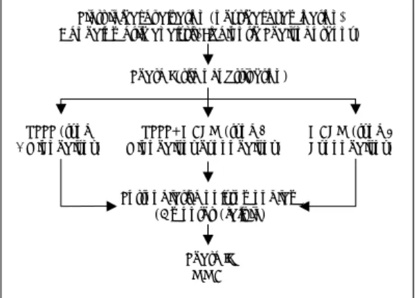
수술의 계획에서 가장 어려운 것은 폐쇄부위의 확인과 수술법의 결정이다. 이학적 검사, Mueller maneuver를 이용한 내시경, 두개골 계측촬영 등의 술전 검사법들을 통해 상기도의 구조와 폐쇄부위를 어느 정도 확인할 수는 있으나 이 역시 확실하지는 않고 최근 apnea level test 라고 불리는 전기적 senser를 이용한 수면 중 폐쇄부위 를 찾는 방법이 도입되었으나 이 역시 많은 불편과 환자들 의 협조가 어렵다. 합리적인 수술방법의 선택은 수술에 의한 외상을 최소화하여 불필요한 수술을 줄이면서 성 공적인 결과를 얻는 것이다. Stanford 대학 수면센터의 Powell과 Riley는 단계적 수술법으로‘Powell-Riley- Stanford protocol’, 즉 phase I과 phase II의 두 가지 수술 단계를 주장하였다(Fig. 1).5) phase I에서는 폐쇄 부위에 따라 비수술, 구개수술과 설근부 수술을 폐쇄부위 에 따라 적용하고 4~6개월의 치유과정 후 수면다원검사 를 다시 시행하여 수술의 결과를 평가한다. 여전히 폐쇄성 수면무호흡증이 남은 환자에게는 phase II로 기도의 재 교신저자:김성완, 130-702 서울 동대문구 회기동 1
경희대학교 의과대학 이비인후과학교실
전화:(02) 958-8474・전송:(02) 958-8470 E-mail:[email protected]
건을 의미하는 상하악전진술(maxillomandibular advan- cement)을 시행할 것을 주장하였다. 단계적 수술의 장 점으로 불필요한 과잉수술을 피하고 phase I을 시행함 으로 phase II의 성공률을 더욱 높일 수 있다고 하였다.
Phase I의 결과는 61%, phase II에서는 95%의 성공률 을 보고하였다.
기관절개술(Tracheostomy)
Pickwickian syndrome 환자에서 상기도폐쇄를 우회 하는 기관절개술의 사용은 폐쇄성 수면무호흡증의 치료에 처음 보고 된 방법이다.6) 가장 효과적인 방법임에도 불 구하고 발성, 미관상 문제, 생활의 불편, 수술 자체의 합 병증 등으로 환자들에게 시행하기는 지극히 어렵다. 또 한 최근에는 자동 기도양압기(automatic positive airway pressure)의 도입으로 응급상황의 환자에서도 검사없이 바로 착용이 가능한 양압술이 개발되어 있어 그 입지는 더욱 없어졌다고 할 수 있다. 현재 기관절개술은 중증의 비만이나 두개안면기형 등으로 수술전후로 고도의 위험 이 있는 중증 수면무호흡증환자에서 일시적인 기도 보 호의 방법으로 사용된다.7)8) 그러나 폐쇄성 수면무호흡 증의 장기간 치료로써의 영구적인 기관절개술은 obesity hyperventilation syndrome을 가진 심한 비만 환자나 비 수술적 또는 수술적 치료 모두에서 실패한 두개안면부 기형 환자에서 적용될 수도 있다.9)10)
비수술(Nasal Surgery)
비폐색과 수면 호흡장애(Sleep-disordered breath- ing) 사이의 관계는 수많은 연구들에 의해서 입증되어 왔 다. 낮 시간의 비폐색과 야간의 비울혈 모두 수면관련 호 흡장애의 위험인자로 알려져 있다.11)12) 그래서 비폐색의 치료는 수면무호흡 환자의 수술에서 중요한 부분이다. 폐 쇄가 발생하는 세 부분은 비익연골/비밸브, 비중격 그리 고 비갑개부분이다. 가장 흔하게 행해지는 비수술은 비 중격수술과 하비갑개수술이다. 비수술의 주요 효과는 비 통기성의 주관적인 향상과 nasal CPAP 장치의 압력을 줄이는 것이다. 폐쇄성 수면무호흡증의 치료에서 비수술 단독으로 시행되었을 때의 효과에 대해서는 의견이 다양 한데, 비폐색과 폐쇄성 수면무호흡증을 가진 50명의 환 자를 대상으로 한 연구에서 비중격 수술 후 경도 폐쇄성 수면무호흡증의 경우는 호흡장애지수가 악화되었고, 중등 도나 중증의 폐쇄성 수면무호흡증 환자는 유의한 호전 이 없었다.13) 하지만 폐쇄성 수면무호흡증이 있는 40명의 환자에서 23명이 비중격성형술을 받은 결과 8명(35%) 에서 호흡장애 지수가 50% 이상 감소한 보고도 있었 다.14) 하지만 거대한 비용종을 제외하고는 비중격 수술 단 독으로는 폐쇄성 수면무호흡증의 치료에 많은 도움을 기 대하기는 힘들 것으로 생각된다.
인두부의 수술과 동반해서 하는 경우 환자가 한번의 전 신마취로 두 가지 수술을 받을 수 있고, 입원 기간을 줄 이는 등의 장점이 있다. 비수술과 구개수구개인두성형술 (uvulopalatopharyngoplasty, 이하 UPPP)을 180명의 환자에서 동시에 시행하였을 때 코골이의 주관적인 향 상이 97%의 환자에서 있었다는 보고가 있었다.15) 그러 나 Mickelson 등은 347명의 환자에서 UPPP만 단독 시 행하였을 때의 합병증 발생률 2%와 비교하여 비중격 수 술이 동반되었을때 14%의 합병증 발생률을 보고하였고 단계적인 수술을 추천하였다.16) 저자의 경험에 비추어 다 른 부위의 수술과 함께 비수술을 시행하여야 하는 경우 중증이상의 폐쇄성 수면무호흡증의 경우는 수술 후 기도 유지를 고려할 때 단계적인 수술이 안전하고, 그 이하의 폐쇄성 수면무호흡증에서 동시에 시행하는 경우에도 출혈 에 의한 시야확보를 위해 비수술을 마지막에 시행하는 것
Presurgical evaluation (physical examination, Cephalometric analysis, Fiberoptic Pharyngoscopy)
Phase I (site of obstruction)
UPPP (type
1 Oropharynx) UPPP+GAHM (type 2
Oropharynx hypopharynx) GAHM (type 3 Hypopharynx)
Postoperative polysomnogram (6 months)(failure)
Phase II MMA
Fig. 1. The Stanford protocol of phased surgery. UPPP:
Uvulopalatopharyngoplsty, MMA:Maxillomandibular advancement, GAHM:Genioglossus advancement/
hyoid myotomy.
이 좋으며, 비패킹은 가능한 한 피하거나 삽입기간을 줄 이고, 통기가 되는 비스텐트를 사용하는 것이 좋을것으로 생각된다.
또한, 비수술은 nasal CPAP을 견디기 힘들어하는 비폐 색을 가진 환자들에게 적용되어왔다. Nasal CPAP에 환 자들이 적응하지 못하는 원인은 다양한 이유가 있으나 높 은 압력을 줘야하는 경우 그 적응도가 떨어지는데 비수술 은 일반적으로 비저항을 줄여 CPAP의 적응도를 높일 수 있을 것으로 생각된다.17) 실제로 Friedman 등은 비폐
색과 폐쇄성 수면무호흡증을 가진 환자 50명을 대상으로 비중격성형술 후 CPAP 압력을 유의하게 감소시킬 수 있음을 보고하였다.13)
구인두 수술(Oropharyngeal Surgery)
구개수구개인두성형술(UPPP)
UPPP는 1981년 Fujita에 의해 처음 소개된 이후 25 년간 폐쇄성 수면무호흡증에 대해 가장 많이 시행된 수술 이다.18) 전통적인 UPPP의 술식은 구인두 입구부를 넓 히기 위하여 구개수 뿐만 아니라 두터운 연구개와 인두조 직을 제거한다. 방법은 구개 편도 절제술을 시행하고 편도 와의 경계를 이루는 점막을 절제한다. 전방과 후방의 편 도지주(tonsillar pillar)를 손질하고(trimming) 후편도지 주 경계의 처진 점막을 제거하고 구개인두근을 앞 그리 고 외측으로 당겨 구개설근에 봉합하여 편도와를 닫는 다. 구개수와 연구개의 후방경계부를 제거하고, 비강쪽 점 막을 앞으로 돌려 구강쪽 점막에 봉합을 한다(Fig. 2). 수 술 후 합병증에 대해 환자 640명을 대상으로 한 meta- analysis에서는 1개월 이상 지속되는 구개인두 부전증 (2%), 술 후 출혈(1%), 비인두 협착증(1%), 음성변화 (1%), 성공적으로 처치된 상기도 폐쇄(0.3%), 상기도 폐 쇄로 인한 사망(1%) 등이 보고되었다. 그러나 논문의 반 이상이 합병증 유무에 대한 언급이 없어 진정한 합병증 Fig. 2. Technique of uvulopalatopharyngoplasty. Sur-
followed by bottom row. Tonsils are removed. Posterior tonsillar pillars are divided vertically from uvula to level of upper pole of tonsillar fossae, rotated across the fossae, and sutured to trimmed anterior tonsillar pillars. Sutures may tack the posterior pillars to the midportion of the fossa. Uvula and posterior soft palate are transected at approximately the upper pole of the tonsillar fossae.
Fig. 3. Friedman classification of palatal position (A) and tonsil size (B). The Friedman palate position is based on visualization of structures in the mouth with the mouth open widely without protrusion of the tongue. Palate grade I allows the observer to visualize the entire uvula and tonsils. Palate grade II allows visualization of the uvula but not the tonsils. Grade III allows visualization of the soft palate but not the uvula. Grade IV allows visualization of the hard palate only. Tonsil size is graded from 0 to 4. Tonsil size 0 denotes surgically removed tonsils. Size 1 implies tonsils hidden within the pillars. Tonsil size 2 implies the tonsils extending to the pillars. Size 3 tonsils are beyond the pillars but not to the midline. Tonsil size 4 implies tonsils extend to the midline.
I II
III IV
0 1 2
3 4
A A A
A B B B B
율이라고 보기는 어렵다.19) UPPP의 합병증에 대한 연구 들에서 상기도 폐쇄에 의한 사망은 대략 1%라고 보고하
였다.20)21) 91명을 대상으로 한 다른 연구에서 UPPP 시
행 1년 후 목의 건조감과 연하 이상을 호소한 환자는 각 각 31%와 10%였다.21)
UPPP는 구인두 폐쇄를 효과적으로 향상시키지만 하 인두 폐쇄는 이 술식에 거의 영향이 없어 단독으로 사용 시 meta-analysis에서는 성공률을 40.7% 정도로 보고 되었다.4) Friedman 등은 UPPP의 성공률을 예측하기 위 해 편도크기, 구개의 위치, 체질량지수에 따라 4단계로 된 Friedman's staging system(Fig. 3, Table 1)을 이 용하여 stage I 80.6%, stage II 37.9%, stage III 8.1%
의 UPPP 성공률을 보고하였다.22) 따라서 결과를 향상
시키고 합병증을 감소시키기 위해 전통적인 UPPP의 변 형된 여러 가지 방법이 개발되어 왔다.
구개수구개피판술(Uvulopalatal flap, 이하 UPF)
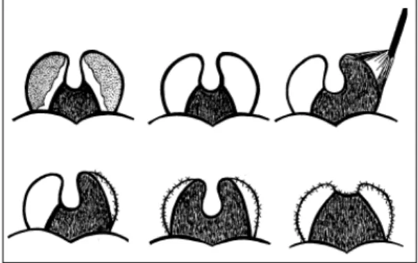
Powell 등이 보고한 UPF는 UPPP의 변형된 방법으 로 구개수를 부분적으로 자른 후 구개수를 당겼을 때 연구 개에 중첩되는 점막만을 절개하여 제거한 후 구개수피판 을 앞으로 당겨 경-연구개 접합부에 매달아 구인두 기 도를 확장시키는 방법이다(Fig. 4).23) 이 방법은 UPPP 시 과도한 조직의 절제로 발생할 수 있는 구개인두부전 증을 예방할 수 있으며, 가역적이라는 장점을 가지고 있다.합병증으로는 출혈, 염증, 혈종 발생에 따른 피판 분리 등 이 보고 되었다. 80명의 전향적 연구에서 술 후 결과에 서는 UPPP와 차이는 없었고, 환자가 호소하는 통증은 UPPP에 비해 유의하게 적었다.
측인두성형술(lateral pharyngoplasty, 이하 LPP)
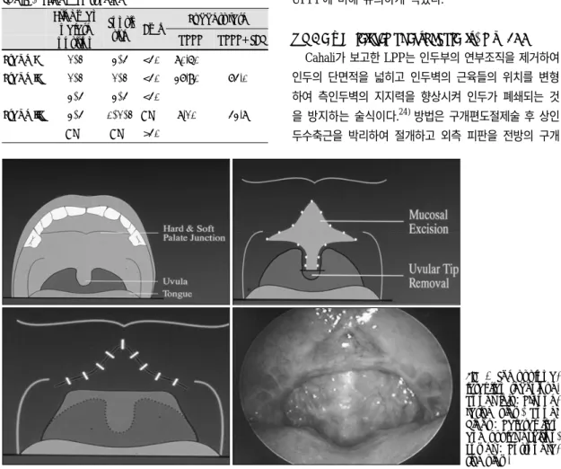
Cahali가 보고한 LPP는 인두부의 연부조직을 제거하여 인두의 단면적을 넓히고 인두벽의 근육들의 위치를 변형 하여 측인두벽의 지지력을 향상시켜 인두가 폐쇄되는 것 을 방지하는 술식이다.24) 방법은 구개편도절제술 후 상인 두수축근을 박리하여 절개하고 외측 피판을 전방의 구개 Table 1. Friedman stagingSuccess rate Friedman
palate position
Tonsil size Bmi
UPPP UPPP+TBR Stage I 1, 2 3, 4 <40 80.60%
Stage II 1, 2 1, 2 <40 37.90% 74.0%
3, 4 3, 4 <40
Stage III 3, 4 0, 1, 2 ANY 8.10% 43.8%
ANY ANY >40
Fig. 4. The uvulopa- latal flap technique.
Upper left:Preope- rative view, Upper Right:Palatal flap and uvular excision, Lower :Postopera- tive view.
설근에 봉합한다. 구개에 상외측으로 절개를 가하여 구개 피판을 만들고, 구개수의 일부도 절제한 후 후편도지주 에도 절개를 가하여 구개피판과 Z-성형술 형태로 봉합 하여 후구개 부위의 허탈을 방지한다(Fig. 5). Cahali의 보고에 의하면 10명의 환자를 대상으로 한 지침연구에서 수술 6개월 후 평균 무호흡-저호흡지수는 41.2에서 9.5 로 감소하였고, 연하장애는 술 후 평균 14.5일간 지속 되었으나 모든 환자가 정상으로 회복하였다.
구개수구개성형술(Uvulopalatoplasty, 이하 UPP)
Friedman 등은 폐쇄성 수면무호흡증 환자들 중 편도 비대와 연구개가 연장되었으나 과다한 인두의 주름이 없는 환자들을 대상으로 한 UPP를 보고하였다.25) 방법 은 편도조직을 고주파를 이용하여 편도표면이 피막까지 낮추어지게 편도조직을 제거하면서 근육의 노출은 피한 후 UPPP과 비슷하게 시행되는데 구개를‘사각형 모양’으 로 만들기 위해 후편도지주에 절개를 가한다. UPPP와 다 른 점은 구개봉합아래에 편도조직이 남는다는 것이다.구개봉합은 2층 구조로 하며 2.0 Vicryl을 이용하여 점막 하층을 봉합하고 3.0 Chromic으로 점막층을 봉합한다
(Fig. 6). 편도비대와 연구개 연장, 과다한 인두주름이 있는 환자들을 대상으로 시행한 UPPP와 비교하여 무 호흡-저호흡지수와 평균산소포화도의 호전 정도는 두 술식간에 차이가 없었으나, UPP를 시행 받은 환자군에 서 통증기간, 마약성 진통제 사용기간, 정상 식이로 복 귀, 이물감 호소 등에서 더 나은 결과를 보였다.
Z-구개성형술(Z-palatoplasty)
Friedman 등은 편도가 없거나 편도선 절제술을 이미 시행 받은 환자들을 대상으로 Z-구개성형술이라는 새로 운 술식을 보고하였다.26) UPPP는 폐쇄성수면무호흡증 치료에 가장 많이 쓰이는 술식이나 편도선수술을 받은 많은 환자들이 매우 좁은 구개궁이 위로 향하여 기도폐 쇄를 보이는 점, 편도절제술을 이미 받은 환자들은 전통 적인 UPPP에 결과가 좋지 못하다는 점,22)27) 그리고 많은 환자들이 UPPP에 의해 좋아지지 않고 오히려 악 화된다는 점 등의 문제가 존재한다.28) 편도선 수술을 받은 환자들에서 후구개지주가 절제되거나 술 후 반흔 조직으로 인해 후인두벽이 당겨져 서로 가까워져 유착이 될 수 있다. Z-구개성형술의 목표는 반흔의 장력을 전
Fig. 5. The lateral pharyngoplasty technique. ① Operative view after left tonsillectomy. (A) palatopharyngeus muscle, (B) palatoglossus muscle, (C) superior pharyngeal constrictor muscle and (D) right tonsil. ② Elevation and section of the left superior pharyngeal constrictor muscle. ③ Anterior suture of the superior pharyngeal constrictor muscle (lateral flap) to the palatoglossus muscle. ④ (A) palatine flap, (B) section of the palatopharyngeus muscle, (**) external palatine vein (right) and ascending palatine artery (left). ⑤ Z-plasty covering the superior part of the tonsillar fossa. (A) palatine flap, (B) upper part of the palatopharyngeus muscle. Incision to remove part of the uvula (dashed line). ⑥ Final aspect of the lateral pharyngoplasty.
1 1 1
1 3 3 3 3
4 4 4
4 5 5 5 5 6 6 6 6
2
2 2
2
방과 측방의 벡터로 바꾸어서 구개와 후인두벽사이의 공 간을 넓히고 인두의 외측 면을 넓힌다. 방법은 구개위에 2개의 피판을 디자인하고 피판의 전면의 점막만을 제거
한 후 정중선에서 구개부분을 두 개로 나눈다. 구개의 전 측면으로 정중선이 경구개와 연구개의 가장자리의 중간 부위에 가도록 하여 2층의 봉합을 시행한다(Fig. 7).
C C C C B
B B B A
A A A
Fig. 6. The uvulopalatoplasty technique. A:Tonsillar coblation. B:The modified uvulopalatoplasty. Incision of the palatal flap after completion of tonsillar coblation. Usually, the flap is beveled, with a longer nasopharyngeal surface of the palate that is rolled over toward the oral surface of the palate to recreate the new free-edge of the palate.
Standard releasing incisions at the corners of the posterior tonsillar pillars may be required to decrease mobilization of the flap. C:The modified uvulopalatoplasty. Two-layer closure of the palate.
Fig. 7. The Z-palatoplasty technique. A:The incision of the palatal flap is marked. B:The mucosa over the palatal flap is removed, exposing the palatal musculature. C:The uvula and palate are split in the midline with a cold knife.
D:The uvular flaps along with the soft palate are reflected back laterally over the soft palate. E:Two-layered closure of the palatal flaps. The submucosal layer is approximated first with 2-0 Vicryl F:Two-layered closure of the palatal flaps-the mucosal closure with 3-0 chromic suture.
B B B B A A A
A C C C C
E E E E D
D D
D F F F F
Z-구개성형술을 받은 환자 25명과 UPPP을 받은 환자 25명의 결과를 비교하였을 때 주관적인 증상의 향상은 두 군에서 같았으나 Z-구개성형술 군에서 객관적인 지 표의 향상이 의미 있게 높았다. 술 후 합병증 등에서는 두 군간의 차이가 없었으나 삶의 질과 술후 연하곤란은 Z-구개성형술 군이 나은 결과를 보였다.
경구개전진 인두성형술(Transpalatal advancement ph- aryngoplasty)
Woodson 등은 구개전진술을 이용한 인두성형술을 보 고하였다.29) Friedman stage 3의 환자들에서 UPPP의 성공률이 8%에 불과하다고 알려져 있다.22) 수술 실패의 원인은 다양하지만 UPPP 부위인 상인두에 주로 발생
한다.30)31) 이 방법은 심한 절제나 연조직의 변경없이 후
상악에 부착하는 연조직과 골을 조작하여 상인두의 기도 를 확장하고 안정화하는 방법이다.32-34) 구개피판은 경 구개의 절골술을 시행할 부위의 1 cm 앞에 만든다(Fig.
8). 피판은 tensor aponeurosis를 확인하고 보존할 때 까지 올린다. 연구개의 이동방법은 2가지가 있는데(Fig.
9), 경구개와 연구개의 분리는 후방 경구개에 tensor aponeurosis가 부착하는 뼈 2~3 mm를 남기고 골절술로 분리하고, 후방 경구개 5~10 mm를 드릴을 이용하여 제 거한다. 골절된 부분들을 다시 봉합하고 UPF 또는 UPPP 을 시행한다. 2005년 Woodson이 Friedman stage 3에 해당하는 환자 74명을 대상으로 한 경구개전진 인두성 형술과 UPPP를 후향적으로 분석한 결과 술 후 수면다 원검사결과가 경구개전진 인두성형술이 더 효과적이라고 보고하였다.29)
하인두수술(Hypopharyngeal Surgery)
이설근전진술(Genioglossus advancement)
하악골과 혀는 기도의 용적결정의 주요 인자다. 이 구 조들을 전방으로 위치시키는 것은 폐쇄성 수면무호흡을 향상시킨다. 이설근 전진술은 genial tubercle을 앞으로 재위치하여 설근육의 긴장도를 향상시키고 수면시의 후 방으로 처지는 것을 방지한다.35) 술전에 두개골계측촬영 (cephalometry)과 panoramic dental X-ray를 시행하 여 수술계획을 세운다. 수술방법은 치은점막 경계부 7~
Fig. 8. Intraoral view of pharyngoplasty with palatal ad- vancement. The palatal flap is constructed with its tip 1 cm. anterior to the level of bone removal (dotted line).
The flap is medial to the greater palatine foramen (stip- pled area, small arrow). A vertical midline incision is ex- tended anteriorly and this allows wider exposure. The location of the palatal drill holes are shown leaving a 5- mm margin of bone. The nasal septum is visible in the midline. The posterior osteotomized segment remains attached to the tensor tendon (large solid arrow). Late- rally, the tensor tendon is incised medial to the hamulus (open arrow).
Soft tissue Osteotomy
Fig. 9. Two methods of pharyngoplasty with palatal ad- vancement. Initially, a soft-tissue technique was per- formed. The attachments to the soft palate to the hard palate were sharply incised, exposing the nasopharynx.
Posterior hard palate bone was removed, and the soft palate was advanced (left). Subsequently, the tech- nique was modified to perform an osteotomy. Distal palatal bone was removed (top small arrow), leaving a 2- to 3-mm bony attachment to the soft palate. Once mobilized, the bony segment and soft palate are ad- vanced and suture tied (right).
8 mm 아래에 절개를 가한 후, 골막하 피판을 들어올려 genial tubercle과 이설근을 확인하고 하악에 직사각형 의 절골을 가하는데 절골은 치근단에서 적어도 5 mm 아래에 시행하여 치아의 이상감각 발생을 최소화하고, 하악의 하연에서 10 mm 위로 시행하여 병적하악골절의 발생을 방지한다. 외측 수직 골절은 송곳니근부 안쪽에 서 시행한다. 절골술을 끝내기 전에 titanium screw를 외측골피질에 고정하여 골조각 조작을 쉽게 할 수 있다.
출혈은 전기소작, bone wax, gelfoam 등을 이용하며, 골조각을 전진시켜서 60~90도 정도 회전 후 고정한다 (Fig. 10). 일반적으로 이 술식은 UPPP 또는 설골 전진 술(Hyoid advancement) 등의 다른 수술과 같이 행하 여져 효과를 극대화한다. 이들의 성공률은 23~77%로 다양하여,36-39) 정확한 성공률을 예측하기는 어렵다. 합 병증으로는 감염, 혈종, 이설근 손상, 하악치의 이상감각, 하악골절 등이 있을 수 있다.
설골 전진술(Hyoid advancement or Hyoid myotomy/
suspension)
설골은 설근부와 인두근육계통과 밀접한 관계가 있어 수면무호흡증의 병태생리에서 해부학적으로 중요한 부분 이다. 수술의 원리는 설골을 갑상연골에 부착하여 앞쪽 으로 재 위치할 수 있어 기도를 확장하는 것이다.40) 이 술 식은 주로 UPPP, 이설근 전진술과 함께 시행하나39)41) 일부에서는 UPPP와만 더불어 시행하기도 한다.42) 방법 은 경부의 설골부에 수평 절개를 하고 설골의 체부의 상설 골 근육들을 절제하고 설골부를 가동화시킨다. 이때 소각 부의 절제를 피하여 상후두신경의 손상을 최소화한다. 설 골은 갑상연골의 상연에 영구봉합한다(Fig. 11). 다른 수
면무호흡 수술들과 함께 시행한 성공률은 23%에서 65%
로 다양하다.38-41) 이 술식의 문제는 목의 피부에 절개가 필요한 것인데 따라서 모든 환자에서 쉽게 받아들일 수 있는 것은 아니다. 합병증으로는 감염, 장액종 그리고 연 하곤란 등이 발생할 수 있다.
설근부 현수법(Tongue base suspension suture with the Repose system)
DeRoew 등43)과 Woodson 등44)은 설근부를 내측 하 악피질에 고정하는 titanium screw와 영구봉합을 이용하 는 새로운 최소 침습적인 외과 kit인 Repose system (InfluENT Inc, Herzalia, Israel)을 보고하였다. 이는 피 부 절개 없이 하악결합의 설측 피질골에 골나사를 위치 시켜 첨부된 proline을 이용하여 설근부를 앞쪽으로 당 겨 봉합해서 기도폐쇄를 방지한다(Fig. 12). 이 방법은 절 개가 필요 없고 가역적이며 20분 안에 시행할 수 있는 장 점이 있으나 전신마취와 술 후 봉합의 재조정이 어려운 단 점이 있다. 단독치료시 성공률은 20%이지만 다른 수술 과 병행시는 60~80% 정도로 보고 된다.43-45)
고주파를 이용한 설근부축소술(Temperature-controlled radiofrequency tongue base reduction)
low wave radiofrequency(RF) 에너지를 사용하여 최 소 침습적인 방법으로 조직을 제거하는 방법으로 상기도 조직에 적용하여 조직의 용적을 감소하고 조직을 경화시 켜 폐쇄성 수면무호흡증을 치료할 수 있다고 보고 되었 다. RF에너지는 바늘전극(needle electrode)를 통해 상 기도조직에 적용되며, 에너지는 전극주위의 조직의 이온
Fig. 10. The genioglossus advancement procedure. A rectangular window of symphyseal bone consisting of the geniotubercle is advanced anteriorly, rotated to allow bony overlap, and immobilized with a titanium screw. (A) Anterior view. (B) Lateral view.
Fig. 11. The hyoid advancement procedure. The hyoid bone is isolated;the inferior body is dissected clean;
and the majority of the suprahyoid musculature remains intact. The hyoid is advanced over the thyroid lamina and immobilized with sutures placed through the sup- erior aspect of the thyroid cartilage.
의 진동을 유발하여 조직의 진동열을 일으킨다. 따라서 전극 자체는 뜨거워지지 않으며 열은 실제로 조직에서 발 생한다. 조직손상은 세포단백질이 변성을 하는 온도인 47℃ 이상에서 일어난다. 혀에서 발생하는 조직손상의 크 기는 전류의 강도와 에너지전달 시간에 따라 다른데,46) 전형적인 조직손상은 전극의 길이의 두 배정도 되는 장 축과 장축의 2/3 정도의 횡축을 가지는 타원형이다. RF 에너지의 발산은 1/반지름에 비례하므로 열의 분산은 제한적이고 과다한 조직손상은 최소화된다. 또한 온도가 90~100℃에 달하면 전극에 숯(char)이 형성되어 임피 던스가 증가되어 전류가 중지된다. 이런 요인들로 인해 RF 절제가 조직손상을 통해 발생할 수 있는 합병증들을 최소화한다.
Powell 등은 설근부의 RF 축소술을 1999년 보고하였
다.47) 구개부수술로 실패한 설근부 폐쇄가 있는 18명의 폐쇄성 수면무호흡증 환자를 대상으로 이 술식을 사용하 여 호흡장애지수가 평균 55% 감소하였으며 7명의 환자 가 호흡장애지수 10 이하로 치료되었다. Li 등은 장기추 적결과(평균 28±4개월) 성공률은 시간에 따라 감소하 나 삶의 질(SF-36)이나 주간과다졸음 등의 증상에는 변 화가 없었다고 하였다.48) 이 후 multi-center study에 서 이 술식이 무호흡-저호흡지수를 호전시키고 CPAP과 유사한 임상적 결과를 보인다는 다양한 결과들이 보고
되었다.49-51) 그러나 이 술식 단독으로는 폐쇄성 수면무
호흡증의 치료에 효과적이지 않아서 다른 수술법들에 보 조적 치료로 이용되는 경우가 많다. 방법은 circumvall- ate papillae 근처의 중앙이나 paramedian의 2~4개의 위치에 각각 800/1000J를 투여한다(Fig. 13). 최소 4주 Fig. 12. Pharyngeal suspension suture with The Repose system device. A:Inserter is placed in the midline floor of the mouth posterior to the orifice of Wharton’s duct. Screw is placed firmly against the mandible, with the screw perpendicular to the lingual cortex, and inserted. B:Suture passer is placed through the stab wound, and a double- looped suture is placed through the tongue lateral to the midline into the hypopharynx. The point of insertion is approximately 1 cm from the midline and 1 cm below the foramen cecum. C:A single strand of the suspension suture is then passed opposite the double loop with the suture passer (*). D:A curved Mayo needle is used to pass the suspension suture across the base of the tongue (**). E:Suspension suture is then passed into the looped strand of suture and pulled anteriorly, finishing all 3 passes. F:Suture is then tied, with care taken to avoid cutting the suture on the incisor teeth. (From Woodson BT, DeRowe A, Hawke M, et al. Pharyngeal suspension suture with Repose bone screw for obstructive sleep apnea. Otolaryngol Head Neck Surg 2000;122:395-401).
A A A
A B B B B C C C C
D D D
D E E E E F F F F
간격으로 반복치료가 가능하나 동일한 위치에 재치료는 피하는 것이 좋고 전심마취나 국소마취 모두에서 가능하 다. 혀의 부종, 농양, 점막 궤양, 통증, 연하곤란 등이 드 물게 발생할 수 있다.
상하악 전진술(Maxillomandibular Advancement, MMA)
상악안면골격의 이상은 폐쇄성 수면무호흡증의 잘 알 려진 위험인자다.18)19) 상하악 전진술은 상악안면 골격의 이상(상악 또는 하악 부전증)이 폐쇄성 수면무호흡증 환자에서 종종 발견된다는 점과 상하악의 발육부전은 기 도용적을 감소시켜 수면 중 기도폐쇄를 유발한다는 점 에 기초하여 초기에 시도되었다. 상하악 전진술은 상악과 하악 모두를 전진시켜 비인두, 구인두, 하인두 기도의 골 격구조를 확장시켜 전체 기도를 확장시킨다. 비인두내시 경과 두개골계측촬영으로 수술 전후를 비교할 수 있는 데 상악하악구조의 전방이동에 의한 기도확장과 상설골 근과 인두 근육의 긴장도와 폐쇄정도를 감소시켜 측인두 벽의 폐쇄를 줄인다.52) 방법은 상악골에 Le Fort I 절골 술을 실시하여 익상와를 분리 후 상악골을 하방골절 시키 고 상악골을 10~12 mm 앞으로 전진시킨다. 상악골은 4개의 플레이트로 고정하며 두개골 피판은 절골 부위에
사용한다. 하악 절골은 양측 시상하악지 절골술을 이용한 다(Fig. 14). 치아가 있는 하악골 부위를 상악골과 같은 거리만큼 전진시켜 교합이 맞도록 하여, 균형 잡힌 미용 적인면 뿐만 아니라 안정적인 치아교합을 유지하면서 최 대한 전진시키는 것이 중요하다. 환자의 술 후 느낌에 대 한 연구에서 환자들은 약간 턱이 나오더라도 거의 불만 을 호소하지 않았다.53) 상하악 전진술은 현재 수면무호흡 의 수술 중 가장 효과적이다. 성공률은 일반적으로 75~
Base of tongue
Pasterior 1/3
Circumvallate papilla
Tip of tongue
Expanded view treatment zone
Horizontal axis
Vertical axis
Tx#1,1.5-2.0 cm apart
1st Treatment 2nd Treatment 3rd Treatment 4th Treatment 5th Treatment 6th Treatment
Fig. 13. Radiofrequency tissue reduction of tongue base. Treatment zone is 2.5 to 3.0 cm2 and circumscribes the circumvallate papilla. Treatment distance between each lesion site is 1.5 to 2.0 cm apart. Two treatment sites are given during a session and are placed as shown. Treatment sites alternate between the vertical and horizontal axis of the tongue until the final treatment is given.
Fig. 14. The maxillomandibular advancement osteo- tomy procedure (lateral view). Le fort I maxillary osteo- tomy with rigid plate fixation and a bilateral sagittal split mandibular osteotomy with bicortical screw fixation. The advancement is at least 10 mm.
i Th ill dib l i d
100%이고,37)38)54)55)
장기간 추적에 의한 보고에서도 90%에 달한다.56)57) 상하악 전진술이 침습적인 방법이지 만 출혈, 감염, 부정교합 등의 합병증율이 생각보다 매우 낮고 기도의 연부조직의 부종을 거의 일으키지 않아 다 른 술식처럼 기도 폐쇄 등의 위험이 상대적으로 적다고 한다.
상하악 확장술(Maxillomandibular Expansion)
높고 좁은 경구개를 가지는 협착된 상악은 비저항을 증 가시키고,58-60) 폐쇄성 수면무호흡증 환자에서 흔히 발견 되는 소견이다.61) 상악 협착증을 가지는 환자들은 종종 하악 협착증을 동반하므로 상악과 하악의 확장은 폐쇄 성 수면무호흡증을 감소시킨다.62) 방법은 상악과 하악에 제한된 절골술을 시행한 후 견인기로 고정한다(Fig. 15).
이 술식은 상하악 전진술에 비하여 덜 침습적이나 치료 기간이 길고 술 후 몇 개월간 견인기를 착용해야한다는 단점이 있다. 치아의 교정치료가 필요한 환자에게 고려 될 수 있을 것으로 생각된다.
수술 전후의 처치
폐쇄성 수면무호흡증 환자들은 수술 후 기도확보에 더 많은 위험이 있다. 목둘레가 46 cm 보다 크고 골격장애 (하악결손과 설골의 심한 하방전위)가 있는 비만 환자 에서 특히 기도 확보가 어렵고 굴곡성 내시경하 삽관 또 는 필요시 기관절개도 고려해야 한다.63) 모든 환자들은 수술 후 수술실에서 깬 상태와 적절한 근육 긴장상태로 회복한 후 삽관을 제거해야한다. 수술 후 첫 24시간 동안 주의 깊은 관찰이 필요한데, 이는 모든 고위험군이 술전 에 확인될 수는 없고 심각한 합병증이 24시간 후에도 발생할 수 있으나 대부분의 주요 합병증은 수술 후 2시 간 이내에 발생하기 때문이다.64)65) 하지만 통상적인 중 환자실에서의 관찰은 모든 환자에게 필요하지는 않다.64) Riley 등은 여러 부위를 동시에 수술하는 경우나 고혈압 과 관상동맥질환 등의 주요 동반 질환이 있는 환자들에 서는 중환자실에서의 관찰을 권장하였다.63)
또한, 가능하면 수술 전 4~6주간 CPAP 치료를 받는 것이 좋은데 특히 중증의 환자는 안전한 수술을 위해 꼭 필요하다. 이의 근거로 nasal CPAP 치료 4~6주 후 자기 공명영상 촬영에서 인두 직경의 증가와 혀용적의 감소가 있다는 보고가 있다.66)67) Rennote 등은 수술 전과 삽관 제거 후에 nasal CPAP의 사용으로 여러 종류의 수술방 법을 안전하게 시행하고 진정제, 마취제, 진통제를 주요 합병증없이 사용할 수 있었다고 보고하였다.68) Powell 등 은 수술 후 REM rebound를 방지하고 수면구조를 정상 으로 전환하기 위해 수술 전 최소한 2주간의 nasal CPAP 치료를 권장하고 술후에는 최소 2주간의 nasal CPAP 또는 nasal CPAP을 사용 못할 경우 35%의 humidified oxygen을 권장하였다.63) 수술 전 CPAP을 사용하지 않 고 수술 후에 사용하는 것은 통증, 불안, 저산소증 등의 합병증이 발생할 수 있고, 적절한 압력의 조정을 결정하 기도 어렵다. CPAP없이 하는 산소치료는 매우 주의해야 하는데 이는 저산소증이 각성을 유발하는데 저산소증이 없다면 무호흡이 오히려 증가할 수 있기 때문이다.
혈압은 주의하여 감시해야 하며, 고혈압은 수술 후 출 혈과 부종 예방을 위하여 적극적으로 치료해야 한다. 수 술 후 무호흡기간 동안 혈압이 크게 동요할 수도 있다.69) Fig. 15. The maxillomandibular expansion procedure.
Upper left and right:Osteotomy is performed above the roots of the maxillary teeth and between the roots of the maxillary central incisors. The distractor at the palate allows for slow expansion of the maxilla. Lower left and right:Osteotomy is performed between the mandibular central incisor teeth. The distractor allows for slow expansion of the mandible. The distractors are usually left in place for 3 months to provide stability of the maxilla and mandible until the newly formed bone is completely ossified.
마약, 마취제와 진통제의 사용은 기도폐쇄의 가능성 때 문에 매우 주의해야 한다. Riley 등은 수술 후 CPAP 치 료를 하면서 중환자실에서는 morphine sulfate 또는 me- peridine HCL 정맥주사, 그리고 일반병실에서는 mepe- ridine HCL 근육주사, 퇴원 후에는 경구 oxycodone을 사용하여 중증도나 비만정도에 관계없이 산소포화도에 영향이 없었음을 보고하였다.63) 또한 비만이 동반된 폐 쇄성 수면무호흡증 환자에서 수술 후 엄격한 체중관리가 환자의 삶의 질을 향상시킨다는 연구가 있어 이들 환자에 서 수술 전 후의 체중관리는 필수적이라 하겠다.70)
결 론
성공적인 수술의 결과는 적절한 수술법뿐만 아니라 적 절한 환자의 선택도 중요하다. 자세한 술 전 검사와 체계 적인 접근을 통해 합리적이고 적절한 수술계획을 세워 수술을 시행하는 것이 수술의 위험성을 최소화하고 수술 결과를 극대화시키는데 필수적이다. 또한 집도의는 고집 스러운 자기만의 술식에서 벗어나 다양한 수술방법의 숙 지를 통해 환자에게 가장 적합한 술식을 적용할 수 있어 야 할 것이다.
중심 단어
:폐쇄성 수면무호흡증・수술적 치료.REFERENCES
1) Rojewski TE, Schuller DE, Clark RW, Schmidt HS, Potts RE. Videoendoscopic determination of the mechanism of obstruction in obstructive sleep apnea. Otolaryngol Head Neck Surg 1984;92:127-31.
2) Sher AE. Obstructive sleep apnea syndrome: a complex disorder of the upper airway. Otolaryngol Clin North Am 1990;23:593-608.
3) Shepard JW Jr, Gefter WB, Guilleminault C, Hoffman EA, Hoffstein V, Hudgel DW, et al. Evaluation of the upper air- way in patients with obstructive sleep apnea. Sleep 1991;
14:361-71.
4) Sher AE, Schechtman KB, Piccirillo JF. The efficacy of sur- gical modifications of the upper airway in adults with obs- tructive sleep apnea syndrome. Sleep 1996;19:156-77.
5) Riley RW, Powell NB, Guilleminault C. Obstructive sleep apnea syndrome: a review of 306 consecutively treated sur- gical patients. Otolaryngol Head Neck Surg 1993;108:117-25.
6) Kuhlo W, Doll E, Franck MD. Erfolgreiche Behandlung eines Pickwick Syndroms durch eine Dauertrachekanuele.
Dtsch Med Wochenschr 1969;94:1286-90.
7) Li KK, Riley RW, Powell NB, Troell RJ, Guilleminault C.
Overview of phase II surgery for obstructive sleep apnea syndrome. ENT J 1999;78:851-7.
8) Li KK, Powell N, Riley R. Postoperative management of the obstructive sleep apnea patients. Oral Maxfac Surg Clin North Am 2002;14:401-4.
9) Haapaniemi JJ, Laurikainen EA, Halme P, Antila J. Long term results of tracheostomy for severe obstructive sleep apnea syndrome. ORL Otoihinolaryngol Relat Spec 2001;63:
131-6.
10) Kim SH, Eisele DW, Smith PL, Schneider H, Schwartz AR.
Evaluation of patients with sleep apnea after tracheotomy.
Arch Otolaryngol Head Neck Surg 1998;124:996-1000.
11) Lofaso F, Coste AD, Ortho MP, Zerah-Lancner F, Delclaux C, Goldenberg F, et al. Nasal obstruction as a risk factor sleep apnea syndrome. Eur Respir J 2002;16:639-43.
12) Young T, Finn L, Palta M. Chronic nasal congestion at night is a risk factor for snoring in a population based cohort study. Arch Intern Med 2001;161:1514-9.
13) Friedman M, Tanyeri H, Lim JW, Landsberg R, Vaidyana- than K, Caldarelli DD. Effect of improved nasal breathing on obstructive sleep apnea. Otolaryngol Head Neck Surg 2000;122:71-4.
14) Caldarelli DD, Cartwright RD, Lilie JK. Obstructive sleep apnea: variations in surgical management. Laryngoscope 1985;95:1070-3.
15) Piche J, Gagnon NB. Snoring, apnea, and nasal resistance.
J Otolaryngol 1996;25:150-4.
16) Mickelson SA, Hakim I. Is postoperative intensive care monitoring necessary after uvulopalatopharyngoplasty? Oto- laryngol Head Neck Surg 1998;119:352-6.
17) Mortimore IL, Bradley PA, Murray JA, Douglas NJ. Uvulo- palatopharyngoplasty may compromise nasal CPAP therapy in sleep apnea syndrome. Am J Respir Crit Care Med 1996;
154:1759-62.
18) Fijita S, Conway W, Zorick F, Roth T. Surgical correction of anatomic abnormalities of obstructive sleep apnea synd- rome: uvulopalatopharyngoplasty. Otolaryngol Head Neck Surg 1981;89:923-34.
19) Sher AE, Schechtman KB, Piccirillo JF. The efficacy of sur- gical modifications of the upper airway in adults with obs- tructive sleep apnea syndrome. Sleep 1996;19:156-77.
20) Esclamado RM, Glenn MG, McCulloch TM, Cummings CW. Perioperative complications and risk factors in the surgical treatment of obstructive sleep apnea syndrome.
Laryngoscope 1989;99:1125-129.
21) Haavisto L, Suonpaa J. Complications of uvulopalatoph- aryngoplasty. Clin Otolaryngol 1994;19:243-47.
22) Friedman M, Ibrahim H, Bass L. Clinical staging for sleep disordered breathing. Otolaryngol Head Neck Surg 2002;
127:13-27.
23) Powell NB, Riley RW, Guilleminault C, Troell RJ. A rever- sible uvulopalatal flap for snoring and obstructive sleep.
Sleep 1996;19:593-9.
24) Cahali MB. Lateral Pharyngoplasty: A New Treatment for Obstructive Sleep Apnea Hypopnea Syndrome Laryngos- cope 2003;113:1961-8.
25) Friedman M, Ibrahim H, Lowenthal S, Vidyasagar R, Joseph NJ. Uvulopalatoplasty (UP2): A Modified Technique for
Selected Patients Laryngoscope, 2004;114:441-9.
26) Friedman M, Ibrahim HZ, Vidyasagar R, Pomeranz J, Jo- seph NJ. Z-palatoplasty (ZPP): A technique for patients without tonsils. Otolaryngol Head Neck Surg 2004;131:
89-100
27) Friedman M, Tanyari H, La Rosa M, Landsberg R, Vaidya- nathan K, Pieri S, et al. Clinical predictors of obstructive sleep apnea. Laryngoscope 1999;109:1901-7.
28) Senior BA, Rosenthal L, Lumley A, Gerhardstein R, Day R.
Efficacy of uvulopalatopharyngoplasty in unselected pa- tients with mild obstructive sleep apnea. Otolaryngol Head Neck Surg 2000;123:179-82.
29) Woodson BT, Robinson S, Lim HJ. Transpalatal advance- ment pharyngoplasty outcomes compare with uvulopalato- pharyngoplasty. Otol Head Neck Surg 2005;133:211-7.
30) Woodson BT, Wooten MR. Manometric and endoscopic localization of airway obstruction following uvulopalatoph- aryngoplasty. Otolaryngol Head Neck Surg 1994;111:38-43.
31) Shepard JW, Thawley SE. Localization of upper airway collapse during sleep in patients with obstructive sleep apnea.
Am Rev Respir Dis 1990;141:1350-5.
32) Woodson BT. Retropalatal airway characteristics in UPPP compared to transpalatal advancement pharyngoplasty. La- ryngoscope 1997;107:735-40.
33) Woodson BT, Toohill RJ. Transpalatal advancement pharyn- goplasty for obstructive sleep apnea. Laryngoscope 1993;
103:269-76.
34) Woodson BT. Acute effects of palatopharyngoplasty on air- way collapsibility. Otolaryngol Head Neck Surg 1999;121:
82-6.
35) Riley RW, Guilleminault C, Powell NB, Derman S. Mandi- bular osteotomy and hyoid bone advancement for obstruc- tive sleep apnea. Otolaryngol Head Neck Surg 1984;92:
127-31.
36) Li KK, Powell NB, Riley RW, Troell RJ, Guilleminault C.
Overview of phase I surgery for obstructive sleep apnea syndrome. ENT J 1999;78:836-45.
37) Lee NR, Givens Jr. CD, Wilson J, Robins RB. Staged sur- gical treatment of obstructive sleep apnea syndrome: a review of 35 patients. J Oral Maxillofac Surg 1999;57:382-5.
38) Bettega G, Pepin J, Veale D, Deschaux C, Raphael B, Levy P. Obstructive sleep apnea syndrome: fifty-one consecutive patients treated by maxillofacial surgery. Am J Respir Crit Care Med 2000;162:641-9.
39) Hsu PP, Brett RH. Multiple level pharyngeal surgery for obstructive sleep apnoea. Singapore Med J 2001;42:160-4.
40) Riley RW, Powell NB, Guilleminault C. Obstructive sleep apnea and the hyoid: a revised surgical procedure. Oto- laryngol Head Neck Surg 1994;111:717-21.
41) Neruntarat C. Genioglossus advancement and hyoid myotomy under local anesthesia. Otolaryngol Head Neck Surg 2003;
129:85-91.
42) Verse T, Baisch A, Hormann K. Multi-level surgery for ob- structive sleep apnea. Preliminary objective results. Laryn- gorhinootologie 2004;83:516-22.
43) DeRowe A, Gunther E, Fibbi A, Lehtimaki K, Vahatalo K, Maurer J, et al. Tongue base suspension with a soft tissue- to-bone anchor for obstructive sleep apnea: preliminary
clinical results of a new minimally invasive technique. Oto- laryngol Head Neck Surg 2000;122:100-3.
44) Woodson BT, DeRowe A, Hawke M, Wenig B, Ross EB Jr, Katsantonis GP, et al. Pharyngeal suspension suture with Repose bone screw for obstructive sleep apnea. Otolaryngol Head Neck Surg 2000;122:395-401.
45) Terris DJ, Kunda LD. Novel surgical techniques for tongue base obstruction associated with sleep apnea. In: Fabiani M, Saponara M, editors. Diagnosis and therapy of snoring and OSAS. The Hague, Netherlands: Kugler Publications;
2000, p.1-4.
46) Powell NB, Riley RW, Troell RJ, Blumen MB, Guillemi- nault C. Radiofrequency volumetric reduction of the tongue.
Chest 1997;111:1348-55.
47) Powell NB, Riley RW, Guilleminault C. Radiofrequency tongue base reduction in sleep-disordered breathing: a pilot study. Otolaryngol Head Neck Surg 1999;120:656-64.
48) Li KK, Powell NB, Riley RW, Guilleminault C. Temperature controlled radiofrequency tongue base reduction for sleep disordered breathing: long-term follow-up. Otolaryngol Head Neck Surg 2002;127:230-4.
49) Woodson BT, Nelson L, Mickelson S, Huntley T, Sher A. A multi-institutional study of radiofrequency volumetric tissue reduction for obstructive sleep apnea syndrome. Otolaryngol Head Neck Surg 2001;125:303-11.
50) Stuck BA, Maurer JT, Verse T, Hormann K. Tongue base reduction with temperature-controlled radiofrequency volu- metric tissue reduction for treatment of obstructive sleep apnea syndrome. Acta Otolaryngol 2002;122:531-6.
51) Fischer Y, Khan M, Mann WJ. Multilevel temperature controlled radiofrequency therapy of soft palate, base of tongue, and tonsils in adults with obstructive sleep apnea.
Laryngoscope 2003;113:1786-91.
52) Li KK, Riley RW, Powell NB, Guilleminault C. Obstructive sleep apnea and maxillomandibular advancement: an ass- essment of airway changes using radiographic and nasoph- aryngoscopic examinationa. J Oral Maxillofac Surg 2002;
60:526-30.
53) Li KK, Riley RW, Powell NB, Guilleminault C. Patient’s perception of the facial appearance after maxillomandi- bular advancement for obstructive sleep apnea syndrome. J Oral Maxillofac Surg 2001;59:377-80.
54) Li KK, Riley RW, Powell NB, Gervacio L, Troell RJ, Guille- minault C. Obstructive sleep apnea surgery: patients’ pers- pective and polysomnographic results. Otolaryngol Head Neck Surg 2000;123:572-5.
55) Li KK, Powell NB, Riley RW, Troell RJ, Guilleminault C.
Overview of phase II surgery for obstructive sleep apnea syndrome. ENT J 1999;78:851-7.
56) Li KK, Powell NB, Riley RW, Guilleminault C. Long-term results of maxillomandibular advancement surgery. Sleep Breath 2000;4:137-9.
57) Conradt R, Hochban W, Brandenburg U, Heitmann J, Peter JH. Long term results after surgical treatment of obstructive sleep apnea by maxillomandibular advancement. Eur Re- spir J 1997;10:123-8.
58) Cistulli PA, Richards GN, Palmisano RG, Unger G, Ber- thon-Jones M, Sullivan CE. Influence of maxillary constric-
tion on nasal resistance and sleep apnea severity in patients with Marfan’s syndrome. Chest 1996;110:1184-8.
59) Timms DJ. Rapid maxillary expansion in the treatment of nocturnal enuresis. Angle Orthod 1990;60:229-33.
60) Kurol J, Modin H, Bjerkhoel A. Orthodontic maxillary ex- pansion and its effect on nocturnal enuresis. Angle Orthod 1998;68:225-32.
61) Kushida C, Efron B, Guilleminault C. A predictive morpho- metric model for the obstructive sleep apnea syndrome. Ann Intern Med 1997;127:581-7.
62) Pirelli P, Saponara M, Guilleminault C. Rapid maxillary ex- pansion in children with obstructive sleep apnea syndrome.
Sleep 2004;27:761-6.
63) Riley RW, Powell NB, Guilleminault C, Pelayo R, Treoll RJ, Li KK. Obstructive sleep apnea surgery: risk management and complications. Otolaryngol Head Neck Surg 1997;117:
648-52.
64) Terris DJ, Fincher EF, Hanasono MM, Fee WE Jr, Adachi K. Conservation of resources: indications for intensive care monitoring after upper airway surgery on patients with ob- structive sleep apnea. Laryngoscope 1998;108:784-8.
65) Rosenberg J, Rasmussen GI, Wojdmann KR, Kirkeby LT, Jorgensen LN, Kehlet H. Ventilatory pattern and associated
episodic hypoxaemia in the late postoperative period in the general surgical ward. Anaesthesia 1999;54:323-8.
66) Mehta Y, Manikappa S, Juneja R, Trehan N. Obstructive sleep apnea syndrome: anesthetic implications in the car- diac surgical patient. J Cardiothorac Vasc Anesth 2000;14:
449-53.
67) Ryan CF, Lowe AA, David LI, Fleetham JA. Magnetic re- sonance imaging of the upper airway in obstructive sleep apnea before and after chronic nasal continuous positive airway pressure therapy. Am Rev Respir Dis 1991;144:
939-44.
68) Rennotte MT, Baele P, Aubert G, Rodenstein DO. Nasal continuous positive airway pressure in the perioperative management of patients with obstructive sleep apnea sub- mitted to surgery. Chest 1995;107:367-74.
69) Shepard JW Jr. Hypertension, cardiac arrhythmias, myo- cardial infarction, and stroke in relation to obstructive sleep apnea. Clin Chest Med 1992;13:437-58.
70) Chang MG, Kim SW, Park KH, Lee IY, Moon JH, Cho JS.
The Effect of the Postoperative Weight Reduction on the Quality of the Life in the Patients with Obstructive Sleep Apnea Syndrome and Obesity. Korean J Otolaryngol 2004;
47:432-6.