Application of a Single-pulsatile Extracorporeal Life Support System for Extracorporeal Membrane Oxygenation
-An experimental study-
Tae Sik Kim, M.D.*, Kyung Sun, M.D.**, Kyu Baek Lee, M.D.***, Sung Young Park, M.S.***
Jae Joon Hwang, M.D.****, Ho Sung Son, M.D.**, Kwang Taik Kim, M.D.**, Hyoung Mook Kim, M.D.**
Background: Extracorporeal life support (ECLS) system is a device for respiratory and/or heart failure treatment, and there have been many trials for development and clinical application in the world. Currently, a non-pulsatile blood pump is a standard for ECLS system. Although a pulsatile blood pump is advantageous in physiologic aspects, high pressure generated in the circuits and resultant blood cell trauma remain major concerns which make one reluctant to use a pulsatile blood pump in artificial lung circuits containing a membrane oxygenator. The study was designed to evaluate the hypothesis that placement of a pressure-relieving compliance chamber between a pulsatile pump and a membrane oxygenator might reduce the above mentioned side effects while providing physiologic pulsatile blood flow. Material and Method: The study was performed in a canine model of oleic acid induced acute lung injury (N=16). The animals were divided into three groups according to the type of pump used and the presence of the compliance chamber. In group 1, a non-pulsatile centrifugal pump was used as a control (n=6). In group 2 (n=4), a single-pulsatile pump was used. In group 3 (n=6), a single-pulsatile pump equipped with a compliance chamber was used. The experimental model was a partial bypass between the right atrium and the aorta at a pump flow of 1.8∼2 L/min for 2 hours. The observed parameters were focused on hemodynamic changes, intra-circuit pressure, laboratory studies for blood profile, and the effect on blood cell trauma. Result: In hemodynamics, the pulsatile group II & III generated higher arterial pulse pressure (47±10 and 41±9 mmHg) than the nonpulsatile group I (17±7 mmHg, p<0.001). The intra-circuit pressure at membrane oxygenator were 222±8 mmHg in group 1, 739±35 mmHg in group 2, and 470±17 mmHg in group 3 (p<0.001). At 2 hour bypass, arterial oxygen partial pressures were significantly higher in the pulsatile group 2 & 3 than in the non-pulsatile group 1 (77±41 mmHg in group 1, 96±48 mmHg in group 2, and 97±25 mmHg in group 3; p<0.05). The levels of plasma free hemoglobin which was an indicator of blood cell trauma were lowest in group 1, highest in group 2,
*고려대학교 의과대학 해부학교실
Department of Anatomy, Korea University Medical College
**고려대학교 의과대학 흉부외과학교실
Department of Thoracic and Cardiovascular Surgery, Korea University Medical College
***고려대학교 의과대학 의공학교실 / 한국인공장기센터
Department of Biomedical Engineering, Korea University Medical College / Korea Artificial Organ Center
****건국대학교 의과대학 흉부외과학교실
Department of Thoracic and Cardiovascular Surgery, Konkuk University Medical College
†본 연구는 보건복지부 의료공학융합기술개발사업의 특정센터연구지원(휴대형완전이식형 인공내분비순환기장치개발 센터: 02-PJ3-PG6- EV09-0001)에 의해 수행되었음.
논문접수일:2003년 10월 14일, 심사통과일:2003년 11월 17일
책임저자 : 선 경 (136-705) 서울 성북구 안암동 5가 126-1번지, 고려대학교 의료원 흉부외과 (Tel) 02-920-5436, (Fax) 02-927-3104, E-mail: [email protected]
본 논문의 저작권 및 전자매체의 지적소유권은 대한흉부외과학회에 있다.
단일 박동형 생명구조장치의 인공폐 적용
-실험연구-
김태식*․선 경**․이규백***․박성영***․황재준****․손호성**․김광택**․김형묵**
서 론
생명구조장치(extracorporeal life support system; ECLS)는 순환과 호흡 보조기능의 개념이 복합된 것으로, 용도는 심부전증의 순환보조 및 급성호흡부전 치료에서부터 심 폐소생술까지 그 적용범위가 다양하다[1-3]. 다른 기계식 순환장치와의 차이는 소형경량화를 통한 이동성 및 현장 성에 있으며, 가슴을 절개하지 않고 순환회로 설치가 가 능하도록 경피 삽입도관의 병행이 중요하다.
회로구성은 구동펌프와 산화기로 이루어지는데, 구동펌 프는 현재 비박동성 원심펌프가 표준으로 선택되고 있으 며 산화기는 막형산화기를 선택하고 있다. 박동형 구동장 치는 생리적인 박동혈류를 제공한다는 측면에서는 유리 하지만, 급격한 회로압 상승과 그에 따른 혈구손상에 대 한 우려가 있어서 특히 막형산화기를 사용하는 인공폐 (extracorporeal membrane oxygenation; ECMO) 회로에서 기 피되어 왔다.
이에 대해 저자 등은 박동형 구동펌프와 막형 산화기 사이에 압력완충장치를 설치함으로써 언급한 부작용을 최소화시키면서 생리적인 박동혈류를 유도할 수 있을 것 이라는 가설을 제시하였다. 따라서 본 연구의 목적은 단 일 박동형 구동펌프를 인공폐의 용도로 사용하였을 때 발 생할 수 있는 문제점을 파악하고, 막형산화기에 의해 예 상되는 회로압 상승과 그에 따른 혈구손상 문제를 감소시 킬 수 있는 방안으로 고안된 압력완충장치(compliance chamber)의 기능을 평가하고자 하였다.
대상 및 방법
1) 실험디자인 및 재료
실험디자인은 대형동물에서 급성 호흡부전모델을 유발 시켜 인공폐 회로(veno-arterial ECMO circuit)를 구성한 후, 실제 임상에서 ECMO 구동펌프로 사용되는 비박동성 원 심펌프(Bio-pump, #BP-80, Medtronic BioMedicus, Minne- and significantly decreased in group 3 (55.7±43.3, 162.8±113.6, 82.5±25.1 mg%, respectively; p<0.05). Other laboratory findings for blood profile were not different. Conclusion: The above results imply that the pulsatile blood pump is beneficial in oxygenation while deleterious in the aspects to high pressure generation in the circuits and blood cell trauma. However, when a pressure-relieving compliance chamber is applied between the pulsatile pump and a membrane oxygenator, it can significantly reduce the high circuit pressure and result in low blood cell trauma.
(Korean J Thorac Cardiovasc Surg 2004;37:201-209) ꠏ
Key words: 1. Extracorporeal life support
2. Extracorporeal membrane oxygenation 3. Extracorporeal circulation
4. Respiratory distress syndrome
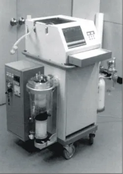
Fig. 1. A prototype of Korean single-pulsatile extracorporeal life support (ECLS) system.
apolis)를 대조군으로 하여 단일 박동형 구동펌프에 압력 완충장치를 부착한 군과 부착하지 않은 군을 비교하는 것 으로 설정하였다. 이 때 단일 박동형 구동펌프는 한국형 생명구조장치의 시제품(prototype)을 이용하였다(Fig. 1, 2).
실험동물은 한국산 잡견(20∼25 kg) 16마리를 대상으로 하였고, 사용된 구동펌프의 종류에 따라 3개 군으로 분류 하였다. 1군(n=6)은 비박동형 원심펌프를 사용하였고, 2군 (n=4)은 단일 박동형 구동펌프를 단독으로 사용하였고, 3 군(n=6)에서는 단일 박동형 구동펌프와 막형산화기 사이 에 압력완충장치(pressure-relieving compliance chamber)를 추가하였다. 사전 예비실험을 통해 구동펌프와 막형산화 기 사이에 Y-자 회로를 구성하여 PVC 튜브 내부에 위치 하는 공기의 압력완충 기능을 확인한 후(Fig. 3A, B), 본 실험에서는 자체 제작한 개량형 압력완충장치를 사용하 였다(Fig. 4A, B).
구동펌프 이외에 회로구성에 사용된 재료는 정맥도관 (Polystan, 28 Fr.), 동맥도관(Stockert, 5.2 mm), PVC순환회 로(Avecor, 10 mm ID×4 M), 막형산화기(Terumo, SX-10) 등이었다.
2) 실험 준비
실험동물의 관리는 고려대학교 의과대학 실험동물 관 리규정에 따랐다. 환경변화에 의한 실험동물의 생리변화 를 최소화하기 위해 구입 후 7일간 전문사육인에 의해 실 험실 환경에서 안정시켰다. 실험시작 전 8시간 동안 금식 시킨 후, 케타민 근육주사로 전처치하였다. 전박정맥에 주 사통로를 확보하여 펜토탈(25 mg/kg)을 정맥주사하여 전 신마취를 유도하였다. 정맥주사통로는 실험 중 수액요법 에도 사용하였다. 기도삽관 후 흡입마취기를 이용하여 기 계환기(흡기 산소분압 0.4, 호흡수 10∼15회/분, 일회 환기 량 15 mL/kg)를 시키면서 동맥혈 산소 분압과 이산화탄소 분압을 정상범위(150∼200 mmHg 및 35∼45 mmHg)로 유 지시켰다. 마취 도중 소변, 출혈 등에 의한 체액손실을 보 충하기 위해서 Ringer's lactate 용액을 3 mL/kg/hr의 속도 로 점적 유지 주사하였다. 마취가 완료되고 혈역학이 안 정된 후 심박수와 심전도를 지속적으로 관찰하면서 경피 천자법으로 우측 대퇴동맥에 20 gauge의 도관을 삽입하여 동맥압 관찰과 채혈 통로로 사용하였다. 실험견의 목을 좌측으로 젖히고 우측 내경정맥을 박리하여 7 Fr. Swan- Fig. 2. Schematic diagram of the ECLS layout.
Oxygenater
Vacuum Actuator
Oxygen Blood Sac
Inlet: 23 Fr catheter Outlet: 19 Fr (Medtronics.co.)
Heat controller
Electro-hydraulic Ventricular assist device
Ganz 카테타를 삽입하였다. 수술은 모든 동물에서 흉골 횡절개를 통해 심장을 노출시킨 다음, 전신 헤파린 처치(3 mg/kg) 후 우심방과 상행대동맥에 도관을 삽입하고 순환 장치에 연결하였다. 이 시점에서 실험동물에 급성 호흡부 전증을 유도하기 시작하였으며, 급성 호흡부전증이 유발 된 후 부분 체외순환(partial cardiopulmonary bypass)을 실 시하였다. 부분순환의 혈류속도는 1.8∼2 L/min를 유지하 였으며, 체온은 자체 제작한 열교환기를 사용하여 항온을
유지하였다. 수술 과정의 마취 및 호흡관리는 마취전문의 가 담당하였다.
3) 급성 호흡부전증의 유발
올레익산(cis-9-Octadecenoic acid, Sigma, St. Louis, USA) 0.1 mL/kg를 동량의 100% 에탄올에 녹여 infusion pump로 Swan-Ganz 카테타를 통해 폐동맥으로 30분 동안 서서히 주 사하였다[4]. 올레익산 정맥주사 시작 후 60분이 되었을 때
Fig. 4. (A) Schematic diagram for location of a pressure-relie- ving compliance chamber, (B) A revised pressure-relieving compli- ance chamber.
A
Single pulse ECLS
Pressure-relieving compliance chamber
RA Aorta
Membrane oxygenator
B
Fig. 3. (A) A schematic diagram for location of a compliance PVC tube using a Y-loop circuit, (B) A compliance PVC tube using a Y-loop circuit (arrow head) was located at between a single- pulsatile pump and a membrane oxygenator in the previous pilot study.
A
Single pulse ECLS
Compliance PVC tube using Y-loop circuit
Air cushion
RA Aorta
Membrane oxygenator
B
각종 관찰지표를 측정하였으며, 이 때 흡기 산소분압 0.4에 서 동맥혈 산소분압이 60 mmHg 이하(PaO2÷FiO2<150)인 경우 급성 호흡부전증 모델이 완성된 것으로 간주하였다.
4) 관찰지표 측정
관찰지표는 순환을 시작한 1시간 및 2시간 후에 관찰한 혈역학 지표(심박수, 동맥혈압 및 맥동압, 심박출량, 중심 정맥압, 좌심방압, 폐동맥압, 폐동맥 쐐기압), 회로압, 동맥 혈 및 혼합정맥혈 가스분석, 혈액검사(CBC, electrolytes, 간기능, 신기능, 혈당), 혈장 유리 헤모글로빈(plasma free hemoglobin) 등에 국한하였다. 모든 측정치는 평균±표준
편차로 표기하였고, 통계처리는 SPSS 7.0 standard version 을 사용하여 oneway ANOVA 테스트 중 Duncan법과 Turkey- HSD 테스트 방법을 사용하여 p<0.05 이하인 경우를 통 계적으로 유의성이 있는 것으로 간주하였다.
결 과
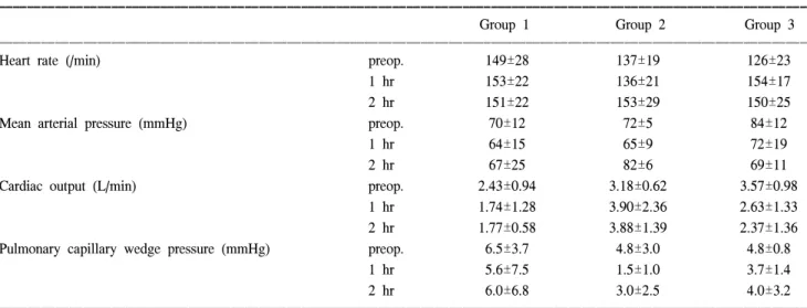
1) 혈역학 지표
맥동압(pulse pressure=systolic pressure-diastolic pressure) 은 박동형 구동펌프를 이용한 2군(47±10 mmHg)과 3군 (41±9 mmHg) 간에 의미있는 차이는 없었고, 비박동형 원 Table 1. Hemodynamic parameters
ꠚꠚꠚꠚꠚꠚꠚꠚꠚꠚꠚꠚꠚꠚꠚꠚꠚꠚꠚꠚꠚꠚꠚꠚꠚꠚꠚꠚꠚꠚꠚꠚꠚꠚꠚꠚꠚꠚꠚꠚꠚꠚꠚꠚꠚꠚꠚꠚꠚꠚꠚꠚꠚꠚꠚꠚꠚꠚꠚꠚꠚꠚꠚꠚꠚꠚꠚꠚꠚꠚꠚꠚꠚꠚꠚꠚꠚꠚꠚꠚꠚꠚꠚꠚꠚꠚꠚꠚꠚꠚꠚꠚꠚꠚꠚꠚꠚꠚꠚꠚꠚꠚꠚꠚꠚꠚꠚꠚꠚꠚꠚꠚꠚꠚꠚ
Group 1 Group 2 Group 3
ꠏꠏꠏꠏꠏꠏꠏꠏꠏꠏꠏꠏꠏꠏꠏꠏꠏꠏꠏꠏꠏꠏꠏꠏꠏꠏꠏꠏꠏꠏꠏꠏꠏꠏꠏꠏꠏꠏꠏꠏꠏꠏꠏꠏꠏꠏꠏꠏꠏꠏꠏꠏꠏꠏꠏꠏꠏꠏꠏꠏꠏꠏꠏꠏꠏꠏꠏꠏꠏꠏꠏꠏꠏꠏꠏꠏꠏꠏꠏꠏꠏꠏꠏꠏꠏꠏꠏꠏꠏꠏꠏꠏꠏꠏꠏꠏꠏꠏꠏꠏꠏꠏꠏꠏꠏꠏꠏꠏꠏꠏꠏꠏꠏꠏꠏ
Heart rate (/min) preop. 149±28 137±19 126±23
1 hr 153±22 136±21 154±17
2 hr 151±22 153±29 150±25
Mean arterial pressure (mmHg) preop. 70±12 72±5 84±12
1 hr 64±15 65±9 72±19
2 hr 67±25 82±6 69±11
Cardiac output (L/min) preop. 2.43±0.94 3.18±0.62 3.57±0.98
1 hr 1.74±1.28 3.90±2.36 2.63±1.33
2 hr 1.77±0.58 3.88±1.39 2.37±1.36
Pulmonary capillary wedge pressure (mmHg) preop. 6.5±3.7 4.8±3.0 4.8±0.8
1 hr 5.6±7.5 1.5±1.0 3.7±1.4
2 hr 6.0±6.8 3.0±2.5 4.0±3.2
ꠏꠏꠏꠏꠏꠏꠏꠏꠏꠏꠏꠏꠏꠏꠏꠏꠏꠏꠏꠏꠏꠏꠏꠏꠏꠏꠏꠏꠏꠏꠏꠏꠏꠏꠏꠏꠏꠏꠏꠏꠏꠏꠏꠏꠏꠏꠏꠏꠏꠏꠏꠏꠏꠏꠏꠏꠏꠏꠏꠏꠏꠏꠏꠏꠏꠏꠏꠏꠏꠏꠏꠏꠏꠏꠏꠏꠏꠏꠏꠏꠏꠏꠏꠏꠏꠏꠏꠏꠏꠏꠏꠏꠏꠏꠏꠏꠏꠏꠏꠏꠏꠏꠏꠏꠏꠏꠏꠏꠏꠏꠏꠏꠏꠏꠏ
Table 2. Changes in blood gas patterns
ꠚꠚꠚꠚꠚꠚꠚꠚꠚꠚꠚꠚꠚꠚꠚꠚꠚꠚꠚꠚꠚꠚꠚꠚꠚꠚꠚꠚꠚꠚꠚꠚꠚꠚꠚꠚꠚꠚꠚꠚꠚꠚꠚꠚꠚꠚꠚꠚꠚꠚꠚꠚꠚꠚꠚꠚꠚꠚꠚꠚꠚꠚꠚꠚꠚꠚꠚꠚꠚꠚꠚꠚꠚꠚꠚꠚꠚꠚꠚꠚꠚꠚꠚꠚꠚꠚꠚꠚꠚꠚꠚꠚꠚꠚꠚꠚꠚꠚꠚꠚꠚꠚꠚꠚꠚꠚꠚꠚꠚꠚꠚꠚꠚꠚꠚ
Group 1 Group 2 Group 3
ꠏꠏꠏꠏꠏꠏꠏꠏꠏꠏꠏꠏꠏꠏꠏꠏꠏꠏꠏꠏꠏꠏꠏꠏꠏꠏꠏꠏꠏꠏꠏꠏꠏꠏꠏꠏꠏꠏꠏꠏꠏꠏꠏꠏꠏꠏꠏꠏꠏꠏꠏꠏꠏꠏꠏꠏꠏꠏꠏꠏꠏꠏꠏꠏꠏꠏꠏꠏꠏꠏꠏꠏꠏꠏꠏꠏꠏꠏꠏꠏꠏꠏꠏꠏꠏꠏꠏꠏꠏꠏꠏꠏꠏꠏꠏꠏꠏꠏꠏꠏꠏꠏꠏꠏꠏꠏꠏꠏꠏꠏꠏꠏꠏꠏꠏ
Arterial PO2 (mmHg) preop. 224±75 208±31 225±66
1 hr 54±14 56±16 48±13
2 hr 77±41 96±48* 97±25*
Arterial PCO2 (mmHg) preop. 41±4 40±3 29±8
1 hr 39±14 42±14 34±5
2 hr 57±31 42±12 41±5
Mixed venous PO2 (mmHg) preop. 50±12 43±7 52±8
1 hr 30±8 43±20 35±7
2 hr 31±8 61±18* 39±15*
Mixed venous PCO2 (mmHg) preop. 67±21 58±7 57±25
1 hr 54±16 49±17 44±12
2 hr 67±32 48±18 49±8
ꠏꠏꠏꠏꠏꠏꠏꠏꠏꠏꠏꠏꠏꠏꠏꠏꠏꠏꠏꠏꠏꠏꠏꠏꠏꠏꠏꠏꠏꠏꠏꠏꠏꠏꠏꠏꠏꠏꠏꠏꠏꠏꠏꠏꠏꠏꠏꠏꠏꠏꠏꠏꠏꠏꠏꠏꠏꠏꠏꠏꠏꠏꠏꠏꠏꠏꠏꠏꠏꠏꠏꠏꠏꠏꠏꠏꠏꠏꠏꠏꠏꠏꠏꠏꠏꠏꠏꠏꠏꠏꠏꠏꠏꠏꠏꠏꠏꠏꠏꠏꠏꠏꠏꠏꠏꠏꠏꠏꠏꠏꠏꠏꠏꠏꠏ
*p<0.05 compared to group 1.
심펌프를 이용한 1군(17±7 mmHg)에 비해 유의하게 높았 다(p<0.001). 심박수, 동맥혈압, 심박출량, 중심정맥압, 좌 심방압, 폐동맥압, 폐동맥 쐐기압은 각 군 사이에 의미있 는 차이를 나타내지 않았다(p=NS)(Table 1).
2) 회로압
막형산화기에 걸리는 회로압은 1군의 경우 222±8 mmHg, 2군의 경우 739±35 mmHg, 3군의 경우 470±17 mmHg였 다(p<0.001).
3) 혈액가스분석
순환 2시간째 박동형 구동펌프를 사용한 2군과 3군에서 동맥혈 산소분압이 비박동형 원심펌프를 사용한 1군에 비 해 유의하게 높았다(1군; 77±41 mmHg, 2군; 96±48 mmHg, 3군; 97±25 mmHg, p<0.05)(Table 2). 정맥혈 산소 분압을 포함한 기타 가스분석 결과는 각 군 사이에 의미
있는 차이를 나타내지 않았다(p=NS).
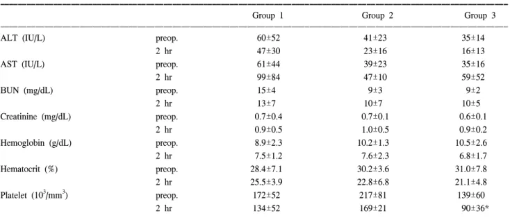
4) 혈액검사 지표
CBC, electrolytes, 간기능(protein, albumin, cholesterol, AST, ALT), 신기능(BUN, creatinine), 혈당치는 각 군 사이 에 의미있는 차이를 나타내지 않았다(p=NS)(Table 3).
5) 혈장 유리 헤모글로빈치
순환 2시간째 혈구손상지표인 혈장 유리 헤모글로빈은 1군에서 가장 낮았고, 2군에서 가장 높았으며, 3군에서 유 의하게 감소하였다(1군; 55.7±43.3 mg%, 2군; 162.8±
113.6 mg%, 3군; 82.5±25.1 mg%, p<0.05)(Table 4).
고 찰
생명구조장치(ECLS)는 주로 응급상황에서 심폐기능을 Table 3. Blood chemistry and cells
ꠚꠚꠚꠚꠚꠚꠚꠚꠚꠚꠚꠚꠚꠚꠚꠚꠚꠚꠚꠚꠚꠚꠚꠚꠚꠚꠚꠚꠚꠚꠚꠚꠚꠚꠚꠚꠚꠚꠚꠚꠚꠚꠚꠚꠚꠚꠚꠚꠚꠚꠚꠚꠚꠚꠚꠚꠚꠚꠚꠚꠚꠚꠚꠚꠚꠚꠚꠚꠚꠚꠚꠚꠚꠚꠚꠚꠚꠚꠚꠚꠚꠚꠚꠚꠚꠚꠚꠚꠚꠚꠚꠚꠚꠚꠚꠚꠚꠚꠚꠚꠚꠚꠚꠚꠚꠚꠚꠚꠚꠚꠚꠚꠚꠚꠚ
Group 1 Group 2 Group 3
ꠏꠏꠏꠏꠏꠏꠏꠏꠏꠏꠏꠏꠏꠏꠏꠏꠏꠏꠏꠏꠏꠏꠏꠏꠏꠏꠏꠏꠏꠏꠏꠏꠏꠏꠏꠏꠏꠏꠏꠏꠏꠏꠏꠏꠏꠏꠏꠏꠏꠏꠏꠏꠏꠏꠏꠏꠏꠏꠏꠏꠏꠏꠏꠏꠏꠏꠏꠏꠏꠏꠏꠏꠏꠏꠏꠏꠏꠏꠏꠏꠏꠏꠏꠏꠏꠏꠏꠏꠏꠏꠏꠏꠏꠏꠏꠏꠏꠏꠏꠏꠏꠏꠏꠏꠏꠏꠏꠏꠏꠏꠏꠏꠏꠏꠏ
ALT (IU/L) preop. 60±52 41±23 35±14
2 hr 47±30 23±16 16±13
AST (IU/L) preop. 61±44 39±23 35±16
2 hr 99±84 47±10 59±52
BUN (mg/dL) preop. 15±4 9±3 9±2
2 hr 13±7 10±7 10±5
Creatinine (mg/dL) preop. 0.7±0.4 0.7±0.1 0.6±0.1
2 hr 0.9±0.5 1.0±0.5 0.9±0.2
Hemoglobin (g/dL) preop. 8.9±2.3 10.2±1.3 10.5±2.6
2 hr 7.5±1.2 7.6±2.3 6.8±1.7
Hematocrit (%) preop. 28.4±7.1 30.2±3.6 31.0±7.8
2 hr 25.5±3.9 22.8±6.8 21.1±4.8
Platelet (103/mm3) preop. 172±52 217±81 139±60
2 hr 134±52 169±21 90±36*
ꠏꠏꠏꠏꠏꠏꠏꠏꠏꠏꠏꠏꠏꠏꠏꠏꠏꠏꠏꠏꠏꠏꠏꠏꠏꠏꠏꠏꠏꠏꠏꠏꠏꠏꠏꠏꠏꠏꠏꠏꠏꠏꠏꠏꠏꠏꠏꠏꠏꠏꠏꠏꠏꠏꠏꠏꠏꠏꠏꠏꠏꠏꠏꠏꠏꠏꠏꠏꠏꠏꠏꠏꠏꠏꠏꠏꠏꠏꠏꠏꠏꠏꠏꠏꠏꠏꠏꠏꠏꠏꠏꠏꠏꠏꠏꠏꠏꠏꠏꠏꠏꠏꠏꠏꠏꠏꠏꠏꠏꠏꠏꠏꠏꠏꠏ
*p<0.05 compared to group 1. AST=Aspartate aminotransferase; ALT=Alanine aminotransferase; BUN=Blood urea nitrogen; Hgb=
Hemoglobin.
Table 4. Blood cell trauma
ꠚꠚꠚꠚꠚꠚꠚꠚꠚꠚꠚꠚꠚꠚꠚꠚꠚꠚꠚꠚꠚꠚꠚꠚꠚꠚꠚꠚꠚꠚꠚꠚꠚꠚꠚꠚꠚꠚꠚꠚꠚꠚꠚꠚꠚꠚꠚꠚꠚꠚꠚꠚꠚꠚꠚꠚꠚꠚꠚꠚꠚꠚꠚꠚꠚꠚꠚꠚꠚꠚꠚꠚꠚꠚꠚꠚꠚꠚꠚꠚꠚꠚꠚꠚꠚꠚꠚꠚꠚꠚꠚꠚꠚꠚꠚꠚꠚꠚꠚꠚꠚꠚꠚꠚꠚꠚꠚꠚꠚꠚꠚꠚꠚꠚꠚ
Group 1 Group 2 Group 3
ꠏꠏꠏꠏꠏꠏꠏꠏꠏꠏꠏꠏꠏꠏꠏꠏꠏꠏꠏꠏꠏꠏꠏꠏꠏꠏꠏꠏꠏꠏꠏꠏꠏꠏꠏꠏꠏꠏꠏꠏꠏꠏꠏꠏꠏꠏꠏꠏꠏꠏꠏꠏꠏꠏꠏꠏꠏꠏꠏꠏꠏꠏꠏꠏꠏꠏꠏꠏꠏꠏꠏꠏꠏꠏꠏꠏꠏꠏꠏꠏꠏꠏꠏꠏꠏꠏꠏꠏꠏꠏꠏꠏꠏꠏꠏꠏꠏꠏꠏꠏꠏꠏꠏꠏꠏꠏꠏꠏꠏꠏꠏꠏꠏꠏꠏ
Plasma free hemoglobin (mg/dL) preop. 20.8±15.8 7.6±5.3 18.9±8.3
2 hr 55.7±43.3 162.8±113.6* 82.5±25.1†
ꠏꠏꠏꠏꠏꠏꠏꠏꠏꠏꠏꠏꠏꠏꠏꠏꠏꠏꠏꠏꠏꠏꠏꠏꠏꠏꠏꠏꠏꠏꠏꠏꠏꠏꠏꠏꠏꠏꠏꠏꠏꠏꠏꠏꠏꠏꠏꠏꠏꠏꠏꠏꠏꠏꠏꠏꠏꠏꠏꠏꠏꠏꠏꠏꠏꠏꠏꠏꠏꠏꠏꠏꠏꠏꠏꠏꠏꠏꠏꠏꠏꠏꠏꠏꠏꠏꠏꠏꠏꠏꠏꠏꠏꠏꠏꠏꠏꠏꠏꠏꠏꠏꠏꠏꠏꠏꠏꠏꠏꠏꠏꠏꠏꠏꠏ
*p<0.05 compared to group 1; †p<0.05 compared to group 2
단기간 보조하는 목적으로 사용한다[5,6]. 체외순환 기법 을 사용하는 다양한 기계순환장치는 그 용도에 따라 명칭 이 다르게 붙여진다. 예를 들어, 심장수술을 할 때 심폐기 능을 전체적으로 보조하는 데 쓰이는 장치는 일반적으로 인공심폐기(heart-lung machine)[7] 혹은 심폐우회장치(cardio- pulmonary bypass; CPB)[8,9]라고 부른다. 만일 호흡부전 치료를 주목적으로 사용하는 경우는 인공폐(artificial lung) [10], 체외 막형 산화장치(extracorporeal membrane oxygena- tion; ECMO)[11], 체외 폐보조(extracorporeal lung assist;
ECLA)[12]라고 부르고, 이 때 심장기능 보조는 필요하지 않다. 심폐정지와 같은 응급상황에서 경피 도관삽관을 통 해 응급 순환보조에 이용되면 심폐보조(cardiopulmonary support; CPS)[13], 체외 심폐소생장치(extracorporeal cardio- pulmonary resuscitation; ECPR)[14] 혹은 이동성 심폐소생 장치(portable cardiopulmonary resuscitation; PCPR)[15-17]라 고 불리게 된다. 만일 혈액펌프 기능만을 가지고 있으면 심실보조기(ventricular assist device; VAD)[18]라고 부르며 혈액산화 기능은 배제된다. 결국 기계순환장치는 우회되 는 순환계의 특성, 산화기의 부착 여부, 도관 삽입방법, 구 동펌프의 형태, 열교환기의 부착 여부 등에 따라 다양한 기능과 명칭을 가지지만, 기본적으로 체외순환이라는 기 법을 사용하는 것이다. 임상에서 생명구조장치는(ECLS)는 인공폐 혹은 체외 막형 산화장치(ECMO)와 거의 동일한 의미로 사용되는 경우가 많다[10]. 그러나 연구자의 관점 에 따라 미국의 Michigan 대학과 같이 위에 언급한 모든 순 환기기장치들을 넓은 의미의 ECLS에 포함시키기도 한다.
ECLS의 기본구성은 구동펌프, 막형산화기, 도관, 연결 관, 열교환기, 기타 측정장치들로 되어 있다. 이 때 막형산 화기를 사용하므로 구동펌프로 비박동형 원심펌프가 선 호된다[6]. 인체 내에서 박동성 혈류가 비박동성 혈류보다 말초혈관저항이 적어 미세순환을 유지하고, 조직 대사를 향상시키며, 부종 형성을 방지하는 등 말초조직의 대사에 더 유리하게 작용한다는 것은 이미 알려진 바이다[19-22].
또한 최근에는 비박동성 혈류에 비해 박동성 혈류가 관상 동맥을 확장시켜 혈류를 증가시킨다는 연구결과도 보고 되고 있다[23,24]. 이렇듯 박동형 구동장치는 생리적인 박 동혈류를 제공한다는 측면에서는 유리하지만, 급격한 회 로압 상승과 그에 따른 혈구손상에 대한 우려가 있어서 특히 막형산화기를 사용하는 인공폐(ECMO) 회로에서 기 피되어 왔다.
이에 대해 저자 등은 박동형 구동펌프와 막형 산화기 사이에 압력완충장치를 설치함으로써 언급한 부작용을
최소화시키면서 생리적인 박동혈류를 유도할 수 있을 것 이라는 가설을 제시하였고, 사전 예비실험을 통해 구동펌 프와 막형산화기 사이에 Y-자 회로를 구성하여 PVC 튜브 내부에 위치하는 공기의 압력완충 효과를 검증하여 보았 다. 그 결과 막형산화기에 걸리는 압력이 효과적으로 떨 어지는 것을 확인하였기에, 본 실험에서는 자체 제작한 개량형 압력완충장치를 적용하게 되었다.
본 연구에 사용된 단일 박동형 구동펌프는 한국형 생명 구조장치의 시제품(prototype)으로서, 구동 메카니즘은 체 외형 좌심실 보조장치(LVAD) 기술을 근간으로 하고 있 다. 생명구조장치에 사용되는 구동펌프의 조건은 좌심실 보조장치보다 순간적으로 더 많은 양의 혈액을 박출시켜 야 하고, 아울러 길이가 긴 도관과 막형 산화기에 의해 발 생하는 회로 내부의 저항을 이겨내고, 막형 산화기 후반 의 혈압 강하를 최소화하여야 한다. 따라서, 기존의 좌심 실 보조장치보다 성능과 안정성을 향상시키고 조작이 간 편하도록 개선된 구조를 가지고 있다. 추가적인 특징으로 는 소형 경량화를 통해 이동성과 현장성이 가능하도록 시 도하고 있으며, 특히 경피 도관 삽입을 통해 응급실이나 중환자실 혹은 심도자실 등에서 즉시 사용이 가능하도록 고안되고 있다. 더 나아가 구급차에 설치하여 병원 밖의 현장에서도 활용할 수 있는 가능성을 기대하고 있다. 실 험실 조건에서 구동펌프의 성능은 다음과 같다. : 최대 혈 류보조량 6 L/min, 최대 후부하(afterload) 500 mmHg, 최대 전부하(preload) -200 mmHg, synchronous operation with R pulse: 100 B.P.M (beat per minute), 전동기 자동속도 조절 기능. 올레익산으로 유발되는 급성 호흡부전 실험동물모 델은 유용한 방법으로 널리 이용되고 있고, 본 연구에서 도 이를 사용하였다[25,26].
실험결과 혈역학 지표에서 순환 전후기간 동안 3군 간 에 차이가 없었다는 것은 한국형 단일 박동형 구동펌프가 외국산 원심펌프에 비하여 혈액순환능력이 뒤지지 않음 을 증명하는 것이다. 또한 혈액검사지표에서도 군 간에 유의한 차이를 나타내지 않았으며, 이는 2시간의 순환보 조에서 말초장기 혈류 유지효과가 대등하다는 것을 시사 한다.
회로압 상승과 혈구손상은 압력완충장치를 사용한 3군 에서 유의하게 감소시킬 수 있었으며, 1군의 비박동형 원 심펌프와 비교해도 통계적인 차이는 없었다. 그러나 전반 적으로 높은 회로압과 혈장 유리 헤모글로빈 수치를 감안 할 때, 단일 박동형 구동펌프를 임상에 적용하기는 아직 어려울 것으로 판단된다.
본 실험에서 특기할 내용은, 박동형 혈류를 사용한 2군 과 3군에서 비박동형 혈류를 사용한 1군에 비해 우수한 혈액산화효과를 보였다는 점이다. 이는 막형산화기에서의 산소교환 효율에 박동형 혈류가 영향을 미친 것으로 해석 이 되는데, 이에 대해서는 알려진 기전이 없기에 추가연 구가 필요할 것으로 본다.
저자 등은 본 실험을 통해 한국형 생명구조장치 개발에 몇 가지 개선방향을 설정하였으며 향후 연구목표로 삼고 자 한다. 첫째, 회로압과 혈구손상을 보다 최소화할 수 새 로운 박동형 구동 메카니즘의 개발; 둘째, 구성 면에서 경 량 소형화의 추진과 보다 나은 사용 편리성을 확보하며 경피접근이 가능한 주변부속기구의 개발; 셋째, 전자 및 기계 면에서 펌프의 구동력 증가, 제어회로의 안정성 증 진, 동기성(synchronization) 부여 등을 고려하고 있다.
결 론
단일 박동형 혈류구동장치는 막형산화기에서 산소교환 효율에 유리한 반면, 단독으로 사용할 경우 회로압과 혈 구세포 손상 측면에서 불리하다는 것을 알 수 있었다. 구 동펌프와 막형 산화기 사이에 압력완충장치를 설치하는 경우, 회로압과 혈구세포 파괴를 유의하게 줄일 수 있다 는 가능성이 확인되었다. 향후 박동형 생명구조장치 개발 을 위해 새로운 구동 메카니즘에 대한 연구가 필요하다.
참 고 문 헌
1. Weber TR, Tracy TF Jr, Connors R, Kountzman B, Penning- ton DG. Prolonged extracorporeal support for nonneonatal respiratory failure. J Ped Surg 1992;27:1100-5.
2. Zapal WM, Snider MT, Hill JD, et al. Extracorporeal mem- brane oxygenation in severe acute respiratory failure. JAMA 242:2193-6.
3. Kanto WP Jr, Shapiro MB. The development of prolonged extracorporeal circulation. In: Zwischenberger JB & Bartlett RH. Extracorporeal cardiopulmonary support in critical care.
Ann Arbor: Extracorporeal life support organization, 1995.
4. Schoene RB, Roberton HT, Thorning DR, Springmeyer SC, Hlastala MP, Cheney FW. Patholophysiological patterns of resolution from acute oleic acid lung injury in the dog. J App Physiol 1984;56:472-81.
5. Zwischenberger JB, Bartlett RH. An introduction to Extra- corporeal life support. In: Zwischenberger JB & Bartlett RH.
Extracorporeal cardiopulmonary support in critical care.
Ann Arbor: Extracorporeal life support organization, 1995.
6. McDonald M, Smedira NG. Adult extracorporeal life sup-
port: In: Franco KL, Verrier ED. Advanced therapy in car- diac surgery. Hamilton: B.C. Decker, Inc., 1999;326-34.
7. Austin JW, Harner DL. The heart-lung machine & related technologies of open heart surgery. Phoenix: Phoenix Medical Communication Medical Publisher, 1986.
8. Kenneth MT. Cardiopulmonary bypass: Principles and man- agement. Baltimore: Chapman and Hall Ltd., 1986.
9. Kim WG, No JL. Principles and practices on cardiopul- monary bypass. Seoul: Korea Medical Book publisher, 1996.
10. Palmer AS, Collins J, Head LR. Development of an implan- table artificial lung. J Thorac Cardiovasc Surg 1973;66:521-5.
11. Zwischenberger JB, Bartlett RH. Extracorporeal cardio- pulmonary support in critical care. Ann Arbor: Extracor- poreal life support organization, 1995.
12. Fumagalli R, Kolobow T, Arosio P, Chen V, Buckhold DK, Pierce JE. Successful treatment of experimental neonatal res- piratory failure using extracorporeal membrane lung assist.
Int J Artif Organs 1986;9:427-32.
13. Mooney MR, Arom KV, Joyce LD, et al. Emergency cardio- pulmonary bypass support in patients with cardiac arrest. J Thorac Cardiovasc Surg 1991;101:450-4.
14. Kurose M, Okamoto K, Sato T, et al. The determinant of severe cerebral dysfunction in patients undergoing emergency extracorporeal life support following cardiopulmonary resus- citation. Resuscitation 1995;30:15-20.
15. Waters DJ, Belz M, Lawse D, Ulstad D. Portable cardiopul- monary bypass: resuscitation from prolonged ice-water submersion and asystole. Ann Thorac Surg 1994;57:1018-9.
16. Kim HM, Lee IS, Baek MJ, et al. Report for development of Korean portable cardiopulmonary bypass machine. I. A com- parison of open-versus closed-chest cardiopulmonary resus- ictation in normal dogs. Korean J Thorac Cardiovasc Surg 1998;31:827-36.
17. Kim HM, Lee IS, Baek MJ, et al. Report for development of Korean portable cardiopulmonary bypass machine. II. Exper- imental study of protable cardiopulmonary bypass for emer- gency cardiopulmonary resusictation after cardiac arrest in normal dogs. Korean J Thorac Cardiovasc Surg 1998;31:
1147-58.
18. Arabia FA, Copeland JG. Bridge to transplantation with left ventricular assist devices and total artificial heart. In: Fran- co KL, Verrier ED. Advanced therapy in cardiac surgery.
Hamilton: B.C. Decker, Inc., 1999;416-29.
19. Milnor WR. Pulsatile blood flow. New Engl J Med 1972;
287:27.
20. Hickey PR, Buckley MJ, Philbin DM. Pulsatile and nonpul- satile cardiopulmonary bypass: Review of a counterprodu- ctive controversy. Ann Thorac Surg 1983;36:720-37.
21. Minami K, Korner MM, Vyska K, Kleesiek K, Knobi H, Korfer R. Effects of pulsatile perfusion on plasma catechol- amine levels and hemodynamics during and after cardiac operations with cardiopulmonary bypass. J Thoracic Cardio-
vasc Surg 1990;99:82-91.
22. Nakano T, Tominaga R, Ichiro N, Hayato O, Yasui H. Pul- satile flow enhances endothelium-derived nitric oxide release in the peripheral vasculature. Am J Physiol Heart Circ Physiol 2000;278;H1098-H1104.
23. Paolocci N, Pagliaro P, Isoda T, Saavedra FW, Kass DA.
Role of calcium-sensitive K+ channels and nitric oxide in in vivo coronary vasodilation from enhanced perfusion pulsa- tility. Circulation 2001;103:119-24.
24. Qiu WP, Hu Q, Paolocci N, Ziegelstein RC, Kass DA. Dif-
ferential effects of pulsatile versus steady flow on coronary endothelial membrane potential. Am J Physiol Heart Circ Physiol 2003;285;H341-H346.
25. Schuster DP. ARDS: clinical lessons from the oleic acid model of acute lung injury. Am J Resp Crit Care Med 1994;149:245-60.
26. Han SB, Baek KJ, Kim JS, Cheon YJ, Sun K. Effects of tidal volume changes in mechanical ventilation management for acute lung injury -An experimental study-. J Korean Soc Traumatol 1999;12:1-8.
=국문 초록=
배경: 생명구조장치(ECLS)는 호흡부전 및 심부전에 적용하는 장치로 세계적으로 많은 연구개발과 임 상시도가 계속되고 있다. 현재까지 생명구조장치의 혈류펌프로는 비박동형 구동장치가 표준으로 사 용되고 있으며, 박동형 구동장치는 생리적인 측면에서 유리하지만 급격한 회로압 상승과 그에 따른 혈구손상에 대한 우려가 있고 특히 막형산화기를 사용하는 인공폐 회로에서 기피되어 왔다. 본 연구 는 인공폐 실험모델에서 단일 박동형 구동펌프와 막형 산화기 사이에 압력완충장치를 설치함으로써 언급한 부작용을 최소화시키면서 생리적인 박동혈류를 유도할 수 있을 것이라는 가능성을 검증하였 다. 대상 및 방법: 실험대상은 올레익산으로 급성 호흡부전증이 유발된 잡견(N=16)을 사용하였다. 실 험군은 사용된 구동펌프의 종류와 압력완충장치의 유무에 따라 3개 군으로 분류하였다. 제1군(n=6)은 대조군으로 원심펌프를 이용하였고, 제2군(n=4)은 단일 박동형 구동펌프를 사용하였으며, 제3군(n=6) 은 단일 박동형 구동펌프에 압력완충장치를 설치하였다. 실험모델은 흉강 절개 후 우심방-대동맥을 우회시키는 부분 체외순환 형태로, 1.8∼2 L/min의 펌프박출량에서 2시간을 구동하였다. 관찰지표는 주로 혈역학 변화, 회로압, 혈액검사 및 혈구세포에 미치는 영향 등에 한정하였다. 결과: 맥동압(pulse pressure)은 박동형 구동펌프를 이용한 2군(47±10 mmHg)과 3군(41±9 mmHg)에서 비박동형 원심펌 프를 이용한 1군(17±7 mmHg)에 비해 유의하게 높았다(p<0.001). 막형산화기에 걸리는 회로압은 1 군의 경우 222±8 mmHg, 2군의 경우 739±35 mmHg, 3군의 경우 470±17 mmHg였다(p<0.001). 순환 2시간째의 동맥혈 산소분압은 1군에 비해 박동혈류를 사용하는 2군과 3군에서 현저하게 높았다(77±
41, 96±48, 97±25 mmHg; p<0.05). 혈구손상지표인 혈장 유리 헤모글로빈치는 1군에서 가장 낮았고, 2군에서 가장 높았으며, 3군에서 유의하게 감소하였다(55.7±43.3, 162.8±113.6, 82.5±25.1 mg%; p<
0.05). 기타 혈액검사치는 특별한 차이가 없었다. 결론: 이상의 결과에서 단일 박동형 구동펌프는 비 박동형 원심펌프에 비해 막형산화기의 산소교환에 유리한 반면, 회로압과 혈구세포 손상 측면에서 불리하다는 것을 알 수 있었다. 그러나 단일 박동형 구동펌프와 막형 산화기 사이에 압력완충장치를 설치하는 경우, 회로압 상승과 혈구세포손상을 유의하게 감소시킬 수 있었다.
중심 단어:1. 생명구조장치 2. 인공폐 3. 체외순환 4. 호흡부전증