INTRODUCTION
Mycoplasma pneumoniae (M. pneumoniae), an atypical mi- croorganism, is a major causative agent of respiratory infection in children1 and is isolated from more than 40% of patients with community-acquired pneumonia.2 M. pneumoniae infection occurs most commonly in children aged 4-5 years, with waves of prevalence every 3-4 years.1
The relationship between M. pneumoniae infection and asth- ma development has been debated for the past 20 years.3 Previ- ous studies have demonstrated that the incidence of M. pneu- moniae infection is higher in patients with chronic stable asth- ma or acute exacerbation than in control subjects.4-6 In addition, M. pneumoniae infection has been shown to correlate with the induction and aggravation of asthma symptoms. Biscardi et al.7 have demonstrated that the recurrence rate of asthma is higher in atopic children aged 2-15 years whose first attack was accom-
Mycoplasma pneumoniae Infection Affects the Serum Levels of Vascular Endothelial Growth Factor and Interleukin-5 in Atopic Children
You-Cheol Jeong, Mun-Soo Yeo, Joo-Hwa Kim, Ha-Baik Lee, Jae-Won Oh*
Department of Pediatrics, Hanyang University College of Medicine, Seoul, Korea
panied by M. pneumoniae infection, compared with those without. This observation suggests that M. pneumoniae infec- tion is related to asthma attacks. Further studies analyzing the expression of various cytokines are needed to understand the relationship between M. pneumoniae infection and asthma.
Vascular endothelial growth factor (VEGF) is a major media- tor that induces airway hypersensitivity to inhalant allergens.
VEGF is secreted from endothelial cells and smooth muscle cells as well as inflammatory cells upon inflammatory reac- tions. It induces angiogenesis, permeability changes, and ede- Allergy Asthma Immunol Res. 2012 March;4(2):92-97.
http://dx.doi.org/10.4168/aair.2012.4.2.92 pISSN 2092-7355 • eISSN 2092-7363
Purpose: Previous studies have outlined mechanisms by which Mycoplasma pneumonia (M. pneumonia) infection may promote allergic lung in- flammation and airway remodeling, and increasing evidence from human studies suggests that atypical bacterial infections contribute to asthma ex- acerbation, chronic asthma, and disease severity with changes in cytokine expression. The present study evaluated changes in serum levels of vas- cular endothelial growth factor (VEGF) and interleukin (IL)-5 in atopic children with Mycoplasma pneumoniae pneumonia. Methods: We recruited a total of 72 children with pneumonia. The patients were divided into 4 groups: atopic children with M. pneumonia pneumonia (group I, n=24), non- atopic children with M. pneumonia pneumonia (group II, n=23), atopic children with viral pneumonia (group III, n=13), and non-atopic children with viral pneumonia (group IV, n=12). Serum levels of IL-5, IL-13, VEGF, and tumor necrosis factor-α were measured at admission and at recovery using enzyme-linked immunosorbent assays. Results: Serum levels of VEGF and IL-5 were elevated in group I compared with the other groups at both admission phase and clinical recovery phase. In group I, serum levels of VEGF and IL-5 were higher at recovery phase than at admission phase (VEGF:
1,102.2±569.4 vs. 874.9±589.9 pg/mL, respectively; IL-5: 150.5±63.9 vs. 120.2±46.7 pg/mL, respectively). Conclusions: The serum levels of VEGF and IL-5 were more increased in atopic children with M. pneumonia pneumonia than in the other groups. In this group, the serum levels of VEGF and IL-5 were more increased at recovery phase than at admission phase. The results of this study suggest that increases in VEGF and IL-5 may con- tribute to the development of hypersensitivity during M. pneumonia infection. These cytokines may act through their respective pro-inflammatory pathways to aggravate the allergic status and induce airway hypersensitivity during M. pneumonia pneumonia in atopic children.
Key Words: Atopy; interleukin-5; Pneumonia, Mycoplasma; vascular endothelial growth factor
This is an Open Access article distributed under the terms of the Creative Commons Attribution Non-Commercial License (http://creativecommons.org/licenses/by-nc/3.0/) which permits unrestricted non-commercial use, distribution, and reproduction in any medium, provided the original work is properly cited.
Correspondence to: Jae-Won Oh, MD, PhD, Department of Pediatrics, Hanyang University Guri Hospital, 249-1 Gyomun-dong, Guri 471-701, Korea.
Tel: +82-31-560-2254; Fax: +82-31-552-9493; E-mail: [email protected] Received: August 19, 2011; Revised: October 11, 2011;
Accepted: October 25, 2011
•There are no financial or other issues that might lead to conflict of interest.
ma formation in the airway wall; all of these play crucial roles in the pathophysiology of asthma. Hypersecretion of VEGF in the airways induces T cell sensitization to inhalant allergens and stimulates the synthesis of co-stimulatory molecules in lung- resident dendritic cells.8 A previous study has indicated that se- rum VEGF levels are significantly higher in M. pneumoniae in- fected-patients with wheezing than in those without wheezing.9 Interleukin (IL)-5 is primarily secreted by Th2 cells and is re- lated to the growth, maturation, differentiation, survival, and activation of eosinophils. Eosinophils accumulate in the lungs of transgenic mice that overexpress IL-5.10,11 Moreover, IL-5 plays a role in the development of asthma. Choi et al.12 have demon- strated that serum levels of VEGF and IL-5 are significantly higher in atopic patients with wheezing than in those without or in control subjects. IL-13, which plays a role in airway re- modeling, induces tissue fibrosis through transforming growth factor-β activation.13 In addition, IL-13 has been shown to in- duce myofibroblast hyperplasia, airway obstruction, and air- way hyperresponsiveness.9 In in vitro experiments, Thavagna- nam et al.14 showed that IL-13 enhances goblet cell hyperplasia and decreases the number of bronchial epithelial cells detected in samples taken from children, suggesting an IL-13-mediated mechanism for the development of asthma. Interferon (IFN)-γ is released by Th1 cells and suppresses Th2 cells, thus main- taining the balance between Th1 and Th2 cells. Stirling and Chung15 have advocated that serum IFN-γ levels are decreased in atopic patients and that there is a significant relationship be- tween the decrease in IFN-γ and the severity of asthma. Lack et al.16 reported that IL-5 levels and the number of eosinophils are both decreased in bronchoalveolar lavage fluid after intrathecal administration of IFN-γ. Tumor necrosis factor (TNF)-α increas- es airway responsiveness and promotes the synthesis of various cytokines such as regulated upon activation, normal T-cell ex- pressed and secreted, IL-8, and granulocyte-macrophage stim- ulating factor, which in turn initiate chronic airway inflamma- tion.15
This study was conducted to investigate the effect of Mycoplas- ma infection on allergic diseases by measuring the serum levels
of VEGF, IL-5, IL-13, IFN-γ, and TNF-α.
MATERIALS AND METHODS Study subjects
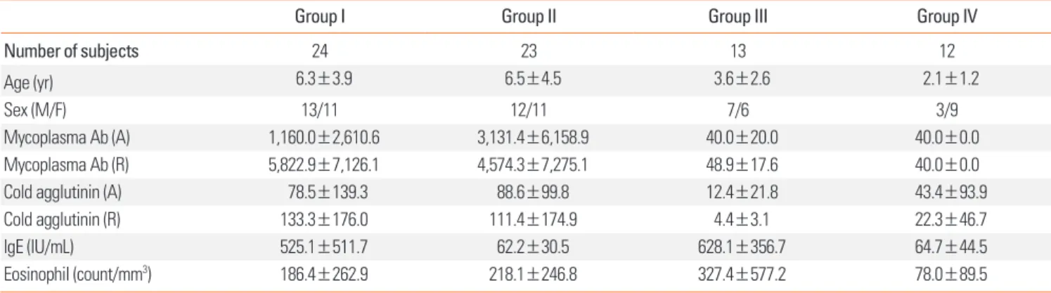
This study enrolled a total of 72 children who were admitted to the Department of Pediatrics at Hanyang University Guri Hospital in Korea, due to pneumonia. This study was performed prospectively from October, 2010 to June, 2011. The mean age of the children was 5.2±3.9 years (range, 2-15 years). All chil- dren underwent evaluation for past/family history of allergic diseases, physical examination, routine laboratory tests, and the measurement of total IgE and the number of eosinophils in serum. The children were divided into 4 groups (Table 1): those with M. pneumoniae pneumonia who had increased serum IgE and past/family history of allergic diseases and (group I, n=24);
those with M. pneumoniae pneumonia who had normal serum IgE and no past/family history of allergic diseases (group II, n=23); those with viral pneumonia who had increased serum IgE and past/family history of allergic diseases (group III, n=
13); and those with viral pneumonia who had normal serum IgE and no past/family history of allergic diseases (group IV, n=12). The protocol and consent forms of this study were ap- proved by the Institutional Review Board at Hanyang Universi- ty. Informed written consent was obtained from all parents and/or participants.
Diagnosis of M. pneumoniae pneumonia
Pneumonia was diagnosed based on clinical symptoms such as fever, cough, dyspnea, and crackles and radiological find- ings. M. pneumoniae as a causative organism of pneumonia was identified by anti-mycoplasma antibody titers, which were measured by the microparticle agglutination method using a commercial kit (Serodia-Myco II, Fujirebio, Tokyo, Japan). Anti- mycoplasma antibody titers were regarded as positive when the they were ≥1:160 at admission phase or when the titers at symptomatic recovery phase were more than four-fold those measured at admission phase. The range of duration from ad-
Table 1. Characteristics of subject in the study
Group I Group II Group III Group IV
Number of subjects 24 23 13 12
Age (yr) 6.3±3.9 6.5±4.5 3.6±2.6 2.1±1.2
Sex (M/F) 13/11 12/11 7/6 3/9
Mycoplasma Ab (A) 1,160.0±2,610.6 3,131.4±6,158.9 40.0±20.0 40.0±0.0
Mycoplasma Ab (R) 5,822.9±7,126.1 4,574.3±7,275.1 48.9±17.6 40.0±0.0
Cold agglutinin (A) 78.5±139.3 88.6±99.8 12.4±21.8 43.4±93.9
Cold agglutinin (R) 133.3±176.0 111.4±174.9 4.4±3.1 22.3±46.7
IgE (IU/mL) 525.1±511.7 62.2±30.5 628.1±356.7 64.7±44.5
Eosinophil (count/mm3) 186.4±262.9 218.1±246.8 327.4±577.2 78.0±89.5
mission to symptomatic recovery was 7-10 days.
Diagnosis of viral pneumonia
Viral pneumonia was diagnosed based on respiratory symp- toms, positive radiographic findings, and positive results of vi- rus polymerase chain reaction (PCR) analysis of nasopharyn- geal secretions. At admission, samples of nasopharyngeal se- cretions were obtained from the nasopharynx by aspiration through catheters after injecting 5 mL of sterile physiological saline solution into mucus traps. Each specimen was indepen- dently screened for the presence of common respiratory virus- es, including influenza viruses, parainfluenza viruses, human coronaviruses, human rhinoviruses, human metapneumovi- ruses, human adenoviruses, and respiratory syncytial viruses, by reverse transcription-PCR using a commercial kit (Seeplex RV 12 ACE Detection, Seegene, Seoul, Korea).
Measurement of serum IgE and number of eosinophils
Serum IgE levels were measured using an IRMA kit (Diagnos- tic Products Co., Los Angeles, CA, USA). The number of periph- eral eosinophils was measured with an automated hematology analyzer (Coulter Counter STKS, Beckman Coulter, Fullerton, CA, USA), using blood samples collected into tubes containing EDTA.
Measurement of serum VEGF, IL-5, IL-13, IFN-γ, and TNF-α Venous blood was taken from each subject, centrifuged at 1,000×g for 10 minute at 4°, and stored at -70°C. IL-5, IL-13, IFN-γ, and TNF-α levels were measured using commercial en- zyme-linked immunosorbent assays (ELISAs) (DBD Inc., San Diego, CA, USA), according to the manufacturer’s instructions.
The detection limit was 2 pg/mL for IL-5, IL-13, IFN-γ, and TNF-α. VEGF was measured using a human VEGF kit (Bio- source, Camarillo, CA, USA). The values measured at admis- sion were compared with those measured at the time of symp- tomatic recovery.
Statistical analysis
Statistical analyses were performed using SPSS version 18.0 (SPSS Inc., Chicago, IL, USA). The data are expressed as means±SD. Statistical comparisons were made with paired t- tests. A P value of <0.05 indicated statistical significance. Para- metric one-way ANOVA with post hoc Tukey correction was used to compare levels of VEGF and IL-5 among the study groups.
RESULTS
Each patient group shows different clinical characteristics, laboratory results, total IgE level, and number of eosinophils (Table 1). In groups I and II, anti-mycoplasma antibody titers were ≥1:160 at admission phase; the titers at the time of symp-
tomatic recovery phase were more than four-fold those at ad- mission phase. In groups III and IV, anti-mycoplasma antibody titers were <1:40 at admission phase. and were not increased at recovery phase. Serum IgE levels were significantly higher in groups I and III compared with groups II and IV. There was no significant difference in the number of eosinophils among the four subject groups.
Serum VEGF levels
Serum VEGF levels measured at admission phase were signif- icantly higher in group I (874.9±589.9 pg/mL) than in groups III and IV (396.9±251.5 and 356.9±310.6 pg/mL, respectively;
P<0.01). At the time of symptomatic recovery phase, serum VEGF levels were significantly higher in group I (1,102.2±569.4 pg/mL) than in groups II, III and IV (659.4±434.8, 431.3±210.6, and 411.7±367.6 pg/mL, respectively) (Fig. 1; Table 2). Serum VEGF levels tended to be higher at recovery phase than at ad- mission phase in all four groups, but only group I showed sig- nificantly increased VEGF levels at the time of symptomatic re- covery phase (1,102.2±569.4 vs. 874.9±589.9 pg/mL, recovery phase vs. admission phase; P<0.05).
Serum levels of IL-5, IL-13, and TNF-α
Serum IL-5 levels were significantly higher in group I (120.2±
46.7 pg/mL) than in groups III and IV (92.5±36.2 and 91.0±
34.9 pg/mL, respectively; P<0.01) at admission phase. Serum IL-5 levels were increased at recovery phase compared with those at admission phase in all four groups, but the increase was significant only in group I (150.5±63.9 vs. 120.2±46.7 pg/
mL, recovery phase vs. admission phase; P<0.05). At the time of symptomatic recovery phase, serum IL-5 was significantly higher in group I (150.5±63.9 pg/mL) than in groups III and IV (98.6±33.1 and 96.3±49.9 pg/mL, respectively; P<0.05) (Fig. 2;
Table 2).
Table 2. The comparison of serum VEGF, IL-5, IL-13, and TNF-α level in the study
Group I Group II Group III Group IV
VEGF(A) 874.9±589.9*,‡ 617.9±420.6 396.9±251.5‡ 356.9±310.6‡ VEGF(R) 1,102.2±569.4*,§ 659.4±434.8§ 431.3±210.6§ 411.7±367.6§ IL-5(A) 120.2±46.7† 101.5±45.1 92.5±36.2 91.0±34.9 IL-5(R) 150.5±63.9†,‡ 112.4±43.3 98.6±33.1II 96.3±49.9II IL-13(A) 218.1±111.8 262.0±142.7 273.3±155.0 215.3±77.8 IL-13(R) 221.4±147.8 240.8±208.5 290.9±173.5 232.8±128.0 TNF-α(A) 84.4±49.3 85.4±60.9 96.3±56.9 82.2±38.4 TNF-α(R) 94.7±47.6 86.7±64.0 105.3±100.9 87.5±46.8
*VEGF(A) vs VEGF(R): P<0.01, †IL-5(A) vs IL-5(R): P=0.01, ‡VEGF(A) group I vs group III, group I vs group IV: P<0.01, §VEGF(R) group I vs group II, group I vs group III, group I vs group IV: P<0.01, IIIL-5(R) group I vs group III, group I vs group IV: P<0.05, Scale for cytokine level: pg/mL.
VEGF, vascular endothelial growth factor; IL, interleukin; TNF, Tumor necrosis factor.
Serum levels of IL-13 and TNF-α were not significantly differ- ent among the four groups, at admission phase or at recovery phase. Serum IL-13 and TNF-α levels did not differ significantly between admission phase and symptomatic recovery phase in any group (Table 2).
DISCUSSION
In this study, serum VEGF levels were significantly higher in group I than in groups II and III, and IV at admission phase.
Our findings, which included atopic children with and without wheezing, are consistent with those of previous studies that in- cluded children with wheezing.9,12 It has been suggested that compared with allergy, M. pneumoniae can more frequently aggravate or initiate asthma symptoms, and that M. pneumoni- ae pneumonia and asthma are significantly correlated in terms of clinical symptoms.17,18 It has also been shown that allergic re- actions are induced or aggravated by IgE-related hypersensitiv- ity and Th2 immune reactions.19-21 M. pneumoniae induces up- per airway infections such as acute pharyngitis and laryngitis more frequently than lower airway infections.22 Clyde23 report- ed that 20% of patients with M. pneumoniae infection were as- ymptomatic, and that less than 5% of all patients with M. pneu- moniae infection had lower airway infections such as pneumo- nia. Based on these results, it is conceivable that M. pneumoni-
ae infection may stimulate or aggravate allergic reactions.
In the present study, VEGF levels were significantly higher in atopic children with M. pneumoniae pneumonia than in those with viral pneumonia. VEGF, an endothelial cell-specific mito- genic peptide, serves as a vascular permeability factor and plays a crucial role in vasculogenesis and angiogenesis. It also induces extravasation of protein-rich plasma from the airway, increases vascular permeability, and causes airway inflamma- tion through edema formation.24,25 Increased VEGF levels with M. pneumoniae infection suggest that M. pneumoniae stimu- lates allergic inflammation, as well as airway vascular angio- genesis and edema formation, consequently aggravating air- way inflammation. In previous studies, the levels of IL-13, IL-4, and TNF-α were increased with M. pneumoniae infection, sug- gesting that this infection can aggravate asthma symptoms.26,27 However, in our study, there were no significant differences in serum IL-13 and TNF-α levels between admission phase and symptomatic recovery phase in any of the four study groups.
This apparent discrepancy may be attributable to differences between the two study cohorts.
In the present study, serum IL-5 levels at admission were high- er in groups I and II (M. pneumonia groups) than in groups III and IV (viral pneumonia groups), although these differences did not reach statistical significance. This result agrees with pre- vious results reported by Esposito et al.28
Fig. 1. Comparison of serum VEGF levels at admission phase (A) and at clinical recovery phase (R) in the four subject groups. The mean serum VEGF level was high- er at clinical recovery phase than at admission phase in group I, whereas the mean serum VEGF levels did not differ significantly between recovery phase and ad- mission phase in the other groups.
VEGF (A) VEGF (R)
3,000 2,500 2,000 1,500 1,000 500 0
P<0.01
Group I
pg/mL
VEGF (A) VEGF (R)
3,000 2,500 2,000 1,500 1,000 500 0
NS
Group II
pg/mL
VEGF (A) VEGF (R)
3,000 2,500 2,000 1,500 1,000 500
0 Group III
NS
pg/mL
VEGF (A) VEGF (R)
3,000 2,500 2,000 1,500 1,000 500
0 Group IV
NS
pg/mL
Serum levels of VEGF and IL-5 were significantly higher at the time of symptomatic recovery phase than at admission phase.
This implies that M. pneumonia infection stimulates bacterial inflammation as well as allergic inflammation, and that inflam- mation reactions related to VEGF and IL-5 persist even after re- covery from M. pneumoniae pneumonia, prolonging allergic inflammation. The increased VEGF levels at the time of symp- tomatic recovery phase in our study suggest the presence of VEGF-induced increases in vascular permeability, airway vas- cular angiogenesis, and edema formation, all of which aggra- vate airway inflammation. Serum IL-5 levels at recovery phase were significantly higher in group I than in the other groups, in- dicating that IL-5-dependent allergic inflammatory reactions are persistently induced by M. pneumoniae infection in atopic children, but not in non-atopic children.
In the present study, serum levels of IL-13 and TNF-α tended to be higher in atopic children with viral pneumonia (group III) compared with the other groups; however, the differences were not significant. We also did not detect differences in serum cy- tokine levels between children with viral pneumonia caused by respiratory syncytial virus or rhinovirus and children infected by other viruses (data not shown).
The serum levels of VEGF and IL-5 were more increased in
atopic children with M. pneumonia pneumonia than in the other groups. In this group, the serum levels of VEGF and IL-5 were more increased at recovery phase than at admission phase.
In conclusion, our study results suggest that M. pneumoniae pneumonia may induce allergic inflammatory reactions as well as bacterial inflammatory reactions and can persistently aggra- vate existing allergic inflammation. Further studies with larger sample sizes are needed to confirm our results. Mechanisms of allergic reactions through other cytokine mediators also merit additional investigation.
ACKNOWLEDGMENTS
This study was supported by the 2010 Hanyang University, College of Medicine, Research Fund (HY-2010-MC) and a Re- search Grant of Alumni of Pediatrics, College of Medicine, Han- yang University.
REFERENCES
1. Kim KW, Kim KE. Mycoplasma and chlamydia infection in Korea.
Korean J Pediatr 2009;52:277-82.
2. Kim JW, Seo HK, Yoo EG, Park SJ, Yoon SH, Jung HY, Han MY. My- Fig. 2. Comparison of serum IL-5 levels at admission phase (A) and at clinical recovery phase (R) in the four subject groups.The mean serum IL-5 level was higher at clinical recovery phase than at admission phase in group I, whereas the mean serum IL-5 levels did not differ significantly between recovery phase and admission phase in the other groups.
IL-5 (A) IL-5 (R)
300 250 200 150 100 50 0
P=0.01
Group I
pg/mL
IL-5 (A) IL-5 (R)
300 250 200 150 100 50 0
NS
Group II
pg/mL
IL-5 (A) IL-5 (R)
300 250 200 150 100 50 0
NS
Group III
pg/mL
IL-5 (A) IL-5 (R)
300 250 200 150 100 50 0
NS
Group IV
pg/mL
coplasma pneumoniae pneumonia in Korean children, from 1979 to 2006-a meta-analysis. Korean J Pediatr 2009;52:315-23.
3. Hansbro PM, Beagley KW, Horvat JC, Gibson PG. Role of atypical bacterial infection of the lung in predisposition/protection of asth- ma. Pharmacol Ther 2004;101:193-210.
4. Esposito S, Blasi F, Arosio C, Fioravanti L, Fagetti L, Droghetti R, Tarsia P, Allegra L, Principi N. Importance of acute Mycoplasma pneumoniae and Chlamydia pneumoniae infections in children with wheezing. Eur Respir J 2000;16:1142-6.
5. Kraft M, Cassell GH, Henson JE, Watson H, Williamson J, Marmion BP, Gaydos CA, Martin RJ. Detection of Mycoplasma pneumoniae in the airways of adults with chronic asthma. Am J Respir Crit Care Med 1998;158:998-1001.
6. Martin RJ, Kraft M, Chu HW, Berns EA, Cassell GH. A link between chronic asthma and chronic infection. J Allergy Clin Immunol 2001;
107:595-601.
7. Biscardi S, Lorrot M, Marc E, Moulin F, Boutonnat-Faucher B, Heil- bronner C, Iniguez JL, Chaussain M, Nicand E, Raymond J, Gen- drel D. Mycoplasma pneumoniae and asthma in children. Clin In- fect Dis 2004;38:1341-6.
8. Brown LF, Detmar M, Claffey K, Nagy JA, Feng D, Dvorak AM, Dvorak HF. Vascular permeability factor/vascular endothelial growth factor: a multifunctional angiogenic cytokine. EXS 1997;79:
233-69.
9. Seo Y, Yu BK, Oh YJ, Lee Y, Yoo Y, Choung JT, Koh YY. Increased vascular endothelial growth factor in children with acute Myco- plasma pneumoniae pneumonia and wheezing. Korean J Pediatr 2008;51:487-91.
10. Mould AW, Ramsay AJ, Matthaei KI, Young IG, Rothenberg ME, Foster PS. The effect of IL-5 and eotaxin expression in the lung on eosinophil trafficking and degranulation and the induction of bronchial hyperreactivity. J Immunol 2000;164:2142-50.
11. Rosenwasser LJ, Rothenberg ME. IL-5 pathway inhibition in the treatment of asthma and Churg-Strauss syndrome. J Allergy Clin Immunol 2010;125:1245-6.
12. Choi IS, Byeon JH, Yoo Y, Lee KC, Choung JT. Increased serum in- terleukin-5 and vascular endothelial growth factor in children with acute mycoplasma pneumonia and wheeze. Pediatr Pulmonol 2009;44:423-8.
13. Lee CG, Homer RJ, Zhu Z, Lanone S, Wang X, Koteliansky V, Shipley JM, Gotwals P, Noble P, Chen Q, Senior RM, Elias JA. Interleukin-13 induces tissue fibrosis by selectively stimulating and activating transforming growth factor beta(1). J Exp Med 2001;194:809-21.
14. Thavagnanam S, Parker JC, McBrien ME, Skibinski G, Heaney LG, Shields MD. Effects of IL-13 on mucociliary differentiation of pedi-
atric asthmatic bronchial epithelial cells. Pediatr Res 2011;69:95- 100.
15. Stirling RG, Chung KF. New immunological approaches and cyto- kine targets in asthma and allergy. Eur Respir J 2000;16:1158-74.
16. Lack G, Bradley KL, Hamelmann E, Renz H, Loader J, Leung DY, Larsen G, Gelfand EW. Nebulized IFN-gamma inhibits the devel- opment of secondary allergic responses in mice. J Immunol 1996;
157:1432-9.
17. MacDowell AL, Bacharier LB. Infectious triggers of asthma. Immu- nol Allergy Clin North Am 2005;25:45-66.
18. Nisar N, Guleria R, Kumar S, Chand Chawla T, Ranjan Biswas N.
Mycoplasma pneumoniae and its role in asthma. Postgrad Med J 2007;83:100-4.
19. Tipirneni P, Moore BS, Hyde JS, Schauf V. IgE antibodies to Myco- plasma pneumoniae in asthma and other atopic diseases. Ann Al- lergy 1980;45:1-7.
20. Yano T, Ichikawa Y, Komatu S, Arai S, Oizumi K. Association of My- coplasma pneumoniae antigen with initial onset of bronchial asth- ma. Am J Respir Crit Care Med 1994;149:1348-53.
21. Wills-Karp M. Immunologic basis of antigen-induced airway hy- perresponsiveness. Annu Rev Immunol 1999;17:255-81.
22. Atkinson TP, Balish MF, Waites KB. Epidemiology, clinical manifes- tations, pathogenesis and laboratory detection of Mycoplasma pneumoniae infections. FEMS Microbiol Rev 2008;32:956-73.
23. Clyde WA Jr. Mycoplasma pneumoniae respiratory disease sympo- sium: summation and significance. Yale J Biol Med 1983;56:523-7.
24. Hirakawa S, Okazaki H, Sayama K, Tohyama M, Hashimoto K. Pos- sible association of vascular endothelial growth factor with the de- velopment of edema in drug-induced hypersensitivity syndrome. J Dermatol 2011;38:292-4.
25. Corrigan CJ, Wang W, Meng Q, Fang C, Wu H, Reay V, Lv Z, Fan Y, An Y, Wang YH, Liu YJ, Lee TH, Ying S. T-helper cell type 2 (Th2) memory T cell-potentiating cytokine IL-25 has the potential to pro- mote angiogenesis in asthma. Proc Natl Acad Sci U S A 2011;108:
1579-84.
26. Kraft M, Cassell GH, Duffy LB, Metze T, Pak J, Gaydos CA, Martin RJ. Mycoplasma and Chlamydia are present in the airways of chronic, stable asthmatics. Am J Respir Crit Care Med 1999;159:
A517.
27. Hassan J, Irwin F, Dooley S, Connell J. Mycoplasma pneumoniae in- fection in a pediatric population: analysis of soluble immune markers as risk factors for asthma. Hum Immunol 2008;69:851-5.
28. Esposito S, Droghetti R, Bosis S, Claut L, Marchisio P, Principi N.
Cytokine secretion in children with acute Mycoplasma pneumoni- ae infection and wheeze. Pediatr Pulmonol 2002;34:122-7.