서론
일반적으로 내경동맥 협착이나 폐색에 의해 해당 혈관영역의 관류압이 감소하게 되더라도, 일정한 뇌관류를 유지하기 위하여 앞 혹은 뒤 교통동맥(anterior- or posterior-communicating ar- tery)을 포함한 윌리스환(circle of Willis)이 일차성(primary) 측 부순환 경로로, 안동맥(ophthalmic artery), 전맥락동맥(anterior choroidal artery), 혹은 뇌표면에서 주행하는 연수막혈관들
(leptomeningeal vessels)이 이차성(secondary) 측부순환 경로 로 작용하여 뇌의 허혈성 손상을 막는 것으로 알려져 있다(1).
이 밖에도 뇌혈관들은 혈관과 두개내 압력의 변화에 따라 비교 적 크기가 큰 전도성(conductance) 혈관부터 세동맥까지의 혈 관벽 평활근 세포의 긴장도를 조절하여 뇌관류를 유지하려 하 는데 이것을 뇌혈관 자동조절능(cerebral autoregulation)이라 일컫는다. 관류압이 감소되는 상황에서도 말단 뇌세동맥들의 내경을 확장시켜 관류압을 유지하려 하기 때문에, 뇌혈류 예비
J Korean Soc Radiol 2014;70(1):1-12 http://dx.doi.org/10.3348/jksr.2014.70.1.1
Received September 5, 2013; Accepted November 26, 2013 Corresponding author: Young Hen Lee, MD Department of Radiology, Ansan Hospital, Korea University College of Medicine, 123 Jeokgeum-ro, Danwon-gu, Ansan 425-707, Korea.
Tel. 82-31-412-5228 Fax. 82-31-412-5224 E-mail: younghen@korea.ac.kr
This is an Open Access article distributed under the terms of the Creative Commons Attribution Non-Commercial License (http://creativecommons.org/licenses/by-nc/3.0) which permits unrestricted non-commercial use, distri- bution, and reproduction in any medium, provided the original work is properly cited.
Purpose: We correlated cerebrovascular reserve in unilateral high grade carotid ar- tery stenosis or occlusion with a type of collateral circulation using acetazolamide- challenged perfusion CT (ACZ-PCT).
Materials and Methods: Among the patients who underwent ACZ-PCT in our in- stitution, we retrospectively selected the patients with unilateral high grade internal carotid artery or middle cerebral artery stenosis (> 70%) or occlusion; we verified the types of their dominant collateral circulation by digital subtraction angiography or 3T MR-angiography; first, the primary collaterals flow through the circle of Wil- lis; second, the secondary collaterals that flow through the opthalmic artery, the basal artery or other external carotid artery. Using ACZ-PCT, we measured the dif- ference in percentage change of cerebral blood flow of the stenotic hemisphere against contralateral normal hemisphere and compared cerebrovascular reserves of lesional hemisphere, according to the type of collaterals.
Results: A total of 28 patients were included. The percentage changes of cerebral blood flow were significantly lower in the stenotic hemisphere than the contralat- eral hemisphere (14.34 ± 36.43% and 34.53 ± 47.82%, p < 0.001), and in the hemi- sphere predominantly supplied by secondary collaterals than primary (7.03 ± 32.71% and 24.37 ± 42.03%, p < 0.05), respectively.
Conclusion: Cerebrovascular reserves in the ipsilateral hemisphere predominantly supplied by secondary collaterals were more impaired than primary collaterals in patients with unilateral high grade carotid stenosis or occlusion.
Index terms Carotid Stenosis
Cerebrovascular Insufficiency Collateral Circulations Perfusion Imaging
Correlation of Cerebrovascular Reserve as Measured by
Acetazolamide-Challenged Perfusion CT with Collateral Circulation in Unilateral High Grade Carotid Stenosis
1심한 편측성 경동맥 협착에서 아세타졸라마이드 부하 뇌관류 CT를 이용하여 측정한 뇌혈류 예비능과 측부순환의 연관성
1Doran Hong, MD
1, Young Hen Lee, MD
1, Hyung Suk Seo, MD
1, Bo-Kyung Je, MD
1, Sang-il Suh, MD
2, Hae Young Seol, MD
2, Nam Joon Lee, MD
3, Jung Hyuk Kim, MD
3, Kyung-Sook Yang, MD
41Department of Radiology, Ansan Hospital, Korea University College of Medicine, Ansan, Korea
2Department of Radiology, Guro Hospital, Korea University College of Medicine, Seoul, Korea
3Department of Radiology, Anam Hospital, Korea University College of Medicine, Seoul, Korea
4Department of Biostatistics, Korea University College of Medicine, Seoul, Korea
ACZ-PCT 시행 1주일 이내에 급성 뇌경색 여부를 판단하기 위 하여 확산강조영상을 포함한 자기공명영상이 시행된 경우였다.
혈관 협착의 정도는 일차적으로 3T 자기공명영상 동맥조영술을 이용하여 결정하였고, 측부순환 평가를 위하여 시행된 디지털 감 산처리 혈관조영술에서 확인하였다. 두개내 혈관에 대한 평가는 3차원 time-of-flight (이하 TOF) 자기공명영상 동맥조영술의 최대 신호강도 영상에서 원위부 내경동맥이나 중대뇌동맥의 근 위분절(M1)을, 두개외 혈관의 경우 조영증강 자기공명영상 동 맥조영술에서 총경동맥 분기부위직후의 근위부 내경동맥을 대 상으로 하여, 최대 혈관협착부위의 내경이 협착이 없는 부위에 비해 육안상 70% 이상 감소하였거나 폐쇄가 확인된 경우를 연 구에 포함시켰다(24). 양측성 협착, 70% 미만의 일측성 협착이 나 반대측 경동맥에 경도 이상의 협착을 보이는 경우와 우세한 측부순환의 형태가 디지털 감산처리 혈관조영술, 그리고 3T 자 기공명영상 동맥조영술에서 확인되지 않은 경우 등은 제외하였 다. 모든 환자에서 혈관의 협착 정도와 위치, 허혈성 증상 유무 와 뇌경색 위험인자에 해당하는 고혈압, 당뇨, 흡연, 고지혈증, 심장혈관질환 유무를 기록하였다. 입원하여 시행한 확산강조영 상에서 급성 뇌경색이 확인되거나 단일 혹은 반복되는 일과성 허 혈 발작 및 해당 혈관 영역의 경색 증상이 있는 경우 유증상으 로, 해당하지 않으면 무증상으로 분류하였다. 본 연구는 임상시 험심사위원회의 심사를 통과하였다.
영상 검사 방법
자기공명영상은 3T 장비(Achieva X-series, Philips Medical Systems, Best, the Netherlands)의 16채널 뇌혈관코일(neuro- vascular coil)을 사용하여 T1, T2 강조영상 및 액체감쇄역전회 복(fluid attenuated inversion recovery)영상, 경사에코영상과 3차원 TOF 자기공명영상 동맥조영술, 경동맥을 포함한 뇌혈 관의 조영증강 자기공명영상 동맥조영술을 포함하였다. 급성 경색 유무를 확인하기 위하여 b값 1000 s/mm2의 확산강조영 상(diffusion weigheted image)과 현성확산계수영상(apparent diffusion coefficient map)이 자기공명 혈관조영술 시행 3~4 일 전에 시행되었으며, 확산강조영상에서 고신호강도, 현성확 산계수영상에서 저신호강도로 일치되는 병변을 급성 뇌경색으 로 정의하였다.
두개내 윌리스환 영역을 포함하는 3차원 TOF 자기공명영상 동맥조영술은 다음과 같은 조건에서 횡단면(axial)영상을 획득 하여 구성하였다: 영상범위(field of view), 25 × 20 cm; 반복 시간/반향시간(repetition time/echo time), 25/3.5 ms; 숙임 각(flip angle), 20˚; 행렬크기(matrix), 716 × 282; 화적소 크 기(voxel size), 0.35 × 0.7 × 1.2 mm; 영상재구성 화소 크 능(cerebrovascular reserve)이 감소되었다는 것은 뇌조직 수준
에서의 말단 혈관들이 이미 최대한 확장하여 뇌관류를 보상하 고 있어 더 이상의 혈관 확장을 기대할 수 없는 상황으로, 향후 뇌졸중이나 일시적 허혈성 발작의 위험이 증가한다는 것이 이전 연구들에서 밝혀져 있다(2-4). 즉, 경동맥 혈관성 질환 환자들 의 허혈성 뇌손상의 임상양상은 협착 자체뿐만 아니라, 측부순 환과 뇌혈관 자동조절능을 통하여 협착 말단부위 혈류를 얼마 나 효과적으로 보상하느냐에 의하여 결정된다(1, 5, 6).
진행된 경동맥 혈관성 질환 환자들에서 발달하는 이차성 측 부순환이 뇌혈류의 증가를 통하여 뇌의 허혈성 손상을 예방하 는데 기여한다고 알려진 것에 대하여(7-9), 일부 연구들은 이차 성 측부순환 존재 자체가 일차성 측부순환 경로의 불완전성을 시사하기 때문에 뇌 혈역동학적 불안정성과 뇌혈류 예비능의 감 소와 연관이 있다고 지적하였다(2, 3, 10-15). 하지만, 이전 연구 들에서 뇌혈류 예비능을 평가하기 위해 사용한 경두개 초음파, 단일광자방출컴퓨터단층촬영(single photon emission computed tomography), 양전자방출단층촬영(positron emission tomog- raphy)에 비하여 해상도가 우수하고, 간편하게 정량적 평가가 가능하며 기저 뇌관류 전산화단층촬영(baseline perfusion CT) 에 비해 더 정확한 것으로 알려진 아세타졸라마이드 부하 뇌관 류 CT (acetazolamide-challenged perfusion CT; 이하 ACZ- PCT)를 이용하여 측부순환의 형태에 따른 뇌혈류 예비능을 평 가하려 한 시도는 없었다(7, 9, 16-23). 이러한 이유로 저자들은 디지털 감산처리 혈관조영술과 3T 자기공명영상 동맥조영술로 우세한 측부순환의 형태가 확인된 심한 일측성 경동맥 협착 환 자들을 대상으로, ACZ-PCT를 이용하여 환자의 측부순환의 형태에 따른 뇌혈류 예비능을 비교하고자 하였다.
대상과 방법
환자 선택
2010년 10월부터 2012년 6월까지 3T 자기공명영상 동맥조영 술에서 경동맥의 협착과 폐색이 발견되어 뇌혈류 예비능을 평가 하기 위하여 ACZ-PCT를 시행한 총 80명의 신경과와 신경외 과 입원 환자 중 다음의 조건을 만족시키는 환자를 후향적으로 선택하였다. 연구에 포함시킨 조건은 1) 디지털 감산처리 혈관조 영술과 3T 자기공명영상 동맥조영술 중에 적어도 한 가지에서 내경동맥이나 중대뇌동맥의 심한 일측성 협착이나 폐색[North American Symptomatic Carotid Endarterectomy Trial (이하 NASCET) 분류법을 기준으로 70% 이상의 중증 협착과 완전 폐색], 2) 디지털 감산처리 혈관조영술과 3T 자기공명영상 동맥 조영술을 통하여 우세한 측부순환의 형태가 확인되었으며, 3)
명영상 동맥조영술)의 평균 간격은 4일(1~10일)이었다.
뇌관류 CT를 시행하여 얻어진 영상은 비반복적 디콘볼루션 (noniterative deconvolution)에 바탕을 둔 뇌관류 소프트웨어 (Extended Brilliance Workspace v 3.0, Philips Medical Sys- tems, Cleveland, OH, USA)를 이용한 후처리 작업을 거쳐 상 대적인 뇌혈류용적(cerebral blood volume), 뇌혈류량(cerebral blood flow; 이하 CBF), 평균통과시간(mean transit time) 및 최고농도도달시간(time to peak) 등의 지도로 재구성되었다 (20). 전대뇌동맥(anterior cerebral artery) 분절과 상시상정맥 (superior sagital sinus)을 각각 동맥입력기능(arterial input function), 정맥출력기능(venous output function)의 참고값으 로 사용하였고, 뇌피질 주위의 혈관은 자동으로 제거하였다.
영상분석
연구에 포함된 환자의 후향적 선정 기준이 되는 혈관 협착 정 도와 측부순환의 평가는 신경영상전문의 1인에 의해, 급성 뇌 경색유무를 판단하기 위한 자기공명영상과 뇌관류 CT의 평가 는 신경영상전문의 1인과 영상의학 전공의 1인의 합의에 의해 이루어졌다. 측부순환의 평가는 디지털 감산처리 혈관조영술이 우선적인 기준이 되어, 디지털 감산처리 혈관조영술을 시행한 환자의 경우, NASCET 분류법을 기준으로 70% 이상의 중증 협착이나 완전 폐색이 확인된 일측성 내경동맥 또는 75% 이상 의 중증 협착이나 완전 폐색이 확인된 중대뇌동맥의 동측 반구 의 측부순환 종류를 파악하였다. 심한 편측성 뇌혈관 협착 및 폐색의 동측 중뇌동맥의 혈행 부위를 공급하는 것이 전교통동 맥, 후교통동맥 또는 전/후교통동맥을 동시 경유하여 확인되면 일차성 측부순환으로, 안동맥, 기저부(basal)동맥, 전대뇌동맥 과 후대뇌동맥(posterior cerebral artery) 또는 외경동맥(ex- ternal carotid cartery) 경유의 연수막혈관인 경우는 이차성 측 부순환으로 간주하였다(25). 디지털 감산처리 혈관조영술을 시 행하지 않은 환자의 경우, 3T 자기공명영상 동맥조영술을 평 가하여 심한 경동맥 협착 혹은 폐색이 확인된 동측에서 윌리스 환을 통한 측부순환이 확인되면 일차성이고, 그 외 안동맥, 뇌 기저부와 뇌피질의 연수막혈관들이 확인되면 이차성 측부순환 으로 기록하였다. 하지만 3T 자기공명영상 동맥조영술의 개선 된 신호 대 잡음비에도 불구하고, 자기공명영상 동맥조영술은 말단 분지의 미세혈관 영역으로 갈수록 민감도가 떨어져 이차 성 측부순환과 같은 미세혈관을 통한 측부순환은 발견되지 않 을 수 있기 때문에, 3T 자기공명영상 동맥조영술에서 측부순 환이 확인 되지 않은 경우는 연구에서 제외시켰다(26, 27).
뇌혈류 예비능의 정량적 분석을 위하여, 아세타졸라마이드 부하 전후의 뇌관류 CT의 기저핵(basal ganglia)이 가장 구별 기(reconstruction voxel size), 0.26 × 0.26 × 0.6 mm; 획
득시간(acquisition time), 5분 10초. 영상과 경부 총경동맥 및 내/외경동맥을 포함하는 조영증강 자기공명영상 동맥조영술은 Gadobutrol (Gadovist, Bayer, Schering Pharma AG, Berlin, Germany)을 사용하였고, 조영제의 정량은 0.1 mmol/kg body weight로 모든 환자에서 자동주입기(Auto Injector, Spectris Solaris MR Injection System, Medrad, Indianola, PA, USA)를 이용하여 전주와정맥(antecubital vein)을 통하여 2 mL/sec의 속도로 주입하였으며 3차원경사에코기법(gradient echo) 기반의 관상면(coronal)영상을 얻었다: 영상범위, 32 cm;
반복시간/반향시간, 500/50 ms; 숙임각, 30˚; 행렬크기, 512
× 512; 화적소크기, 0.62 × 0.62 × 0.9 mm; 영상재구성 화 소 크기, 0.44 × 0.44 × 0.45 mm; 획득시간, 1분 10초.
환자의 자기공명영상 동맥조영술 최대 신호강도 영상에서 육 안상 70% 이상 협착이 확인된 환자를 대상으로 경동맥 협착의 위치와 정도, 측부순환 유무를 파악하기 위하여 디지털 감산처 리 혈관조영술(Axiom Artis, Siemens, Erlangen, Germany) 을 시행하였다. 모든 환자는 대퇴동맥을 통한 Seldinger 법을 이용하여 5프랜치 도관을 삽입하였고, 비이온성 조영제(Visi- paque 320, GE healthcare, Carrigtwohill, Ireland)를 초당 3 mL로 각각 5~10 mL 주입하여 양측 내경동맥, 외경동맥 및 추골동맥을 검사하였다.
ACZ-PCT는 64채널 다중검출전산화단층촬영기(Brillia- nce64; Philips Medical Systems, Cleveland, OH, USA)에서 고식적인 축상면 영상을 얻었다: 32 × 1.25 mm 검출기 조준 (detector collimiation), 5 mm 절편두께(slice thickness), 튜브 전압 80 kVp, 튜브전류 200 mAs, 영상범위 250 mm의 조건에 서 1회 회전당 0.42초의 스캔(scan)속도로 안와이개선부터 두 정골부위까지 촬영. 요골 정맥을 통하여 총 50 mL의 조영제 (Ultravist 300, iodine 300 mg/mL, Bayer-Schering Pharma, Berlin, Germany)를 초당 5.0 mL로 주입한 후 6초 뒤에 시작 하여 toggling table technique을 이용하여 시상면 방향으로 전 체 80 mm(40 mm × 2 locations = 80 mm)가 포함되었고, 스캐닝은 축상면에서 전후 방향으로 1.8초의 간격으로 테이블 이 이동하면서 반복하였으며, 총 16개의 절편에서 각 3.8초 간 격으로 총 15번의 스캐닝이 연속적으로 반복되어 결과적으로 5 mm 절편두께를 가지는 총 16개의 영상단면이 구성되었다.
요골 정맥을 통하여 1000 mg의 아세타졸라마이드(Diamox;
다이아목스 주, 한국와이어스, 서울, 한국)를 생리식염수 10 mL와 혼합하여 3분 동안 주입하였으며, 주입 전과 주입 20분 후에 각각 동일한 조건으로 스캐닝을 시행하였다(18). ACZ- PCT와 동맥조영술(디지털 감산처리 혈관조영술 혹은 자기공
모야병 환자들을 대상으로 시행된 뇌혈류 예비능에 관한 선행 연구에서 뇌혈류량변화율이 평균통과시간에 비하여 측부순환 의 혈관조영술적 단계(angiographic stage)와 연관성이 높아 뇌 혈역동학을 반영하는 데 더 신뢰성이 있는 지표로 보고된 점에 근거를 두고 있다(18-20).
뇌혈류변화율 = CBF (아세타졸라마이드 부하 후) - CBF (아세타졸라마이드 부하 전)
× 100 CBF (아세타졸라마이드 부하 전)
통계 분석
환자들의 심한 편측성 혈관 협착 혹은 폐색이 확인된 반구와 정상 혈관에 의해 혈류 공급이 가능한 반대측 반구의 뇌혈류변 화율에 유의한 차이가 존재하는지에 대하여 Wilcoxon signed rank test로 검정하였다. 또한 환자에서 보이는 일차성 혹은 이 차성 측부순환 중 우세한 형태에 따라 두 군으로 나누어 뇌혈류 이 잘 되는 레벨에서 양측 중대뇌동맥의 피질 혈류 분포 영역
(cortical flow territories)에 거울 대칭적인(mirrored) 4개의 관심구역(regions of interest; 이하 ROI)을 설정하고 각각에서 CBF를 기록하여 그 평균값을 해당 반구의 대표 CBF로 정하였 다(Fig. 1). 뇌관류 CT에서 ROI의 위치에 대한 표준화는 현재 까지 이루어지지 않은 상태이나 ROI의 크기가 커질수록 뇌백질 이 포함되어 CBF 측정값을 저하시킬 가능성이 있기 때문에 저 자들은 가급적 뇌백질이 ROI에 포함되지 않도록 주의를 기울 였다. 이와 더불어 동일한 환자에서 20분의 시간 간격을 두고 뇌관류 CT를 시행할 때 동맥 입력 혈관, ROI의 위치가 최대한 일치하도록 하였으며, 동일한 영상재구성 간격과 후영상처리기 법을 사용하였다.
뇌혈류 예비능의 지표로 아세타졸라마이드 부하 전과 부하 20분 뒤에 얻은 뇌혈류량변화율을 사용하였고, 이는 이전 모야
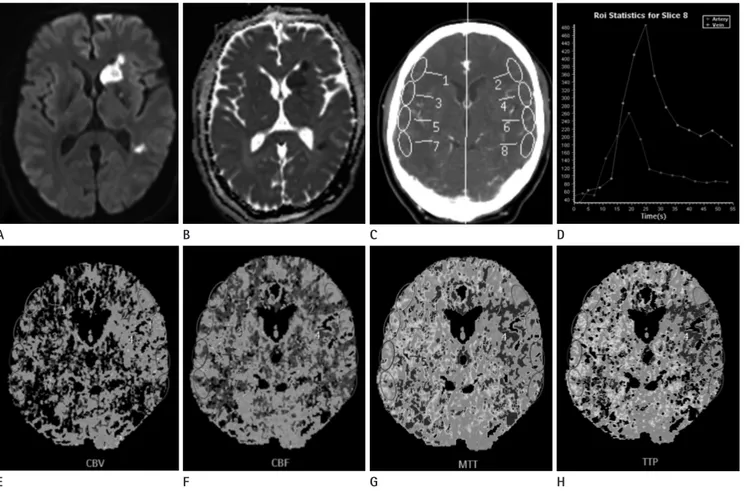
Fig. 1. Locations of regions of interest (ROI) in cerebral perfusion CT.
A, B. Diffusion weighted image (A) and apparent diffusion coefficient map (B) show acute infarction at left basal ganglia in a 50-year male patient.
C. Four ROIs in both middle cerebral artery (MCA) territories of perfusion CT before and after acetazolamide infusion are demonstrated at the lev- el of basal ganglia.
D. This is a representative arterial input and venous outflow time-attenuation curves at left MCA cortical flow territory. This time-attenuation curve is then used to calculate the perfusion CT parameters.
E-H. Examples of cerebral blood volume (CBV), cerebral blood flow (CBF), mean transit time (MTT), and time to peak enhancement (TTP) on perfu- sion CT before acetazolamide infusion at the level of basal ganglia (right to left).
E A
F B
G C
H D
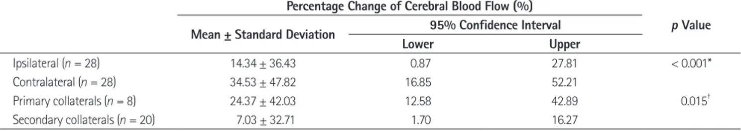
환에서 7.03 ± 32.71%(95% 신뢰구간: 1.71~16.27)로 일차성 측부순환 24.37 ± 42.03%(95% 신뢰구간: 12.58~42.89)에 비하여 통계적으로 유의하게 낮았다(Mann-Whitney test, p = 0.015)(Figs. 2, 3, Table 3).
고찰
뇌동맥의 심한 협착이나 폐색은 말단 부위의 관류압을 저하 시키지만, 저하된 혈류량을 보상하기 위해 측부순환과 자동조 절 반사성 혈관 확장이 생리학적 예비능으로 기여하고 있기 때 문에 협착의 정도만으로는 혈역동학적손상(hemodynamic compromise)을 예측하기 어렵다(1, 5). 즉, 측부순환과 자동 량변화율 값에 대하여 Mann-Whitney test를 시행하여, 각 군의
뇌혈류 예비능에 차이가 있는지를 평가하였다. SPSS 프로그램 (version 18, SPSS Inc., Chicago, IL, USA)을 이용하여 통계분 석을 하였으며, 통계적 유의성은 p < 0.05를 기준으로 하였다.
결과
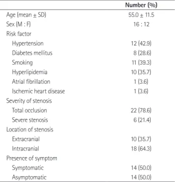
연구에 포함된 환자는 총 28명으로 평균연령은 57.0 ± 11.5 세(33~79세), 성별은 남자 16명, 여자 12명이었으며, 모두 ACZ-PCT를 부작용 없이 성공적으로 시행하였다. 환자들의 뇌 경색 위험인자, 허혈성 증상유무, 경동맥 협착 정도와 위치, 그 리고 측부순환을 확인한 방법은 다음과 같다(Table 1). 우세한 측부순환의 형태에 따라 전체 환자를 두 군으로 나누었고, 각 군의 특성은 다음과 같다(Table 2). 일차성과 이차성 측부순환 이 각각 8명과 20명에서 우세하였으며, 두 군 간의 성비, 혈관 협착의 정도, 증상유무는 통계적 차이가 없었으나(p > 0.05), 이차성 측부순환이 우세한 군에서 두개내 혈관협착이 유의하게 많았고(p = 0.011), 평균연령은 유의하게 낮았다(p = 0.004).
심한 경동맥 협착 및 폐색 혈관 측의 뇌혈류 예비능을 평가 하기 위하여, 같은 환자에서 협착 혈관 측과 혈관조영술을 통 하여 정상 혈관이 확인된 반대측 중뇌동맥피질영역의 아세타졸 라마이드 부하 전후의 뇌혈류량변화율을 계산하여 비교하였다.
심한 경동맥 협착 및 폐쇄 혈관 측과 반대쪽 정상 혈관 측에서 얻은 뇌혈류량변화율의 평균값은 각각 14.34 ± 36.43%와 34.53 ± 47.82%로 측정되어, 통계적으로 유의한 차이가 있었 다(Wilcoxon signed rank test, p < 0.001)(Table 3).
측부순환 형태에 따른 뇌혈류 예비능을 비교하기 위하여, 협 착 혈관 쪽만을 대상으로 일차성 측부순환이 우세한 군과 이차 성 측부순환이 우세한 군의 중뇌동맥피질부위 뇌혈류량변화율 의 평균을 비교하였다. 뇌혈류량변화율의 평균은 이차성 측부순
Table 1. Demographics and Risk Factors of Patients
Number (%)
Age (mean ± SD) 55.0 ± 11.5
Sex (M : F) 16 : 12
Risk factor
Hypertension 12 (42.9)
Diabetes mellitus 8 (28.6)
Smoking 11 (39.3)
Hyperlipidemia 10 (35.7)
Atrial fibrillation 1 (3.6)
Ischemic heart disease 1 (3.6)
Severity of stenosis
Total occlusion 22 (78.6)
Severe stenosis 6 (21.4)
Location of stenosis
Extracranial 10 (35.7)
Intracranial 18 (64.3)
Presence of symptom
Symptomatic 14 (50.0)
Asymptomatic 14 (50.0)
Table 2. Patient Characteristic in Primary and Secondary Collateral Circulation
Primary Collateral (n = 8) Secondary Collateral (n = 20) p Value Methods to determine type of collateral circulation
(DSA + 3T MRA : 3T MRA only) 3 : 5 18 : 2
Age (years) 66.5 ± 7.23 53.2 ± 10.8 0.004*
Gender (male : female) 5 : 3 11 : 9 1.000†
Severity of stenosis (occlusion : severe stenosis) 8 : 0 14 : 6 0.141†
Location of stenosis (extracranial : intracranial) 6 (proximal ICA) : 2 (distal ICA) 4 (proximal ICA) :
16 (distal ICA;2, MCA M1;14) 0.011†
Presence of symptom (symptomatic : asymptomatic) 5 : 3 9 : 11 0.678†
Note.-p value < 0.05 is statistically significant.
*Student t-test.
†Fisher’s exact test.
DSA = digital subraction angiography, ICA = internal cerebral artery, MCA = middle cerebral artery, MRA = MR angiography
Table 3. Comparison of Quantitatively Measured Cerebrovascular Reserve Using Acetazolamide Perfusion CT: Percentage Change of Cerebral Blood Flow
Percentage Change of Cerebral Blood Flow (%)
p Value Mean ± Standard Deviation 95% Confidence Interval
Lower Upper
Ipsilateral (n = 28) 14.34 ± 36.43 0.87 27.81 < 0.001*
Contralateral (n = 28) 34.53 ± 47.82 16.85 52.21
Primary collaterals (n = 8) 24.37 ± 42.03 12.58 42.89 0.015†
Secondary collaterals (n = 20) 7.03 ± 32.71 1.70 16.27
Note.-Data are mean ± standard deviation.
*Wilcoxon-signed rank test.
†Mann-Whitney test
Fig. 2. Percentage change of cerebral blood flow (CBF) in patient with left proximal middle cerebral artery occlusion with secondary collaterals. A 55-year-old woman without evidence of ischemic symptom.
A. There is no definable abnormal sign on the initial brain MRI T2 weighted image.
B, C. Left middle cerebral artery is occluded just distal to distal internal cerebral artery bifurcation on time of flight MR angiography (B) and digi- tal subtraction angiography (DSA) (C).
D. Secondary collaterals from ipsilateral anterior cerebral artery and external carotid artery are noted on DSA.
E. Relative preservation of CBF is shown in left frontotemporal cortex on baseline perfusion study, the value are 52.48 mL/100 g/min and 50.92 mL/100 g/min in occlusive and mirrored hemisphere, respectively.
F. But there is paradoxical response in stenotic area on post-acetazolamide study. Percentage change of CBF is -8.44%, suggesting significant decreased cerebrovascular reserve as compared with 21.70% in contralateral cortex.
E B
D A
F C
PRE-ACZ CBF POST-ACZ CBF
컴퓨터단층촬영, 관류 전산화단층촬영술, 관류 자기공명영상 (perfusion MR) 등이 이용되었다(12, 18, 28-33). 이 중 과탄산 혈증을 유발하여 중뇌대동맥의 직경을 측정하는 방식의 경두 개 초음파가 쉽게 적용이 가능하고 인체에 해가 없다는 점 그리 고 CBF와 직접 관련이 있는 기저부 동맥들의 속도나 파형을 실 시간 측정하여 혈관운동반응성(vasomotor reactivity)을 평가 할 수 있다는 장점 때문에 많이 시행되었지만, 낮은 해상도와 더불어 주로 근위부 혈관에만 국한된 혈관운동반응성을 반영 한다는 점에서 협착 말단부위의 상황을 평가하기에 한계가 있 다(12, 30, 31). 이밖에 Tc99m HMPAO의 방사성 핵종을 이 용하는 단일광자방출컴퓨터단층촬영술은 뇌혈류 예비능을 평 가하고 뇌혈관 개통술 시행 여부를 결정하는 데에 가장 널리 인 조절 반사성 혈관 확장을 통하여 혈류를 얼마나 효과적으로 보
상하느냐에 의하여 허혈성 뇌손상의 임상양상이 결정되기 때문 에, 이들에 대한 종합적인 평가는 급성뿐만 아니라 만성 경동맥 협착 및 폐색 환자의 임상양상을 예측하고 치료를 결정하는 데 결정적이다(1, 5, 6).
뇌혈관의 협착과 측부순환에 대한 평가에서 디지털 감산처리 혈관조영술이 직경이 매우 작고 혈류 속도가 느린 이차성 측부 순환까지 진단하는 데 탁월하기 때문에 침습적임에도 불구하 고 표준으로 여겨져 왔으나, 진단된 측부순환에 따른 말단 뇌조 직 관류상태와 같은 기능적 평가에 제한적이라는 점이 지적되 어 왔다(5). 따라서 뇌관류 상태 및 뇌혈관 예비능에 대한 평가 를 위해 경두개 초음파, 양전자방출단층촬영술, 단일광자방출
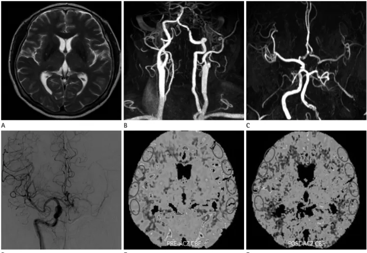
Fig. 3. Percentage change of cerebral blood flow (CBF) in patient with left proximal internal cerebral artery (ICA) occlusion with primary collater- als. A 71-year-old woman without ischemic symptom.
A. There is no demonstrable abnormal finding on the initial brain MRI T2 weighted image.
B, C. Total occlusion of left proximal ICA is demonstrated on contrast-enhanced MR angiography (MRA) (B). However, left middle cerebral artery (MCA) is visualized on her time of flight MRA through anterior communicating artery (C).
D. Ipsilateral anterior cerebral artery, MCA and their cortical branches distal to occluded left ICA are visualized on digital subtraction angiography, suggesting primary collaterals.
E, F. Cerebral blood flow maps of baseline (E) and post-acetazolamide (F) cerebral perfusion study show relative preserved cerebrovascular re- serve in left frontotemporal cortex. The percentage change of CBF is 32.88% and 35.87% is measured in mirrored hemisphere.
E B
D A
F C
PRE-ACZ CBF POST-ACZ CBF
계적으로 유의한 차이를 보이며 감소되어 있었다. 이러한 소견은 협착혈관 측 말단부위의 혈관들이 자동조절 반사성 혈관 확장기 전을 작동시켜 뇌관류를 유지하고 있다는 것을 시사하며, 이전 연구에서 보고한 아세타졸라마이드 주입 이후 정상 혈관 측 반구 에서 관찰된 뇌혈류백분율의 증가의 범위(약 30~60%)에 부합 하고(34), 협착 혈관 측과 정상 혈관 측의 아세타졸라마이드 주 입 전후의 뇌혈류량변화율의 유의한 차이와도 일치한다(32).
더불어 우세한 측부 순환의 형태에 따라 비교한 아세타졸라 마이드 부하 전후의 뇌혈류량변화율이 이차성 측부순환이 우세 한 군에서 윌리스환 중심의 일차성 측부순환이 우세한 군에 비 하여 의미 있게 감소되어 있음을 확인하였다. 이와 같이 이차성 측부순환이 우세한 영역의 아세타졸라마이드 자극에 의한 혈 류량 증가 정도가 비정상적 범위로 평가하는 10% 미만으로 측 정된 것은 말단 혈관의 반응성 혈관 확장이 이미 진행되어 추가 적인 혈관확장에 제한이 있음을 시사하며(35), 그 원인이 이차 성 측부순환에 의한 보상성 혈류공급이 일차성에 비하여 원활 하지 않음에 있을 것으로 추정할 수 있다. 이러한 결과는 경동 맥의 심한 협착이나 폐쇄에서 발달하는 이차성 측부순환과 뇌 혈류 예비능의 연관성에 대한 기존의 논란에 대하여, 두개내 경 동맥 죽상경화증 환자를 대상으로 측부순환의 종류와 뇌관류 의 저하 정도는 관련이 없다고 보고한 일부 연구와는 상반되며 (9, 36), 이차성 측부순환과 혈역동학적 불안정성의 관련성을 주장한 연구들과 일치한다(2, 10-13).
한편, 본 연구에 포함된 측부순환의 형태에 따른 각 환자들 의 두개내와 두개외 혈관 협착 위치의 빈도가 통계적으로 유의 한 차이를 보인 점을 감안하면(p = 0.011)(Table 2), 심한 경동 맥의 협착 및 폐색환자의 뇌혈류예비능은 일차성 측부순환이 우세한 두개외 혈관협착에 비해, 이차성 측부순환이 우세한 두 개내 혈관협착에서 상대적으로 저하되어 있음을 시사한다. 이 러한 소견은 경두개 초음파를 이용한 선행 연구의 결과와도 일 치한다(37).
양전자방출단층촬영술을 이용하여 뇌혈류 예비능을 평가한 이전 연구들은 안동맥 또는 연수막 혈관을 통한 측부순환이 관 찰되는 영역의 증가된 산소추출률(oxygen extraction fraction) 을 통하여 뇌관류상태가 심하게 감소되어 있음을 주장하였고 (12, 13), 그 외의 연구들에서도 다양한 종류의 정량적 분석을 통하여 이차성 측부순환이 우세한 영역에서의 혈역동학적 불안 정을 제시하였다(2-4, 10, 11, 14, 15). 최근에는 모야모야 질 환 환자들의 대표적인 측부순환 경로에 해당하는 연수막혈관 들의 증식이 활발할수록 뇌혈류 예비능의 감소가 진행되고 있 음이 보고되었다(11, 14, 15, 19). 따라서 다른 관류 영상에 비 하여 접근성과 공간적 해상도가 뛰어난 ACZ-PCT를 이용하여 정되는 검사로, CBF의 정량적 분석이 가능하고 양전자방출단
층촬영술에서 제시된 혈역동학적 상황과도 일치된 결과를 보이 나, 사용하는 방사선 핵종의 특성상 검사가 이틀에 걸쳐 이루어 져야 하며 CT나 MRI에 비해 해부학적 정보를 제시하지 못한 다는 단점이 있다(28).
이에 비하여 뇌관류 CT는 시간경과에 따른 조영제의 혈관이 나 혈관외 부분 또는 세포외 부분의 분포 변화를 측정하고 수 학적인 계산을 이용하여 조직의 관류인자를 적량적으로 계산 할 수 있으며, 기존의 양전자방출단층촬영술과 단일광자방출 컴퓨터단층촬영술에 비해 우수한 공간해상력과 접근성의 장점 을 가지고 있다(18, 32, 33). 그러나 뇌혈관 협착 환자에서 뇌 혈류 예비능을 예측하는 것은 기저 뇌관류 CT만으로는 제한적 인 것으로 알려져 있어, 자동조절 보상기전에 의하여 이미 확장 된 혈관영역을 제외한 뇌조직의 세동맥을 단기적(short-term) 으로 확장시켜 뇌혈류의 증가를 유도하는 아세타졸라마이드를 주입하고 20분 뒤 뇌관류 CT를 추가적으로 시행하여 기저 뇌 관류 CT와 비교하는 아세타졸라마이드 부하 뇌관류 CT (ACZ-PCT)가 도입되었다. ACZ-PCT는 30분 내외의 비교적 짧은 시간 내에 검사가 가능하고 99m Tc-HMPAO 양전자방출 단층촬영술의 결과와 매우 높은 일치도를 보여, 만성 경동맥 폐 쇄 환자의 뇌혈류 예비능 평가에 이용되고 있다(19-23). 그러나 현재까지 ACZ-PCT를 이용하여 경동맥 협착환자의 측부순환 의 형태에 따른 뇌혈류 예비능을 평가하려는 시도는 없었으며, 과거 연구들은 측부순환을 확인하는 데 주로 자기공명 혹은 전 산화단층촬영 혈관촬영을 이용하여 일부 이차성 측부순환이 연 구과정에서 배제되었을 가능성이 높다(7, 9, 11). 측부순환의 형태에 따른 뇌혈류 예비능을 정량적으로 비교한 가장 최근 연 구에서는 두개내 경동맥 폐쇄환자의 측부순환을 디지털 감산 혈관촬영을 이용하여 일차성과 이차성으로 구분하였지만, 아세 타졸라마이드를 주입하지 않은 기저관류 전산화단층촬영의 인 자들만을 비교하는 데 그쳤다(10). 본 연구에서는 이전 선행연 구의 제한점을 극복하기 위하여, ACZ-PCT를 시행한 환자들에 서 측부순환이 흔히 관찰되는 것으로 알려진 심한 편측성 경동 맥 협착과 폐쇄 환자들만을 연구의 대상으로 결정하였고, 양측 경동맥의 협착 정도와 우세한 측부순환의 형태를 평가하는 데 최적표준(gold standard)이 되는 디지털 감산처리 혈관조영술 위 주로 평가하였다. 디지털 감산처리 혈관조영술이 시행되지 않은 환자들에서는 3T 자기공명영상 동맥조영술이 이차성 측부순환 의 발견에 민감도가 낮음을 고려하여(26, 27), 완전한 윌리스환 과 기저혈관이 명백히 보이는 경우에만 연구에 포함시켰다.
이번 ACZ-PCT를 이용한 뇌혈류 예비능의 정량적 측정연구 에서 정상혈관측과 비교한 협착혈관측의 뇌혈류량변화율은 통
Chimowitz MI; Warfarin-Aspirin Symptomatic Intracranial Disease (WASID) Investigators. Collateral circulation in symptomatic intracranial atherosclerosis. J Cereb Blood Flow Metab 2011;31:1293-1301
2. Garrett MC, Komotar RJ, Starke RM, Merkow MB, Otten ML, Connolly ES. Radiographic and clinical predictors of hemodynamic insufficiency in patients with athero-occlu- sive disease. J Stroke Cerebrovasc Dis 2008;17:340-343 3. Gupta A, Nair S, Schweitzer AD, Kishore S, Johnson CE,
Comunale JP, et al. Neuroimaging of cerebrovascular dis- ease in the aging brain. Aging Dis 2012;3:414-425 4. Gupta A, Chazen JL, Hartman M, Delgado D, Anumula N,
Shao H, et al. Cerebrovascular reserve and stroke risk in patients with carotid stenosis or occlusion: a systematic review and meta-analysis. Stroke 2012;43:2884-2891 5. Romero JR, Pikula A, Nguyen TN, Nien YL, Norbash A,
Babikian VL. Cerebral collateral circulation in carotid ar- tery disease. Curr Cardiol Rev 2009;5:279-288
6. Vernieri F, Pasqualetti P, Passarelli F, Rossini PM, Silvestrini M. Outcome of carotid artery occlusion is predicted by cerebrovascular reactivity. Stroke 1999;30:593-598 7. Lima FO, Furie KL, Silva GS, Lev MH, Camargo EC, Singhal
AB, et al. The pattern of leptomeningeal collaterals on CT angiography is a strong predictor of long-term functional outcome in stroke patients with large vessel intracranial occlusion. Stroke 2010;41:2316-2322
8. Drakou AA, Koutsiaris AG, Tachmitzi SV, Roussas N, Tsironi E, Giannoukas AD. The importance of ophthalmic artery hemodynamics in patients with atheromatous carotid ar- tery disease. Int Angiol 2011;30:547-554
9. van Everdingen KJ, Visser GH, Klijn CJ, Kappelle LJ, van der Grond J. Role of collateral flow on cerebral hemodynamics in patients with unilateral internal carotid artery occlu- sion. Ann Neurol 1998;44:167-176
10. Cheng XQ, Tian JM, Zuo CJ, Liu J, Zhang Q, Lu GM. Quanti- tative perfusion computed tomography measurements of cerebral hemodynamics: correlation with digital subtrac- tion angiography identified primary and secondary cere- bral collaterals in internal carotid artery occlusive disease.
Eur J Radiol 2012;81:1224-1230
11. Kawashima M, Noguchi T, Yakushiji Y, Takase Y, Matsushi- ma T. Leptomeningeal collateral and cerebral hemody- 체계적인 뇌혈류 예비능 평가를 시도한 본 연구를 통하여, 뇌혈
류 예비능의 저하가 확인된 이차성 측부순환이 우세한 편측 경 동맥 협착 또는 폐색 환자에서 혈관문합수술이나 우회수술 등 의 적극적인 치료의 필요성을 제시할 수 있다.
이번 연구의 제한점은 첫 번째, ACZ-PCT를 시행하면서 증 가되는 피폭 방사선 양이다. 본 연구에 포함된 환자가 뇌관류 CT를 1회 시행할 때의 선량길이곱(dose-length product)은 1580~1709 mGy*cm로 측정되었으며, 아세타졸라마이드 주입 전후에 같은 영상 조건으로 두 번 검사가 시행된다는 점을 고려 하면 ACZ-PCT를 시행한 개별 환자의 총 피폭 선량은 평균 7.2~7.8 mSV에 달한다. 이런 피폭선량은 최근 발표된 다른 연구의 평균값 4.9~6.3 mSV와 비교하였을 때 일회당 선량은 낮았지만, 총 선량이 상당히 높고 방사선에 취약한 것으로 알려 져 있는 안구와 근접해 있다는 점에서 ACZ-PCT 시행 여부를 결정할 때 반드시 고려해야만 하는 문제점이다(38, 39). 이러 한 이유 때문에 혈관 협착이 확인되지 않은 환자에서 ACZ- PCT를 시행할 수 없어, 본 연구에서는 정상혈관 측의 뇌혈류 예비능을 ACZ-PCT를 시행한 환자들 중 혈관협착이 없는 반 대쪽 반구에서 후향적으로 선택하여 평가할 수밖에 없어, 선택 오류(selection bias)와 적은 수의 연구대상이 불가피하였다. 한 편, ACZ-PCT의 비교적 높은 환자 피폭 선량을 최소화하기 위 하여, 갠트리(gantry)의 경사도를 조정하여 안구 피폭을 줄이거 나, 반복적 재구성(iterative reconstruction)과 같은 기법이 시도 되는 것이 요구된다(39, 40).
두 번째, 이번 연구를 통하여 밝혀진 측부순환 종류에 따른 심 한 경동맥 협착 및 폐색의 동측 뇌반구의 뇌혈류 예비능의 통계 적 차이에 대한 임상적 의의를 평가하기 위해서는 긴 기간의 전 향적인 경과관찰이 추가적으로 이루어져야 한다. 이번 연구에서 는 허혈성 증상의 유무만을 평가하였는데, 뇌혈류 예비능과 허 혈성 경색의 위험도 층화(stratification)를 위하여 증상의 중증도 (severity)를 고려한 연구설계가 향후 연구에서 필요할 것이다.
결론적으로 다른 관류 영상에 비하여 접근성과 공간적 해상 도가 뛰어나며 정확한 ACZ-PCT를 이용한 뇌혈류 예비능 비 교 연구를 통하여 심한 경동맥 협착 및 폐색 측의 뇌혈류의 보 상성 공급이 일차성 측부순환에 비하여 이차성 측부순환이 우 세한 경우에 유의하게 감소되어 있음을 확인하였고, 이것은 이 차성 측부순환의 혈역동학적 불안정을 시사하기 때문에 향후 치료시기와 방법을 결정하는 데 적극적으로 고려되어야 한다.
참고문헌
1. Liebeskind DS, Cotsonis GA, Saver JL, Lynn MJ, Cloft HJ,
of acetazolamide-challenged CT perfusion with single- photon emission CT in Moyamoya patients. AJNR Am J Neuroradiol 2008;29:1658-1663
21. Smith LM, Elkins JS, Dillon WP, Schaeffer S, Wintermark M.
Perfusion-CT assessment of the cerebrovascular reserve: a revisit to the acetazolamide challenges. J Neuroradiol 2008;35:157-164
22. Lui YW, Tang ER, Allmendinger AM, Spektor V. Evaluation of CT perfusion in the setting of cerebral ischemia: pat- terns and pitfalls. AJNR Am J Neuroradiol 2010;31:1552- 1563
23. Eicker SO, Turowski B, Heiroth HJ, Steiger HJ, Hänggi D. A comparative study of perfusion CT and 99m Tc-HMPAO SPECT measurement to assess cerebrovascular reserve ca- pacity in patients with internal carotid artery occlusion.
Eur J Med Res 2011;16:484-490
24. Lell M, Fellner C, Baum U, Hothorn T, Steiner R, Lang W, et al. Evaluation of carotid artery stenosis with multisection CT and MR imaging: influence of imaging modality and postprocessing. AJNR Am J Neuroradiol 2007;28:104-110 25. Liebeskind DS. Collateral circulation. Stroke 2003;34:2279-
2284
26. Ozsarlak O, Van Goethem JW, Maes M, Parizel PM. MR an- giography of the intracranial vessels: technical aspects and clinical applications. Neuroradiology 2004;46:955- 972
27. McVerry F, Liebeskind DS, Muir KW. Systematic review of methods for assessing leptomeningeal collateral flow.
AJNR Am J Neuroradiol 2012;33:576-582
28. Bokkers RP, Wessels FJ, van der Worp HB, Zwanenburg JJ, Mali WP, Hendrikse J. Vasodilatory capacity of the cerebral vasculature in patients with carotid artery stenosis. AJNR Am J Neuroradiol 2011;32:1030-1033
29. Alexandrov AV, Nguyen HT, Rubiera M, Alexandrov AW, Zhao L, Heliopoulos I, et al. Prevalence and risk factors as- sociated with reversed Robin Hood syndrome in acute ischemic stroke. Stroke 2009;40:2738-2742
30. Valdueza JM, Draganski B, Hoffmann O, Dirnagl U, Ein- häupl KM. Analysis of CO2 vasomotor reactivity and vessel diameter changes by simultaneous venous and arterial Doppler recordings. Stroke 1999;30:81-86
31. Sharma VK, Tsivgoulis G, Ning C, Teoh HL, Bairaktaris C, namics in patients with ICA and MCA steno-occlusion.
Neurol Res 2011;33:307-313
12. Hofmeijer J, Klijn CJ, Kappelle LJ, Van Huffelen AC, Van Gijn J. Collateral circulation via the ophthalmic artery or leptomeningeal vessels is associated with impaired cere- bral vasoreactivity in patients with symptomatic carotid artery occlusion. Cerebrovasc Dis 2002;14:22-26
13. Yamauchi H, Kudoh T, Sugimoto K, Takahashi M, Kishibe Y, Okazawa H. Pattern of collaterals, type of infarcts, and haemodynamic impairment in carotid artery occlusion. J Neurol Neurosurg Psychiatry 2004;75:1697-1701
14. Kaku Y, Iihara K, Nakajima N, Kataoka H, Fukushima K, Iida H, et al. The leptomeningeal ivy sign on fluid-attenuated inversion recovery images in moyamoya disease: positron emission tomography study. Cerebrovasc Dis 2013;36:19-25 15. Kawashima M, Noguchi T, Takase Y, Ootsuka T, Kido N,
Matsushima T. Unilateral hemispheric proliferation of ivy sign on fluid-attenuated inversion recovery images in moyamoya disease correlates highly with ipsilateral hemi- spheric decrease of cerebrovascular reserve. AJNR Am J Neuroradiol 2009;30:1709-1716
16. Andaluz N, Choutka O, Vagal A, Strunk R, Zuccarello M.
Patient selection for revascularization procedures in adult Moyamoya disease based on dynamic perfusion comput- erized tomography with acetazolamide challenge (PCTA).
Neurosurg Rev 2010;33:225-232; discussion 232-233 17. Mendelowitsch A, Taussky P, Rem JA, Gratzl O. Clinical
outcome of standard extracranial-intracranial bypass sur- gery in patients with symptomatic atherosclerotic occlu- sion of the internal carotid artery. Acta Neurochir (Wien) 2004;146:95-101
18. Vagal AS, Leach JL, Fernandez-Ulloa M, Zuccarello M. The acetazolamide challenge: techniques and applications in the evaluation of chronic cerebral ischemia. AJNR Am J Neuroradiol 2009;30:876-884
19. Kang KH, Kim HS, Kim SY. Quantitative cerebrovascular reserve measured by acetazolamide-challenged dynamic CT perfusion in ischemic adult Moyamoya disease: initial experience with angiographic correlation. AJNR Am J Neuroradiol 2008;29:1487-1493
20. Rim NJ, Kim HS, Shin YS, Kim SY. Which CT perfusion pa- rameter best reflects cerebrovascular reserve?: correlation
lateralization and misery perfusion in patients with carotid occlusion. Stroke 1999;30:1025-1032
37. Uzunca I, Asil T, Balci K, Utku U. Evaluation of vasomotor reactivity by transcranial Doppler sonography in patients with acute stroke who have symptomatic intracranial and extracranial stenosis. J Ultrasound Med 2007;26:179-185 38. Arandjic D, Bonutti F, Biasizzo E, Ciraj-Bjelac O, Floreani M,
Giustizieri M, et al. Radiation doses in cerebral perfusion computed tomography: patient and phantom study. Radi- at Prot Dosimetry 2013;154:459-464
39. Zhang D, Cagnon CH, Villablanca JP, McCollough CH, Cody DD, Stevens DM, et al. Peak skin and eye lens radiation dose from brain perfusion CT based on Monte Carlo simu- lation. AJR Am J Roentgenol 2012;198:412-417
40. Ma J, Zhang H, Gao Y, Huang J, Liang Z, Feng Q, et al. Iter- ative image reconstruction for cerebral perfusion CT using a pre-contrast scan induced edge-preserving prior. Phys Med Biol 2012;57:7519-7542
Chong VF, et al. Role of multimodal evaluation of cerebral hemodynamics in selecting patients with symptomatic ca- rotid or middle cerebral artery steno-occlusive disease for revascularization. J Vasc Interv Neurol 2008;1:96-101 32. Chen A, Shyr MH, Chen TY, Lai HY, Lin CC, Yen PS. Dynamic
CT perfusion imaging with acetazolamide challenge for evaluation of patients with unilateral cerebrovascular ste- no-occlusive disease. AJNR Am J Neuroradiol 2006;27:
1876-1881
33. Orrison WW Jr, Snyder KV, Hopkins LN, Roach CJ, Ringdahl EN, Nazir R, et al. Whole-brain dynamic CT angiography and perfusion imaging. Clin Radiol 2011;66:566-574 34. Yonas H, Darby JM, Marks EC, Durham SR, Maxwell C. CBF
measured by Xe-CT: approach to analysis and normal val- ues. J Cereb Blood Flow Metab 1991;11:716-725
35. Eskey CJ, Sanelli PC. Perfusion imaging of cerebrovascular reserve. Neuroimaging Clin N Am 2005;15:367-381, xi 36. Derdeyn CP, Shaibani A, Moran CJ, Cross DT 3rd, Grubb RL
Jr, Powers WJ. Lack of correlation between pattern of col-
심한 편측성 경동맥 협착에서 아세타졸라마이드 부하 뇌관류 CT를 이용하여 측정한 뇌혈류 예비능과 측부순환의 연관성
1홍도란
1· 이영흔
1· 서형석
1· 제보경
1· 서상일
2· 설혜영
2· 이남준
3· 김정혁
3· 양경숙
4목적: 심한 편측성 경동맥 협착 또는 폐색 환자에서 측부순환의 형태에 따른 뇌혈류 예비능 상태를 아세타졸라마이드 부 하 뇌관류 CT를 이용하여 비교하였다.
대상과 방법: 본원에서 아세타졸라마이드 부하 뇌관류 CT를 시행한 환자들 중, 디지털 감산처리 혈관조영술과 3T 자기공 명영상 동맥조영술을 통하여 내경동맥 및 중뇌동맥의 중등도 이상의 편측성 협착 및 폐색을 보이고, 앞 혹은 뒤 교통동맥을 포함한 윌리스환을 통한 일차성, 이외의 안동맥, 기저부동맥, 전대뇌동맥과 후대뇌동맥, 또는 외경동맥 경유의 이차성 측부 순환 중 우세한 측부순환의 형태가 확인된 환자들을 후향적으로 선택하였다. 아세타졸라마이드 부하 전과 후의 뇌관류 CT 에서 뇌혈류량변화율을 측정하여, 병변 측 반구와 정상 측 반구의 변화율 차이를 조사하였다. 그리고 전체 환자를 측부순환 의 형태에 따라 두 군으로 나누어 두 군 간에 병변 측 반구의 뇌혈류 예비능에 차이가 있는지를 조사하였다.
결과: 총 28명(일차성 8명, 이차성 20명)의 뇌혈류량변화율은, 협착이 없는 반대측 반구와 비교하여 협착 혈관 측 반구에 서(14.34 ± 36.43%와 34.53 ± 47.82%, p < 0.001) 그리고 일차성보다 이차성 측부순환이 우세한 반구(24.37 ± 42.03%와 7.03 ± 32.71%, p < 0.05)에서 각각 유의하게 낮았다.
결론: 아세타졸라마이드 부하 뇌관류 CT를 이용하여 측정한 뇌혈류 예비능은 일차성에 비하여 이차성 측부순환이 우세 한 반구에서 감소되어 있었다.
1고려대학교 의과대학 안산병원 영상의학과, 2고려대학교 의과대학 구로병원 영상의학과,
3고려대학교 의과대학 안암병원 영상의학과, 4고려대학교 의과대학 의학통계학교실