Received: April 24 2020, Revised: April 24 2020, Accepted: May 29 2020
Corresponding author: Yoon Ghil Park, Department of Rehabilitation Medicine, Gangnam Severance Hospital, Rehabilitation Institute of Neuromuscular Disease, Yonsei University College of Medicine, 211 Eonju-ro, Gangnam- gu, Seoul 06273, Korea
Tel: +82-2-2019-3493, Fax: +82-2-2019-4807 E-mail: [email protected]
Copyrights ⓒ The Korean Dysphagia Society, 2021.
Relationship between Swallowing Function, Diet Level and Pulmonary Function in Post-Stroke Patients
Myungeun Yoo, M.D., Hyo Jeong Lee, M.D., Eu Jeong Ko, M.D., Jinyoung Park, M.D., Yoon Ghil Park, M.D., Ph.D.
Department of Rehabilitation Medicine, Gangnam Severance Hospital, Rehabilitation Institute of Neuromuscular Disease, Yonsei University College of Medicine, Seoul, Korea
Objective: To identify the relationship between dysphagia, dietary level, and pulmonary function in post-stroke patients.
Methods: Thirty-six post-stroke patients with dysphagia, who were hospitalized from June 2017 to October 2017 in the Department of Rehabilitation Medicine at a tertiary hospital, were analyzed retrospectively. The video-fluoroscopic swallowing study (VFSS) and videofluoroscopic dysphagia scale (VDS) were used to assess dysphagia. The vital capacity (VC) and peak cough flow (PCF) were used to assess the pulmonary function. Upon admission, the patients were di- vided into three groups according to their dietary level (tube feeding, dysphagia diet, and general diet). The corre- lation between dysphagia and pulmonary function was analyzed using an independent t-test test with the optimal points, and the relationship between the diet level and pulmonary function was evaluated using a one-way analysis of the variance.
Results: Significant correlations between the pulmonary function and sub-items of VDS were found in “oral transit time” with VC, “vallecullar residue” and “aspiration” with PCF, and “triggering of pharyngeal swallow”, “VDS total score”
with VC and PCF. The dietary levels upon admission had a significant correlation with VC and PCF. The VC among groups divided according to three diet levels showed statistically significant differences.
Conclusion: This study revealed the relationship between the pulmonary function and dysphagia in post-stroke patients. Moreover, the pulmonary function correlated with dietary level, even though it was not confirmed that it affected dietary levels. The clinical importance of the pulmonary function in post-stroke patients with dysphagia should be emphasized. In addition, a large-scale study is needed to determine the correlation between the pulmo- nary function and swallowing difficulty. (JKDS 2021;11:25-34)
Keywords: Peak cough flow, Vital capacity, Dysphagia, Diet, Stroke
INTRODUCTION
Swallowing is a complex neuromuscular process, regulated by the swallowing center at the brainstem
1.
Stroke induces abnormal swallowing physiology by
reducing lingual control, leading to pharyngeal dys-
synergia, or delayed swallowing reflex, which causes
pharyngeal pooling
2. It is estimated that 28-65% of
post-stroke patients, with a prevalence of aspiration pneumonia of 18-19%
4,5.
Several studies have reported the relationship be- tween dysphagia and pulmonary function, such as the development of pulmonary complications due to dys- phagia
6and the development of dysphagia in patients with chronic obstructive pulmonary disease (COPD)
7,8. Furthermore, a difference in peak cough flow (PCF) between stroke patients with and without dysphagia has been observed
9. Moreover, a significant decrease in vital capacity (VC) and inspiratory reserve volume in post-stroke patients with dysphagia has been re- ported
10.
Despite of numerous studies on the relationship between dysphagia and pulmonary function, the mechanism which stage of the swallowing is related to the pulmonary function has not been clarified.
Furthermore, less attention has been given to the relationship between pulmonary function and diet level. In this study, we hypothesized that the lower the pulmonary function, the higher the risk of as- piration in post-stroke patients. Additionally, we hypothesized that a lower diet level would correlate to lower pulmonary function in these patients. To support these hypotheses, we assessed the correlation between parameters of a videofluoroscopic dysphagia scale (VDS) and VC or PCF. In addition, it was as- sessed whether there is a difference in pulmonary function according to the diet level, which was de- cided by severity of dysphagia.
MATERIALS AND METHODS
1. Subjects
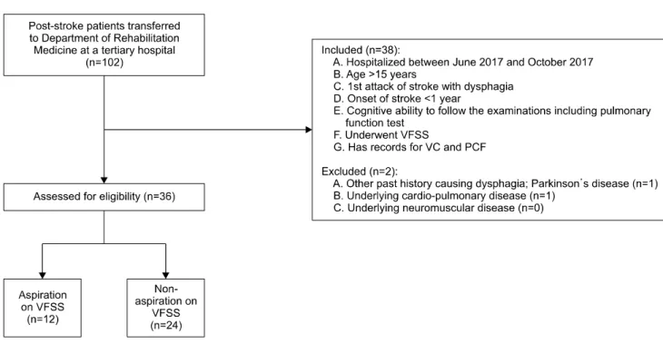
A retrospective analysis was conducted on 102 post-stroke patients who were hospitalized between June 2017 and October 2017 in the Department of Rehabilitation Medicine of a tertiary hospital. Of 102 post-stroke patients, 38 patients met the following inclusion criteria: (1) age ≥15 years; (2) first attack of ischemic or hemorrhagic stroke confirmed by
ability to follow the examinations including pul- monary function test; (5) underwent VFSS for swal- lowing function; and (6) underwent VC and PCF for pulmonary function, as shown in the flowchart.(Fig.
1) Of the 38 patients who met the inclusion criteria, two patients met at least one of the following ex- clusion criteria: (1) history of previous dysphagia (n=1); (2) history of cardiopulmonary disease, such as thoracotomy, COPD, or pulmonary granuloma (n=1);
and (3) history of neuromuscular diseases, such as muscular dystrophy, spinal muscular atrophy, or Guillain-Barre syndrome (n=0).(Fig. 1) Ultimately, 36 post-stroke patients were eligible for the analysis based on the abovementioned criteria. This study was approved by the Institutional Review Board of Gangnam Severance Medical Center (3-2019-0409).
2. Materials
Swallowing function was evaluated using the VFSS, and pulmonary function was assessed with VC and PCF. VFSS was evaluated within 1 week after ad- mission, and VC and PCF were measured prior to VFSS. The patients were divided into two groups (aspiration or non-aspiration) according to VFSS re- sults. The diet levels of the patients were compared at admission and discharge. Demographic data, in- cluding sex, age, days since onset, location of brain lesion, paralyzed side, and tracheostomy status, were collected. Cognition was assessed using the Korean Mini-Mental Status Examination (K-MMSE), and func- tional status was assessed by Functional Independ- ence Measure (FIM) at admission. Both cognition and functional status were assessed at admission.
1) Evaluation of dysphagia
To assess swallowing function, a VFSS was con-
ducted within 1 week after admission. The VFSS was
conducted by two experienced physiatrists, who were
assisted by one technician. The VFSS diet was pre-
pared as a semisolid, a solid and liquid bolus (5 and
15 ml, sequentially) mixed with barium (Solotop Sus-
pension 140). The liquid, semisolid, and solid diets
Fig. 1. Flow diagram.
PCF: peak cough flow, VC: vital capacity, VFSS: video-fluoroscopic swallowing study.
corresponded to level 0 (thin drink), 4 (puréed food), and 7 (regular food), respectively, according to the International Dysphagia Diet Standardization Initi- ative (IDDSI) framework
11. While in a seated position, patients were fed the abovementioned diets using a spoon or syringe. The results of each VFSS were re- corded and encoded into a video file at 30 frames/
sec. An experienced physiatrist reviewed all of the VFSS videos and analyzed them using the Penetration- Aspiration Scale (PAS) and the videofluoroscopic dys- phagia scale (VDS). The VDS, developed by Han et al.
in 2008, has a score range of 0-100
12. A higher score indicates more severe dysphagia.
2) Evaluation of pulmonary function
Pulmonary function was assessed using VC and PCF. While in a seated position, a patient’s nose and lips were fitted into a facial mask to seal the breath, and they were instructed to completely exhale with a constant flow and then to inhale deeply. VC was measured using a portable spirometer (Micro®, Micro Medical Ltd. England), and results were divided by height and age-related prediction value using per- centage. To measure PCF, patients were asked to
forcefully exhale into a peak flow meter (Assess®, Respironics, United States). These measurements were performed by a professional physiatrist who had no relationship with this study, and who performed pro- cedures according to the standards of the American Thoracic Society/European Respiratory Society
13.
Six patients in this study had a tracheostomy tube.
Their pulmonary function was measured with either a spirometer or a peak flow meter, which was con- nected to the proximal tip of the tracheostomy tube according to the procedure described in Winck et al.
14. The pulmonary function test was conducted as one of the critical elements of post-stroke evaluation.
3) Classification of diet levels
Diet levels were divided according to severity of
dysphagia using the IDDSI Functional Diet Scale
(IDDSI-FDS)
15,16, consisting of liquid level (not ap-
plicable, level 4, 3, 2, 1 according to viscosity) and
food level (puréed diet as level 4, minced and moist
diet as level 5, soft diet as level 6, and general solid
diet as level 7). The combination of each food level
and liquid level determines the level of IDDSI-FDS
between 0-8. Patients were divided into three groups
food level 4-6 and liquid level not applicable-1) and general solid diet (level 7,8: food level 7-8 and liquid level 1). Furthermore, the dysphagia diet group was subdivided into 3 groups as follows; level 1 (food level 4 and liquid level not applicable to 4), level 2-3 (food level 4-5 and liquid level 3), level 4-6 (food level 5-6 and liquid level 2-1).
3. Statistical analysis
The baseline characteristics of patients were analyzed using a chi-square test for categorical variables (sex, lesions, paralyzed side, tracheostomy status) and a t-test for continuous variables (age, days since onset, FIM, K-MMSE). To determine the risk factors for aspiration, a logistic regression analysis was perform- ed with the following variables: age, sex, lesions, paralyzed side, tracheostomy status, K-MMSE, and pulmonary function (VC, PCF). The cutoff values of VC (%) and PCF (L/min) to present aspiration on VFSS were obtained using the receiver operating charac- teristic (ROC) curve. The patients were grouped as above and at or below the cutoff values of VC and PCF. Intergroup differences of VDS sub-items and total score were analyzed using an independent t-test.
Pearson correlation coefficients were calculated be- tween VDS total score and pulmonary function. A one-way analysis of variance (ANOVA) analysis was performed to analyze the significant difference be- tween VC and PCF in the more than three diet groups.
Statistical analyses were conducted using SAS version 9.4 (SAS Institute, Cary, NC, United States), and the significance level was set at P<0.05.
RESULTS
1. Baseline characteristics
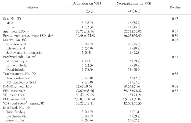
The characteristics of these groups are shown in Table 1. Using VFSS, 12 patients were in the aspir- ation group, and 24 patients were in the non- aspiration group. There was no significant difference in general characteristics between the aspiration and
the prevalence of aspiration (P=0.03).
2. Optimal cutoff values of VC and PCF for aspiration
The optimal cutoff point of VC for aspiration on VFSS, acquired from the maximal Youden’s index, was ≤47.8% (area under the ROC curve [AUC]=0.86 [0.72-0.99], P<0.05, sensitivity=1.00 [1.00-1.00], speci- ficity=0.75 [0.59-0.97]). For PCF, the cutoff point was
≤155 L/min (AUC=0.73 [0.43-1.00], P<0.05, sensitivity=
0.75 [0.5-1.00], specificity=0.69 [0.25-0.97]).
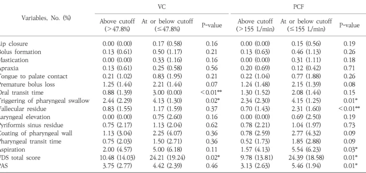
3. Difference in VDS sub-items and PAS between the two groups divided by the cutoff value of VC and PCF for aspiration Kolmogorov-Smirnov test was performed as a normality test, and VDS total score evaluated liquid and solid showed normality (P=0.051, P=0.052, re- spectively). In terms of VDS sub-items, the independ- ent t-test showed intergroup differences in the sub-items of “oral transit time”, “triggering of pha- ryngeal swallow” and “VDS total score” between the two groups according to the cutoff value of VC for aspiration (P<0.01, P=0.02, P=0.02, respectively). There were significant intergroup differences in sub-items of “triggering of pharyngeal swallow”, “aspiration”,
“vallecular residue” and “VDS total score” between the two groups when divided by the cutoff value of PCF for aspiration (P=0.01, P=0.03, P<0.01, P=0.01, re- spectively).(Table 2) In terms of PAS, the independent t-test showed a significant intergroup difference be- tween the two groups according to the cutoff value of PCF for aspiration, however, no significant differ- ence according to the cutoff value of VC (P<0.01, P=0.46).(Table 2)
4. Relationship between VDS score and VC or PCF
The VDS score evaluated with liquid showed a
weak correlation with VC (r=−0.37, P=0.03). The
lower the VDS score with liquid, the higher the VC.
Table 1. Demographic characteristics of patients.
Variables
Aspiration on VFSS Non-aspiration on VFSS
P-value
12 (33.3) 24 (66.7)
Sex, No. (%) 0.47
Male 8 (66.7) 13 (54.2)
Female 4 (33.3) 11 (45.8)
Age, mean±SD, y 56.75±19.94 62.54±16.07 0.39
Period from onset, mean±SD, day 110.58±111.52 86.63±92.59 0.50
Lesions, No. (%) 0.12
Supratentorial 5 (41.7) 18 (75.0)
Infratentorial 6 (50.0) 5 (20.8)
Supra- and infratentorial 1 (8.3) 1 (4.2)
Paralyzed side, No. (%) 0.41
Rt. hemiplegia 1 (8.3) 7 (29.2)
Lt. hemiplegia 4 (33.3) 5 (20.8)
Quadriplegia 7 (58.3) 12 (50.0)
Tracheostomy, No. (%) 0.38
Tracheostomised 3 (25.0) 3 (12.5)
Not tracheostomised 9 (75.0) 21 (87.5)
K-MMSE, mean±SD 23.67±8.62 20.54±7.16 0.28
FIM, mean±SD 69.00±35.66 59.13±24.22 0.33
VC, mean±SD 50.43±27.85 61.13±21.21
PCF, mean±SD 230.83±148.41 209.17±98.82
VDS total score
†, mean±SD 30.25±18.11 12.60±10.36
Diet level, No. (%)
Tube feeding 5 (41.7) 2 (8.3)
Dysphagia diet 5 (41.7) 7 (29.2)
General diet 2 (16.6) 15 (62.5)
FIM: functional independence measure, K-MMSE: Korean Mini-Mental Status Examination, VFSS: video-fluoroscopic swallowing study, PCF: peak cough flow, VC: vital capacity, VDS: videofluoroscopic dysphagia scale.
†