ISSN: 2233-601X (Print) ISSN: 2093-6516 (Online)
Received: October 24, 2016, Revised: December 2, 2016, Accepted: December 5, 2016, Published online: February 5, 2017
Corresponding author: Mohammad Ali Hosseinian, Department of General Surgery, Emam Hosein Hospital, Shahid Beheshti University of Medical Sciences, Arabi Ave, Daneshjoo Blvd, Velenjak, Tehran 198396-3113, Iran
(Tel) 98-9127244717 (Fax) 98-2188644213 (E-mail) [email protected]
© The Korean Society for Thoracic and Cardiovascular Surgery. 2017. All right reserved.
This is an open access article distributed under the terms of the Creative Commons Attribution Non-Commercial License (http://creativecommons.org/
licenses/by-nc/4.0) which permits unrestricted non-commercial use, distribution, and reproduction in any medium, provided the original work is properly cited.
Evaluation of Complications after Surgical Treatment of Thoracic Outlet Syndrome
Mohammad Ali Hosseinian, M.D. 1 , Ali Gharibi Loron, M.D. 1,2 , Yalda Soleimanifard, M.D. 1
1
Department of General Surgery, Emam Hosein Hospital, Shahid Beheshti University of Medical Sciences,
2
Shahed University School of Medicine
Background: Surgical treatment of thoracic outlet syndrome (TOS) is necessary when non-surgical treatments fail. Complications of surgical procedures vary from short-term post-surgical pain to permanent disability. The outcome of TOS surgery is affected by the visibility during the operation. In this study, we have compared the complications arising during the supraclavicular and the transaxillary approaches to determine the appro- priate approach for TOS surgery. Methods: In this study, 448 patients with symptoms of TOS were assessed.
The male-to-female ratio was approximately 1:4, and the mean age was 34.5 years. Overall, 102 operations were performed, including unilateral, bilateral, and reoperations, and the patients were retrospectively evaluated. Of the 102 patients, 63 underwent the supraclavicular approach, 32 underwent the transaxillary approach, and 7 underwent the transaxillary approach followed by the supraclavicular approach. Complications were evaluated over 24 months. Results: The prevalence of pneumothorax, hemothorax, and vessel injuries in the transaxillary and the supraclavicular approaches was equal. We found more permanent and transient bra- chial plexus injuries in the case of the transaxillary approach than in the case of the supraclavicular ap- proach, but the difference was not statistically significant. Persistent pain and symptoms were significantly more common in patients who underwent the transaxillary approach (p<0.05). Conclusion: The supra- clavicular approach seems to be the more effective technique of the two because it offers the surgeon better access to the brachial plexus and a direct view. This approach for a TOS operation offers a better surgical outcome and lower reoperation rates than the transaxillary method. Our results showed the supraclavicular approach to be the preferred method for TOS operations.
Key words: 1. Thoracic outlet syndrome 2. Thoracic outlet
3. Intraoperative complications
Introduction
Thoracic outlet syndrome (TOS) refers to com- pression of the subclavian vessels and nerves of the brachial plexus in the region around the neck and the collarbone [1,2]. Non-operative treatment is the first-line treatment, and surgical treatment is per- formed as a treatment of choice for patients in whom
conservative therapy has failed. Moreover, patients with a non-work-related etiology of TOS, such as traumatic TOS, respond better to TOS operations [3].
Excision of the cervical rib is a surgical technique ex- plained by Coote [4] in 1861. Adson and Coffey [5]
described an anterior scalenotomy technique in 1962.
Clagelt advocated the resection of the first rib via a
posterior approach [6,7]. A transaxillary approach for
https://doi.org/10.5090/kjtcs.2017.50.1.36
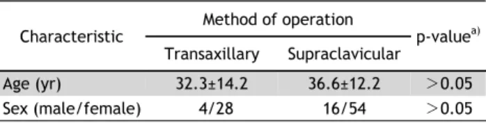
Table 1. Patient demographics Characteristic
Method of operation
p-value
a)Transaxillary Supraclavicular
Age (yr) 32.3±14.2 36.6±12.2 >0.05
Sex (male/female) 4/28 16/54 >0.05
Values are presented as mean±standard deviation.
a)
Based on the Fisher exact test.
such a resection was introduced by Roos [8] in 1966.
A TOS operation should be well thought-out, and an accurate diagnosis should be made before consider- ing decompression surgery to be the final solution [9]. Complications of TOS surgery include the re- currence of symptoms, brachial plexus, phrenic nerve, long thoracic nerve, complete or transient paralysis, subclavian artery and vein injuries, axillary artery thrombosis, hemothorax, pneumothorax, chylothorax, permanent damage to the brachial plexus, severe se- quelae such as causalgia and weak ness of the hand muscles, sensory deficits, autonomic dysfunction, and at times even death [10-12]. Further, some brachial plexus injuries require reoperation [13,14].
The supraclavicular approach allows better access to the brachial plexus [15], but recently, the trans- axillary approach has been used widely. Surgical re- sults correlate directly with an identification of re- sidual anomalous anatomical structures, such as a fi- brous band extending from the first rib and tenting up the subclavian artery and the brachial plexus [13].
This study evaluates the clinical outcomes of these 2 methods.
Methods
This retrospective long-term study examines pa- tients who presented with symptoms of TOS between 2004 and 2011. Patients who complained of pain and numbness in the arm, forearm, or hand are included.
TOS is one of the most intriguing clinical entities. A thorough history with specific attention to previous traumas to the shoulder region and prolonged use of the upper extremities was taken. Careful physical ex- aminations including a visual inspection for asymme- try or deformity, percussion, or symptoms induced by pressure to the supraclavicular area, as well as a complete neurologic examination with a range of mo- tion and strength tests were performed. Symptoms worsened when the patients’ arms were positioned overhead. The ulnar-nerve current velocity in these patients was below 60 m/sec [16]. A magnetic reso- nance imaging evaluation of the neck for discopathy indicated normal intervertebral disk anatomy.
In this study, a total of 448 patients were studied;
of these, 357 were females and 91 were males.
Recalcitrant functional impairment after 6 months of non-surgical treatment and progressive neurologic
dysfunction were observed in 69 patients; therefore, they met our institute’s criteria for surgical therapy [16]. Four patients presented with vascular symp- toms including coolness, pallor, and diminished pulse (arterial TOS) in 1 patient and swelling, uncom- fortable heaviness, and distended superficial veins (venous TOS) in 3 patients. The rest of the patients were diagnosed with neurologic TOS; they reported an array of symptoms including numbness, pain, weakness, and paresthesia. The classical form of neu- rologic TOS is interosseous, thenar, and hypothenar weakness and/or atrophy, as well as antebrachial cu- taneous hypoesthesia. Nerve conduction studies have revealed unobtainable or decreased median and ulnar antebrachial cutaneous sensory nerve action poten- tials in TOS patients. The C8–T1 nerves have been found to be the most affected. The ulnar-nerve cur- rent velocity in these patients was below 60 m/sec [17]. Bilateral TOS was diagnosed in 26 patients, which increased the number of operations to 95.
Sixty-three operations using the supraclavicular ap- proach were performed on 48 females and 15 males.
Thirty-two operations were performed using the transaxillary approach on 28 females and 4 males.
Further details are presented in Table 1.
A brief description of the surgical technique is pre- sented below. In the supraclavicular approach, an in- cision was made in the supraclavicular fossa, 2 cm above the clavicle. The supraclavicular nerve was identified and mobilized. The scalene muscle and the brachial plexus were easily palpated after the omo- hyoid division and the elevation of the supraclavicular fat pads. Then, the lateral portion of the sternoclei- domastoid was divided. The phrenic nerve and the subclavian artery were noted, and the anterior sca- lene muscle was divided. The brachial plexus was gently mobilized, the long thoracic and its branches identified, and the middle scalene muscle divided.
Then, the lower trunk of the brachial plexus and the
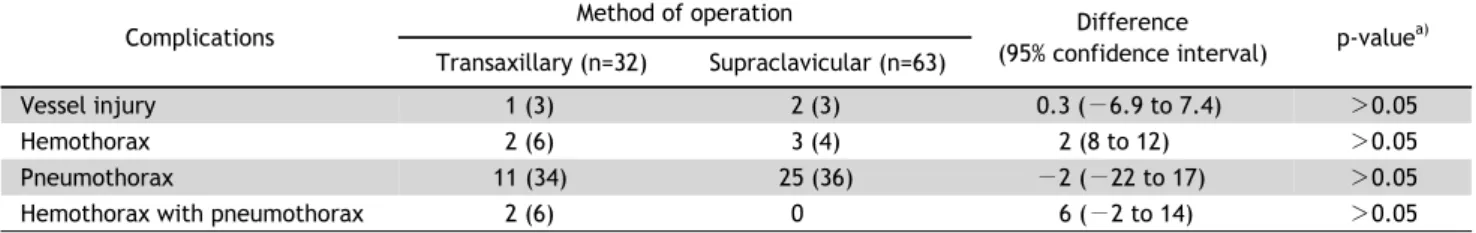
Table 2. Surgical complications other than brachial plexus Complications
Method of operation Difference
(95% confidence interval) p-value
a)Transaxillary (n=32) Supraclavicular (n=63)
Vessel injury 1 (3) 2 (3) 0.3 ( −6.9 to 7.4) >0.05
Hemothorax 2 (6) 3 (4) 2 (8 to 12) >0.05
Pneumothorax 11 (34) 25 (36) −2 (−22 to 17) >0.05
Hemothorax with pneumothorax 2 (6) 0 6 ( −2 to 14) >0.05
Values are presented as number (%), unless otherwise stated.
a)
Based on the Fisher exact test.
Table 3. Brachial plexus complications Complications
Method of operation Difference (95%
confidence interval) p-value
a)Transaxillary (n=32) Supraclavicular (n=63)
Transient paralysis due to T1 root compression 2 (6) 3 (4) 2 ( −8 to 12) >0.05
Permanent paralysis due to T1 root compression 1 (3) 0 3 ( −3 to 9) >0.05
Remaining pain and symptoms after 6 months (failure rate and need for reoperation)
8 (25) 0 −22 (7.5 to 36.2) <0.05
Values are presented as number (%), unless otherwise stated.
a)